[level-membership-for-dermatology-category]
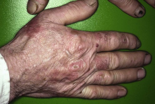
Porphyria cutanea tarda
Specific investigations


First-line therapies
 Serial phlebotomies
 Chloroquine, hydroxychloroquine
Second-line therapies
 Deferoxamine (desferrioxamine)
 Erythropoietin
Third-line therapies
 Antiretroviral therapy
 Interferon-α
 Vitamins E and C
 Plasmapheresis, plasma exchange
 High-flux hemodialysis
 Enteric sorbents (cholestyramine, activated charcoal)
 Metabolic alkalinization by oral sodium bicarbonate
 Cimetidine
 Photothermolysis
Removal of plasma porphyrins with high-flux hemodialysis in porphyria cutanea tarda associated with end stage renal disease.
Carson RW, Dunnigan EJ, DuBose TD Jr, Goeger DE, Anderson KE. J Am Soc Nephrol 1992; 2: 1445–50.
High-flux hemodialysis may remove porphyrins more effectively than conventional hemodialysis.
[/level-membership-for-dermatology-category][not-level-membership-for-dermatology-category]
Porphyria cutanea tarda
