Published on 18/03/2015 by admin
Filed under Dermatology
Last modified 22/04/2025
This article have been viewed 3148 times
Agustin Martin-Clavijo and John Berth-Jones
Evidence Levels: A Double-blind study B Clinical trial ≥ 20 subjects C Clinical trial < 20 subjects D Series ≥ 5 subjects E Anecdotal case reports
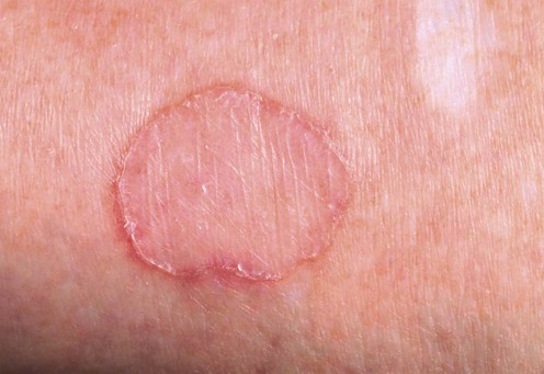
The porokeratoses are a group of disorders of keratinization characterized by lesions with a peripheral keratotic ridge, manifesting histologically as a cornoid lamella. Terminology and classification are debated, but the main recognized forms are: (1) disseminated forms, of which disseminated superficial actinic porokeratosis (DSAP) is predominant; (2) porokeratosis of Mibelli (PM); (3) palmoplantar porokeratosis (porokeratosis palmaris, plantaris et disseminata – PPPD); (4) linear porokeratosis (LP). An autosomal dominant mode of inheritance has been reported in the disseminated form. Overexpression of the p53 tumor suppression protein has been identified in the cornoid lamella. Porokeratotic lesions are progressive and carry malignant potential, especially large long-standing lesions and the linear variants. In addition, the lesions can cause pruritus and represent a cosmetic problem for some patients.
The family history should be reviewed and the patient’s immune function assessed, particularly with the disseminated forms. Discontinuation of immunosuppression has led to resolution of lesions in some patients.
Treatment of porokeratoses may be indicated, not only for cosmetic benefit and symptomatic relief, but also to reduce the risk of malignancy. Optimal therapy is dependent on the type and extent of porokeratosis, and the level of concern over malignant progression. Management should include avoidance of irradiation (UV or X-rays) and observation for signs of malignant transformation (squamous cell carcinoma, basal cell carcinoma, Bowen’s disease).
The lesions are usually asymptomatic. When present, pruritus associated with disseminated lesions is often responsive to topical corticosteroids. The palmoplantar variant may cause functional disability due to pain and discomfort.
Localized disease responds to ‘surgical’ methods such as cryotherapy, CO2 laser, curettage and cautery, or excision, but these can result in significant scarring, especially when the lesions are numerous.
Topical 5-fluorouracil, imiquimod, and vitamin D analogs can be helpful, but only partial responses are likely in DSAP. Inflammatory reactions are likely when using 5-fluorouracil or imiquimod and indicate a greater likelihood of response. With some caution, these modalities can be used under occlusion, treating one area at a time. In the authors’ experience results are inconsistent, even with occlusion.
Systemic retinoids have been effective in localized and systemic disease, but there have been reports of exacerbation of pre-existing lesions. Recurrence is common on discontinuation of therapy, and a long-term maintenance dose may be required. This modality might also reduce the risk of malignant transformation.
There is one report of genital DSAP responding partially to topical diclofenac, but a subsequent case series demonstrated very limited benefit. There are also reports of the effectiveness of topical retinoids, dermabrasion, pulsed dye laser, Nd : YAG laser, corticosteroids, and topical photodynamic therapy. Treatments used in combination have included CO2 laser and tacalcitol, CO2 laser and photodynamic therapy, and 5-fluorouracil and imiquimod.
Skin biopsy
Dermoscopy
Assessment of immune function
Delfino M, Argenziano G, Nino M. J Eur Acad Dermatol Venereol 2004; 18: 194–5.
Dermoscopic examination of DSAP showed a characteristic central scar-like area with a single or double ‘white track’ structure at the margin. The histopathologic correlate of the linear structure was shown to be the cornoid lamella.
Dereli T, Ozyurt S, Osturk G. J Dermatol 2004; 31: 223–7.
Eight patients with 20 lesions received treatment with 30-second cycles of cryospray followed by sharp dissection of the lesion border. Most lesions resolved after one treatment; two required one further treatment.
Limmer BL. Arch Dermatol 1979; 115: 582–3.
Twenty-one lesions of porokeratosis in 11 patients were treated with cryotherapy, resulting in a cure rate of 90.5%. The lesions were pared prior to treatment. There was no evidence of recurrence over an average follow-up period of 22 months.
Gonçalves JC. Arch Dermatol 1973; 108: 131–2.
Six patients with facial lesions were treated with 5% fluorouracil ointment three times daily. The treatment was maintained for 8 to 10 days after a strong inflammatory response occurred. There was no recurrence at 9-month follow-up.
Shelley WB, Shelley ED. Cutis 1983; 32: 139–40.
Resolution of porokeratosis was observed after 3 weeks of daily application of 5% 5-fluorouracil cream. There was no recurrence at 5-month follow-up.
Agarwal S, Berth-Jones J. Br J Dermatol 2002; 146: 338–9.
A 3 cm lesion of PM on the leg was initially treated with topical imiquimod 5% cream five times a week for 3 months, with no improvement. Subsequent treatment with imiquimod 5% cream five times a week under occlusion with an adhesive polythene dressing was successful. There was no recurrence at 1 year.
Arun B, Pearson J, Chalmers R. Clin Exp Dermatol 2011; 3: 509–11.
One patient with DSAP responded to imiquimod 5% cream five times a week for 6 weeks.
Bohm M, Luger TA, Bonsmann G. J Am Acad Dermatol 1999; 40: 479–80.
DSAP responded to 5 months of topical daily treatment with 0.0004% tacalcitol and remained clear on alternate-day maintenance therapy.
Harrison PV, Stollery N. Clin Exp Dermatol 1994; 19: 95.
Three patients were treated with topical calcipotriol daily for 6 to 8 weeks. An overall improvement of 50–75% was noted and maintained for up to 6 months in two patients.
Garg T, Ramchander Varghese B, Barara M, Nangia A. Dermatol Online J 2011; 17: 3.
A patient with generalized LP was treated with acitretin 0.5 mg/kg. There was marked flattening of the lesions after 6 weeks, but not complete resolution after 5 months of treatment.
Bundino S, Zina AM. Dermatologica 1980; 160: 328–36.
Two patients with DSAP were treated with 50–75 mg daily of etretinate, with significant clinical improvement after 5 weeks; 25 mg daily was sufficient to maintain the results. Recurrence was observed 3 to 4 weeks after cessation of treatment.
Kariniemi A, Stubb S, Lassus A. Br J Dermatol 1980; 102: 213–14.
Treatment with 50–100 mg daily of etretinate led to significant clinical improvement and resolution of pruritus within 40 days. A dose of 25 mg on alternate days was required to maintain the results. After 6 months of treatment the patient developed follicular hyperkeratosis with tiny keratin horns on the skin of both forearms.
McCallister RE, Estes SA, Yarbrough CL. J Am Acad Dermatol 1985; 13: 598–603.
A patient with familial PPPD received treatment with 1 mg/kg daily of isotretinoin. Significant clinical improvement was noted after 3 months of treatment. Two months after discontinuation of treatment a gradual recurrence was observed.
Grover C, Goel A, Nanda S, Khurana N, Reddy BS. J Dermatol 2005; 32: 1000–4.
One patient with LP was treated with topical tretinoin and 5-fluorouracil. Both agents were efficacious, but tretinoin was better tolerated.
Agrawal SK, Gandhi V, Madan V, Bhattacharya SN. Int J Dermatol 2003; 42: 919–20.
Once-daily application of topical tretinoin 0.1% gel led to resolution of lesions within 4 months.
Rabbin PE, Baldwin HE. J Dermatol Surg Oncol 1993; 19: 199–202.
CO2 vaporization resulted in better cosmetic and functional improvement than split skin grafting in one patient.
Merkle T, Hohenleutner U, Braun-Falco O, Landthaler M. Clin Exp Dermatol 1992; 17: 178–81.
CO2 laser vaporization of mainly flexural lesions resulted in regression of lesions with slight atrophic scarring and lessening of pruritus. No further treatment was required in a 12-month follow-up period.
Liu HT. Dermatol Surg 2000; 26: 958–62.
A patient’s face and arms were treated four times, 1 month apart, resulting in marked improvement, but not complete clearance of the lesions.
Alster TS, Nanni CA. Cutis 1999; 63: 265–6.
This is a case of LP that responded to a series of 585 nm pulsed dye laser treatments.
Lolis MS, Marmur ES. J Cosmet Laser Ther 2008; 10: 124–7.
This patient received three treatments with Q-switched ruby laser (694 nm) with good improvement.
Chrastil B, Glaich AS, Goldberg LH, Friedman PM. Arch Dermatol 2007; 143: 1450–2.
Two patients received three to six courses of fractioned photothermolysis (erbium-doped fiber laser). Both patients reported 50% improvement.
Cavicchini S, Tourlaki A. J Dermatol Treat 2006; 17: 190–1.
This case demonstrated a striking improvement in response to two treatments, 1 week apart, using methyl aminolevulinate cream 160 mg/g applied with occlusion for 3 hours before illumination with a red light (Aktilite) 37 J/cm2.
Nayeemuddin FA, Wong M, Yell J, Rhodes LE. Clin Exp Dermatol 2002; 27: 703–6.
A report on three patients treated with 20% aminolevulinic acid cream under occlusion for 5 hours prior to illumination with 100 J/cm2 of broadband red light (Waldmann 1200). Resolution of two lesions of DSAP was observed in one case, but the response could not be reproduced in this or two other cases.
Spencer JM, Katz BE. Arch Dermatol 1992; 128: 1187–8.
No recurrence observed at 15-month follow-up, but the lesion healed with slight hyperpigmentation and mild hypertrophy in a 79-year-old Filipino woman.
Cohen PR, Held JL, Katz BE. J Am Acad Dermatol 1990; 23: 975–7.
An excellent cosmetic result. No recurrence or scarring observed after 8 months.
Teixeira SP, de Nascimento MM, Bagatin E, Hassun KM, Talarico S, Michalany N. Dermatol Surg 2005; 31: 1145–8.
This is a case of DSAP treated with a combination of a 70% glycolic peel and a 5% 5-fluorouracil solution every 2 weeks for 4 months. The result was improvement in the appearance and texture of the treated areas and reduced dyskeratosis and epidermal atypia.
Verma KK, Singh OP. J Dermatol Sci 1994; 7: 71–2.
A familial case of progressive porokeratosis received pulses of 100 mg dexamethasone in 5% dextrose intravenously on 3 consecutive days in a month. No new lesions appeared after the first pulse, and clinical improvement was noted after four pulses. There was an 80% improvement after 18 pulses. The patient was then lost to follow-up.
Marks S, Varma R, Cantrell W, Chen SC, Gold M, Muellenhoff M, Elewski B. J Eur Acad Dermatol Venereol 2009; 23: 42–5.
An open label, multicenter trial; 17 patients were treated with 3% diclofenac applied twice daily for 3 to 6 months to a target area. The areas treated progressed less than the untreated areas.
Vlachou C, Kanelleas A, Martin-Clavijo A, Berth-Jones J. J Eur Acad Dermatol Venereol 2008; 22: 1343–5.
Eight patients with DSAP were treated with 3% diclofenac gel (Solaraze gel) twice daily for at least 6 months. At 6 months a partial improvement was seen in two cases, but no improvement in the others.
Venkatarajan S, LeLeux TM, Yang D, Rosen T, Orengo I. Dermatol Online J 2010; 16: 10.
A patient with PM was treated with 5-fluorouracil twice a day and 5% imiquimod cream once a day for 12 weeks, leading to resolution of the lesion.
Kim HS, Baek JH, Park YM, Kim HO, Lee JY. Ann Dermatol 2011; 23: S211–13.
Two patients were treated with CO2 laser to the cornoid lamella followed by photodynamic therapy. There was marked response after four treatments.
Treatment of Skin Disease Comprehensive Therapeutic Strategies 4e
WhatsApp us


 Cryotherapy
 5-Fluorouracil
 Imiquimod
 Vitamin D3 analogs
 Systemic retinoids
 Topical retinoids
 CO2 laser
 Nd : YAG laser
 Pulsed dye laser
 Ruby laser
 Fractional photothermolysis
 Photodynamic therapy
 Dermabrasion
 Fluor-hydroxy pulse peel
 Corticosteroids
 Diclofenac 3% gel
 5-Fluorouracil with imiquimod cream
 Photodynamic therapy with CO2 laser