[level-membership-for-dermatology-category]
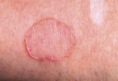
Porokeratoses


 Cryotherapy
Second-line therapies
 5-Fluorouracil
 Imiquimod
 Vitamin D3 analogs
Third-line therapies
 Systemic retinoids
 Topical retinoids
 CO2 laser
 Nd : YAG laser
 Pulsed dye laser
 Ruby laser
 Fractional photothermolysis
 Photodynamic therapy
 Dermabrasion
 Fluor-hydroxy pulse peel
 Corticosteroids
 Diclofenac 3% gel
 5-Fluorouracil with imiquimod cream
 Photodynamic therapy with CO2 laser
[/level-membership-for-dermatology-category][not-level-membership-for-dermatology-category]
Porokeratoses
[level-membership-for-dermatology-category]
[/level-membership-for-dermatology-category][not-level-membership-for-dermatology-category]
