Published on 19/03/2015 by admin
Filed under Dermatology
Last modified 22/04/2025
This article have been viewed 3195 times
Miguel Sanchez
Evidence Levels: A Double-blind study B Clinical trial ≥ 20 subjects C Clinical trial < 20 subjects D Series ≥ 5 subjects E Anecdotal case reports
Pinta (carate, azul, mal de pinto, empeines, lota, tina) and yaws (pian, frambesia, parangi, paru, buba) are non-venereal ‘endemic’ spirochetal infections caused respectively by Treponema carateum and Treponema pallidum subspecies pertenue. Pinta was almost exclusively found in inhabitants of rural, overcrowded, poverty-stricken regions of Mexico, the Caribbean, and the northern part of South America, and has been reported most recently from scattered areas in the Brazilian rainforest. Whereas yaws was prevalent in indigent persons living in tropical, rural, medically under-served areas with high humidity and rainfall within Central Africa, Southeast Asia, Central and northeast South America and some Pacific Islands, outbreaks have recently been reported from Papua New Guinea and Guyana. Most patients are children and young adults who acquire the infections by direct contact of abraded skin with another person’s exudative infected lesions.
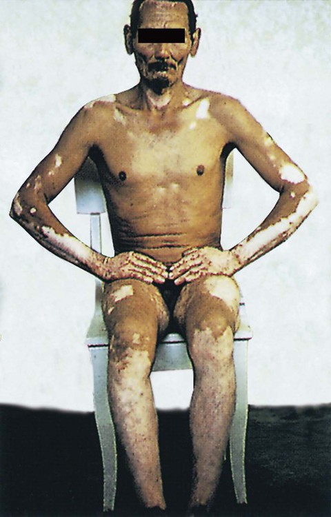
Pinta and yaws are very rare, and familiarity with the types of lesion is essential to differentiate them from syphilis, psoriasis, leprosy, and leukodermas, in order to establish an appropriate treatment plan. As in syphilis, both infections have three distinct clinical stages. In pinta signs or symptoms are limited to the skin and lymph nodes, but yaws can also affect the skeletal system and mucous membranes. The primary stage of pinta develops after an incubation period of 15 days to months (usually 2 to 4 weeks). Following exposure, one to three erythematous papules erupt, usually on the face or extremities, and grow into erythematous scaly plaques that may become hypochromic or light blue in the center. Non-tender regional lymphadenopathy may appear. The secondary stage of pinta usually follows within 2 to 5 months (sometimes years later), with the appearance of erythematous papules (pintids) that enlarge to form psoriasiform plaques, which may remain for years. The plaques, which may be annular or circinate, progress through a range of colors from copper-brown to slate blue or black. Some may be hypochromic. Lymphadenopathy may be present. The tertiary stage is characterized by depigmented patches on the wrists, ankles, elbows, and within old lesions. These develop between 3 months and 10 years after the onset of the secondary stage. At this point patients have a combination of hyperpigmented, hypochromic, achromic, dyschromic, and polychromic patches of different sizes, imparting a mottled appearance to the skin. In 80% of cases serologic tests become reactive two to three months after the onset of the primary lesion, and are always reactive in late lesions.
In yaws the primary stage develops after an incubation period of 10 days to 3 months, with the appearance, usually on a lower extremity, of an erythematous, occasionally pruritic papulonodule (the ‘mother yaw’) that enlarges up to 5 cm in circumference and ulcerates with exuberant granulation tissue imparting a framboesiform appearance. The secondary stage usually ensues over 10 to 16 weeks, but may be as long as 2 years after the onset of the primary stage, with an eruption of reddish, weeping, crust-covered papules (‘daughter yaws’) similar to, but smaller than, the primary lesion. In some patients the clinical findings are similar to those of secondary syphilis, with scaly papules and plaques, hypertrophic condyloma lata resembling lesions on body folds, or mucous patches such as lesions on mucous membranes. Nodules around the joints are common. Some patients suffer with painful osteoperiostitis of the forearm or leg and polydactylitis of the hand or foot. In approximately 10% of infected patients the disease progresses to the tertiary stage with infiltrated plaques and nodules that ulcerate, leaving deep ulcers with raised granulomatous edges. Skeletal changes include chronic hypertrophic osteoperiostitis which most commonly affects the tibiae (saber shins) or the superior nasal processes of the maxillae. This latter process triggers disfiguring progressive exostosis of new bone (goundou) which, in 5 to 20 years, results in massive destruction and perforation of the nose and the palate (rhinopharyngitis mutilans or gangosa).
The recommended treatment of pinta and yaws is a single intramuscular injection of 1.2 million units of benzathine penicillin G in adults, adolescents, and older children, and 0.6 million units in children under 10 years of age. Patients cease to be infectious within 24 hours. In pinta primary and secondary lesions heal in 4 to 12 months, but achromic lesions persist indefinitely. Penicillin-allergic patients over 8 years of age are treated with a 15-day course of tetracycline 250 mg four times daily or doxycycline 50 mg twice daily. Erythromycin should be reserved for penicillin-allergic children under 8 years of age (8 mg/kg four times daily) and for pregnant women (500 mg four times daily).
Serologic tests (cardiolipin flocculation assays, treponemal-specific assays)
Dark-field microscopy
Direct fluorescent antibody test
Histopathology
Konan YE, M’Bea KJ, Coulibaly A, Tetchi EO, Kpebo DO, Ake O, et al. Sante Publique 2007; 19: 111–18.
The presence of treponemes on the serous exudate of lesions examined under dark-field microscopy with negative direct fluorescent antibody test which specifically detects T. pallidum suggests non-venereal treponematoid infection. No serological test is yet able to distinguish infection with any of the endemic treponematoses from each other or from venereal syphilis.
Antal GM, Lukehart SA, Meheus AZ. Microbes Infect 2002; 4: 83–94.
Available serologic tests cannot distinguish yaws or pinta from syphilis. Penicillin treatment often does not lead to seroreversal. Some cases of yaws and pinta may be misdiagnosed as syphilis.
Pecher SA, Azevedo EB. Med Cutan Iber Lat Am 1987; 15: 239–42.
Treponemes are found on silver impregnation between epidermal cells in primary, secondary, and late-stage hyperpigmented lesions, but not in late hypopigmented patches.
Harper KN, Liu H, Ocampo PS, Steiner BM, Martin A, Levert K, et al. FEMS Immunol Med Microbiol 2008; 53: 322–32.
Only minor genetic differences have been found between the subspecies that cause venereal syphilis and yaws, after completion of the T. pallidum genome project. A sequence variation in the arp gene allows differentiation of ssp. pallidum from the non-venereal subspecies.
Smais D, Norris SJ, Weinstock GM. Infect Genet Evol 2012; 12: 191–202.
Genome comparisons between pallidum and non-pallidum treponemes revealed genes with potential involvement in human infectivity, whereas comparisons between pallidum and pertenue treponemes identified genes possibly involved in the high invasivity of syphilis.
Genome analyses also shed light on treponemal evolution and on chromosomal targets for molecular diagnostics of treponemal infections.
WHO Scientific Group. Technical Report Series No. 674. Geneva: World Health Organization, 1982.
Intramuscular benzathine penicillin is the recommended treatment for all patients with pinta or yaws. All household cases and contacts should also be treated in areas where 5% of the population is infected. In areas with 5–10% rates of infection, treatment should be administered to all children under 15 years of age; in areas with higher infection rates the entire population should be treated with penicillin.
Ketchen DK. Ann NY Acad Sci 1952; 55: 1176–85.
This study on Mexican patients with pinta found that, after treatment with penicillin, skin lesions healed completely in all patients with primary lesions, in 69% with secondary lesions, and in 40% with tertiary lesions.
Engelkens HJ, Vuzevski VD, Stolz E. Clin Dermatol 1999; 17: 143–52.
This review stresses that penicillin at previously recommended doses remains the treatment of choice.
Backhouse JL, Hudson BJ, Hamilton PA, Nesteroff SI. Am J Trop Med Hyg 1998; 59: 388–92.
Of 39 children, 28% developed clinical and/or serologic evidence of relapse after treatment with the recommended dose of intramuscular benzathine penicillin. All but three responded to further penicillin treatment. Resistance of yaws to penicillin has also been reported from Ecuador.
Scolnik D, Aronson L, Lovinsky R, Toledano K, Glazier R, Eisenstadt J, et al. Clin Infect Dis 2003; 36: 1232–8.
Clinical cure of yaws lesions was achieved in 94% of 17 children with administration of oral penicillin, 50 mg/kg daily in four divided doses for 7 to 10 days.
Benzathine penicillin G treatment may not be possible in remote areas as the drug requires refrigeration at a temperature of 2–8°C until 7 days before use, at which time it should be stored at <30°C.
Mass treatment programs designed to eradicate endemic trepanematoses expose uninfected people to adverse effects and may promote antibiotic resistance. Treatment programs that exclusively target clinically active cases can significantly reduce the prevalence of disease.
Morand JJ, Simon F, Garnotel E, Mahe A, Clity E, Morlain B. Med Trop 2006; 66: 15–20.
The infection is highly sensitive to penicillin and resistance has not been reported. Endemic control through treatment of the entire treponemal reservoir with single-dose penicillin is highly successful.
Brown ST. Rev Infect Dis 1985; 7: 318–26.
This article reviews studies of treatment for pinta and yaws. Penicillin is the drug of choice. Tetracycline is also concluded to be effective, but studies were done on patients with yaws. Reports of treatment with erythromycin were few. Doxycycline and minocycline appear to be useful alternatives.
Koff AB, Rosen T. J Am Acad Dermatol 1993; 29: 519–35.
Tetracycline or doxycycline is the treatment of choice in older children and adults who are allergic to penicillin.
Farnsworth N, Rosen T. Clin Dermatol 2006; 24: 181–90.
Penicillin remains the drug of choice, but the tetracycline class of antibiotics and erythromycin also appear to be effective.
Asiedu K, Amouzou B, Dhariwal A, Karam M, Lobo D, Patnaik S, et al. Bull WHO 2008; 86: 499A.
Despite once successful attempts to eradicate yaws through campaigns that involved active case finding, treatment of cases and contacts, and community mobilization, yaws is re-emerging in poor, rural, and marginalized populations of Africa, Asia, and South America.
Treatment of Skin Disease Comprehensive Therapeutic Strategies 4e
WhatsApp us


 Penicillin
 Tetracycline
 Doxycycline
 Minocycline
 Erythromycin