Physiologic determinants of cardiac output

Published on 13/02/2015 by admin

Filed under Anesthesiology

Last modified 22/04/2025

Print this page

rate 1 star rate 2 star rate 3 star rate 4 star rate 5 star
Your rating: none, Average: 1 (1 votes)

This article have been viewed 1851 times

Physiologic determinants of cardiac output

Amorn Vijitpavan, MD

Cardiac output (CO) is the quantity of blood that the heart pumps per minute. CO in a normal 70-kg individual with a heart rate (HR) of 70 to 80 beats/min is 5 to 6 L/min, but it decreases by approximately 25% when the individual is resting in the supine position and may increase approximately eightfold with exertion. The cardiac index (CI) normalizes a person’s CO for body surface area (BSA):

< ?xml:namespace prefix = "mml" />CI (L·min1·m2)=CO/BSA

image

A normal CI varies between 2.5 and 3.5·L·min-1·m-2. The two major determinants of CO are stroke volume (SV) and HR:

CO (L/min)=SV×HR

image

Stroke volume

The SV is the amount of blood ejected by the ventricle with each contraction. A normal SV is 70 to 80 mL. Determinants of SV include preload, afterload, and contractility.

Preload

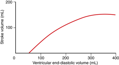
Preload is directly proportional to end-diastolic myocardial fiber length, often represented as end-diastolic volume (EDV, with a normal value of ∼120 mL). During normal cardiac muscle contraction, the sarcomere length is relatively short, but if EDV increases, the length of the sarcomere increases, and during subsequent contractions, more force is generated, the maximum rate of pressure development (dP/dt) increases, and SV increases proportionately (Figure 34-1).

Many factors affect preload, including primarily venous tone (which, in turn, is affected by total blood volume), intrathoracic pressure, body position, pulmonary vascular resistance, and atrial contraction. Impaired venous return (from, for example, peripheral vasodilation, hemorrhage, positive-pressure ventilation) decreases SV and CO. Although total blood volume is important to preload, the distribution of that blood volume between the extrathoracic and intrathoracic compartments is more important. The venous system has a very high capacity, and assuming no loss of volume from dehydration or hemorrhage, a large amount of blood can be shifted centrally from the periphery.

Ventricular volume can be measured using multiple techniques, including echocardiography, angiography, and scintigraphy, but, except for echocardiography, most are not helpful for clinically managing a patient. Transesophageal echocardiography is useful perioperatively to estimate EDV, but it has multiple limitations. Therefore, left ventricular end-diastolic pressure (LVEDP) is frequently used as a surrogate for EDV on the basis of a nonlinear, end-diastolic, pressure-volume relationship. More commonly, left atrial pressure, pulmonary artery occlusion pressure (PAOP), right atrial pressure (RAP), or central venous pressure (CVP) is used to estimate LVEDP and LVEDV.

The reliability of these cardiac pressures in estimating ventricular preload depends on ventricular compliance, the integrity of the cardiac valves, and intrathoracic pressure. Ventricular compliance (the distensibility of the chamber in response to changes in volume) is affected by coronary ischemia, ventricular hypertrophy, pericarditis, and cardiac tamponade, among others, all of which can result in a weak correlation between pressure and volume of the left ventricle. If compliance is decreased, then small increases in ventricular volume may be associated with large increases in ventricular pressure. PAOP and pulmonary artery pressure are most commonly used to estimate LV preload; CVP provides the poorest estimation of LV preload.

Afterload

Afterload is defined as the impedance to ejection, the force that resists muscle shortening during myocardial contraction. Systemic vascular resistance (SVR) accounts for about 95% of the resistance to ejection (the remainder is due to characteristics of the left ventricle and the LV outflow tract and aortic valve) and is often used clinically to estimate afterload.

SVR=80×(MAPRAP)/CO

image

where MAP is the mean arterial pressure and RAP is the right atrial pressure. Normal SVR is 900 to 1500 Dynes∙sec-1∙cm-5. Wood units are also used, most commonly in measuring pulmonary vascular resistance, and are calculated using the same equation but without multiplying ((MAP − RAP)/CO) by 80. Blood pressure is a poor estimation of afterload.

Afterload, as defined by ventricular wall stress, is represented by the Laplace law:

T=Pr/2h

image

where T is the tension in the LV wall, P is pressure, r is the radius, and h is wall thickness. From the Laplace law, it is apparent that ventricular volume, LV wall thickness, and systolic intraventricular pressure are primary determinants of afterload.

Intraventricular pressure has an important effect on afterload. A dilated thin-walled ventricle generates significantly greater wall stress than does a thicker-walled smaller ventricle. A failing ventricle will dilate and significantly increase afterload, which significantly reduces CO. Reducing afterload is an important goal in managing congestive heart failure.

Contractility

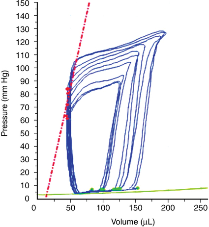
Contractility refers to the intrinsic ability of the myocardium to generate force at given end-diastolic fiber length and is closely related to the availability of intracellular calcium. Contractility is relatively easy to understand conceptually but difficult to define; measurements of cardiac performance include the dP/dt (Figure 34-2), isolated papillary muscle shortening, and the work generated by isolated or whole-heart preparations, but these definitions are not clinically useful.

No specific value represents normal contractility. Contractility may be assessed through echocardiography, angiography, and scintigraphy. A more clinically useful index of contractility is ejection fraction, the slope of the plot of SV against EDV. Although affected by changes in preload, afterload, and HR, ejection fraction is one of the most reliable and sensitive parameters of ventricular performance.

A change in contractility is considered to be a change in the contractile force of the heart in the presence of unchanged diastolic dimensions and pressure. Thus, it is a change in the myocardial force-velocity relation. Catecholamines, digitalis, and calcium ions increase contractility. The adrenergic nervous system exerts the most important influence on contractility. Hypoxia, acidosis, ischemia, and certain drugs (e.g., calcium channel blockers and β-adrenergic receptor blocking agents) decrease contractility.