Percutaneous Neuromodulation Therapy

Published on 06/06/2015 by admin

Filed under Physical Medicine and Rehabilitation

Last modified 22/04/2025

Print this page

rate 1 star rate 2 star rate 3 star rate 4 star rate 5 star
Your rating: none, Average: 0 (0 votes)

This article have been viewed 2915 times

16 Percutaneous Neuromodulation Therapy

After using his own version of acupuncture for several years to treat complaints ranging from migraine headache to alcohol withdrawal to cancer pain, Craig decided in the late 1970s that he would develop a new system based on his belief that the effectiveness of electroacupuncture depended on its influence on the central nervous system. The new system was to be based on placing needles into the vicinity of nerves that conducted impulses away from the affected areas. The earliest recorded workshops called his technique “dermatomal electroacupuncture”.

The system evolved through the years as Craig manipulated the key components of treatment, fine-tuning such important aspects as needle location, overall montage of needles employed, and stimulus frequency and duration. In parallel, other scientists advanced our understanding of the neurophysiology of chronic pain, elucidating the role of various neurotransmitters and the impact of electrical stimulation on the behavior of various nerve pathways. By the mid-1990s, dermatomal electroacupuncture had become percutaneous electrical nerve stimulation (PENS), which soon became Craig-PENS to honor Craig’s contribution. Besides indicating what the technique involved (percutaneous stimulation of nerves with electrical impulses), PENS emphasized the demarcation of this technique from acupuncture. Many practitioners continue to use PENS or Craig-PENS to describe what they do.

A new millennium demanded a new acronym. At the turn of the century, Craig and colleagues renamed the technique Percutaneous Neuromodulation Therapy (PNT). This new moniker moved beyond description to explanation. PENS defines the technical performance of the method, but PNT explicates the neurophysiology behind why it works. Moreover, a new term was needed to distinguish an effective chronic pain treatment from a neurosurgical procedure involving implantable stimulating electrodes. This chapter will discuss PNT as developed by Craig and his associates.

A word about acupuncture: although Craig studied acupuncture and initially called his technique dermatomal electroacupuncture, PNT should be distinguished from electroacupuncture. Unlike electroacupuncture, PNT does not involve needling of specific, traditional acupuncture points. In fact, as will be described later, placement of needles in PNT intends to influence dermatomal (or myotomal or sclerotomal) nerve pathways rather than acupuncture meridians. And PNT does not rely on the concept of “qi” or require any sort of traditional Chinese medicine (TCM) diagnosis. However, like electroacupuncture, PNT does involve needle insertion and electrostimulation, so the results of neurophysiologic studies of electroacupuncture also reflect the neuromodulatory impact of PNT. The importance of differentiating PNT from electroacupuncture lies here: one need not undertake the study of acupuncture to to understand and practice PNT effectively.

How PNT Works

As its name suggests, PNT is believed to work by modulation of central pain pathways using peripheral nerve stimulation. A body of neurophysiology research supports this presumption, and ongoing work continues to reveal details of the complex interactions that underlie the perception of pain.

Peripheral nerve endings known as nociceptors sense pain and transmit signals to the dorsal root ganglion of the spinal cord. Localized, sharp, stinging pain, generally of short duration, reaches the cord by way of thicker, myelinated A-delta fibers. Dull, aching, burning pain, longer in duration and slower in onset, travels along smaller, nonmyelinated C fibers. When the pain signal reaches the dorsal horn, various neurotransmitters activate spinal neurons that send axons across the spinal cord and up fiber tracts that eventually reach the medulla, midbrain, and thalamus. From the thalamus, neurons project to the prefrontal cortex, the motor cortex, the somatosensory cortex, the limbic system, and the hypothalamus. Pain information also finds its way to the pons, cerebellum, and basal ganglia through pathways that remain to be fully elucidated.

Besides the modulation that can be brought about during the ascent of pain signals, descending inhibitory pathways activated in the midbrain, brainstem, and numerous other areas (among them the amygdala, the hypothalamus, the arcuate nucleus, and the periaqueductal gray matter) can modify the transmission of these impulses. PNT aims to influence the balance within this very complex network, enhancing the inhibitory drives and suppressing the excitatory message.

The transmission of pain signals throughout these pathways depends on the balance of neurotransmitters whose release depends, in turn, on a variety of factors, including the frequency of stimulation of the peripheral nerves. Rather than reviewing the various forms of gate theory, originally proposed by Melzack and Wall,1 we will focus here on the influence of different frequencies of electrostimulation on neurotransmitter release in the central nervous system because this neuromodulation underlies the therapeutic effectiveness of PNT. Much of this knowledge derives from electroacupuncture and neurosurgery research, as noted earlier.

Early work by Han and colleagues from Beijing Medical University demonstrated that analgesia induced by electroacupuncture of different frequencies is mediated by different opioid receptors.2 Analgesia induced by low-frequency (2 Hz) electroacupuncture resulted from activation of mu- and delta-opioid receptors, whereas analgesia induced by high-frequency (100 Hz) electroacupuncture derived from activation of kappa-opioid receptors. Intermediate frequency stimulation (2 to 15 Hz) activated all three types of receptors in the spinal cord of rats.

Further work by Han and colleagues3 showed that low-frequency (2 Hz) peripheral stimulation produces a significant increase in enkephalin release into the lumbar cerebrospinal fluid (CSF), whereas high-frequency (100 Hz) peripheral stimulation increases dynorphin release. This validated earlier work by Mayer and colleagues4 and Pomeranz and Chiu.5 Wang and associates went on to establish that a more potent analgesia could be established by asynchronous electroacupuncture stimulation—alternating between low-frequency (2 Hz) and high-frequency (100 Hz) stimulation—than could be attained by synchronous electroacupuncture stimulation, combining low and high-frequency stimulation at the same time.6 The clinical application of these findings will become evident later in this chapter.

The endorphins (enkephalin and dynorphin) are only part of an ever-elaborating story of pain neurotransmission. In the last few years, research has suggested a role in electroacupuncture (and, thus, PNT) for cocaine and amphetamine-regulated transcript (CART) peptide,7 arginine vasopressin,8 serotonin, catecholamine, and spinal Fos expression,9 interleukin-1-beta,10 corticotrophin-releasing hormone.11 No doubt more neurotransmitters and neurochemicals will play important parts in the transmission and perception of pain.

In summary, pain perception is complex and incompletely understood. How PNT modulates the perception of pain remains unclear. Nevertheless, based on this knowledge from basic research and the principles realized through careful clinical study, PNT can be used effectively to address a broad range of chronic painful conditions.

The PNT Technique

In a typical treatment session, ten 32-gauge stainless steel acupuncture needles are inserted to a depth of 2 to 4 cm into the soft tissues and/or paraspinous muscles in and around the dermatomal distribution that covers the painful site(s). The 10 needle probes are then connected to five bipolar leads, with each lead connected to one positive and one negative probe in a pattern designed to direct the stimulus along the path of the nerve supplying the dermatome. The probes are then stimulated at an alternating frequency of 15 Hz and 30 Hz for 30 minutes, with the intensity of the stimulation adjusted to produce a gentle tapping sensation without muscle contraction. Treatment sessions can occur once to several times per week. In published trials, treatments have ranged from 2 to 12 weeks, but the overall duration of treatment in the real clinical setting depends on the patient’s response.

The details of the typical treatment session have been defined through a number of clinical trials investigating specific aspects of the treatment. Here we will describe research supporting the location of the needle probes, the stimulation montage, the stimulation frequency used, and the duration of electrical stimulation.

Location of Needles

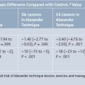
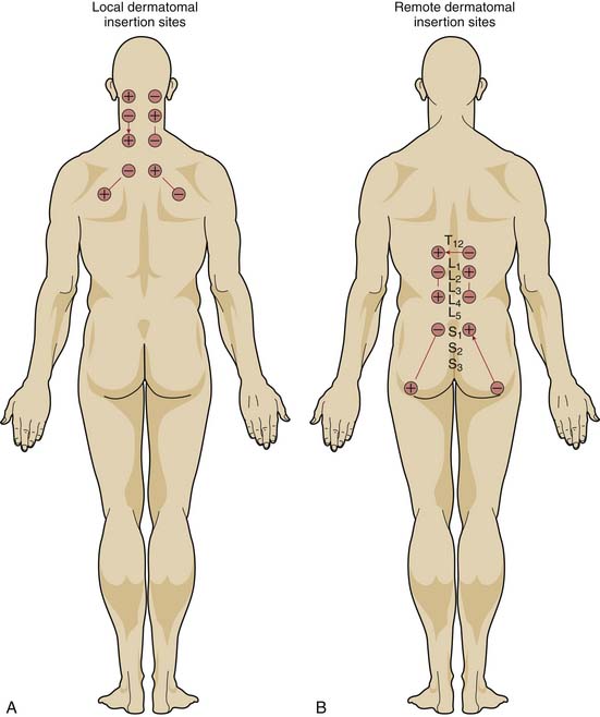
If the mechanism for analgesia relies primarily on increases in analgesic-like neurotransmitters within the central nervous system induced by peripheral nerve stimulation, varying the level of spinal stimulation should yield similar analgesic effects. Alternatively, if central neuromodulatory changes are primarily responsible for the analgesic effects, then stimulation of peripheral nerves in or near the affected area should prove more effective than stimulation of distant peripheral nerves. White, Craig, and others clarified the effect of the location of electrical stimulation on the acute analgesic response to PNT in a crossover study of 68 patients with nonradiating neck pain.12

When PNT was applied to needles located in the dermatomal distribution of the neck pain, visual analog pain scores decreased significantly more than when PNT was applied to needles located in the low back region or when needles were inserted without electrical stimulation. PNT applied to local needles also brought significant improvements in physical activity, quality of sleep, psychological well-being, and the need for oral analgesic medications, compared with PNT applied to distant needles or unstimulated needles. Moreover, unlike the other two treatments, PNT with the local needles showed a cumulative improvement over the course of therapy (Fig. 16-1).

At least in patients with neck pain, therefore, electrical stimulation at the dermatomal levels corresponding to the local pathology provides a greater analgesic effect than does stimulation at remote dermatomes, and electrical stimulation is required to provide the optimal hypoalgesic effect. In order for PNT to be most effective, the practitioner must identify the dermatomes that overlap and surround the painful area and place the needle probes within these dermatomes.

Stimulation Montage

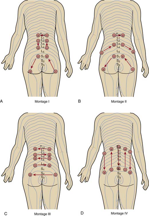
Having placed the needles into the appropriate dermatomes, does it matter how one connects the stimulator leads? In other words, does one specific pattern of electrical stimulation (i.e., montage) provide greater relief than another specific pattern? Work by White and associates demonstrates that montage matters.13

The investigators evaluated the effect of four different montages on the acute analgesic response to PNT when applied at the same dermatomal levels in a crossover study of 72 patients with low back pain (Fig. 16-2). Although all four montages provided significant improvements in pain visual analog scores and in the physical component summary (PCS) and the mental component summary (MCS) of the SF-36, one montage—in which the flow of electrical stimulation paralleled the dermatomes on both sides—produced significantly greater improvements. The cumulative effects over the course of 2 weeks of treatment were also superior with this montage than with the other patterns.

image

Figure 16-2 The needle insertion positions and electrode pairing patterns used for the four different PNT montages. Montage I (A) was the standard montage used in all of the earlier PNT studies involving patients with low back pain.

(Adapted from White PF, Ghoname EA, Ahmed HE, et al: The effect of montage on the analgesic response to percutaneous neuromodulation therapy. Anesth Analg 2001;92:483-487.)

The researchers speculated that the stimulation pattern used with this montage produced more effective electrical stimulation of the involved myotomes and/or sclerotomes and concluded that this montage should be employed in the initial treatment. If this pattern fails, however, they advised resorting to another montage. When initiating PNT, therefore, the best montage employs needles within and around the affected dermatome and electrical stimulation bilaterally between needles in the same dermatome.

Stimulus Frequency

From the animal and human data already described, it would appear that stimulus with alternating frequencies should afford greater pain relief than stimulus with a single frequency. Ghoname and colleagues tested this hypothesis by comparing the effect of three different frequencies on the analgesic response to PNT therapy in 68 patients with low back pain resulting from degenerative lumbar disc disease. Patients were treated with PNT therapy at 4 Hz, alternating 3-second pulses of 15 Hz and 30 Hz, and 100 Hz, as well as sham-PNT (0 Hz), in random order, three times weekly for 2 weeks, with 1 week off between each sequence.

All three frequencies (4 Hz, 15/30 Hz, and 100 Hz) produced significant decreases in pain scores immediately after the treatment and over the 2-week treatment period, as well as significant improvements in physical activity, sleep quality, and need for oral analgesic medications. The overall improvements were significantly greater after 15/30 Hz PNT than after 4 Hz or 100 Hz PNT.

During the 15/30 Hz sequence, patients reported significantly greater improvements in their sense of well-being, compared with the other sequences, and more patients preferred the 15/30 Hz therapy (40%) than preferred the 4 Hz (28%), 100 Hz (30%), or sham (2%) treatments.

Whereas these findings led to the adoption of using a mixed frequency (in this case, alternating 15 Hz and 30 Hz with the stimulation pulses switched on and off every 3 seconds), the optimal combination of frequencies may vary with different patients. The important conclusion is that low and high frequencies in tandem likely stimulate a more diverse mix of inhibitory neurotransmitters than does either the use of a single frequency or the simultaneous application of more than one frequency. PNT using a pair of frequencies applied asynchronously provides optimal pain relief.

Duration of Electrical Stimulation

The needles should be placed within and around the affected dermatome, the stimulus should be applied pair-wise to needles within a single dermatome, and the stimulus should consist of two frequencies (one higher, one lower) used asynchronously. But how long should the electrical stimulation last?

Hamza and coworkers addressed this question in a crossover study of 75 patients with low back pain.14 All patients received electrical stimulation for 0, 15, 30, or 45 minutes in a randomized sequence over the course of an 11-week period (2 weeks of treatment with 1 week off between treatments). The researchers assessed pain scores, health status, sleep quality, and daily oral analgesic requirements.

Electrical stimulation for 15, 30, and 45 minutes significantly improved all measures of pain relief over results obtained with 0 minutes of electrical stimulation. After completion of the sixth treatment, mean improvements in the degree of pain, physical activity, and sleep quality and reductions in the need for oral analgesics were significantly greater with 30- and 45-minute treatment intervals than with the 15-minute treatment interval.

Improvements in the physical and mental components of the health survey (SF-36) were greater after the 30-minute interval than after the 45-minute interval, but the differences did not reach statistical significance. Clearly, however, adding 15 minutes afforded no improvement over the 30 minutes of electrical stimulation. These findings support the use of 30 minutes of PNT, at least in patients with low back pain.

Clinical Applications of PNT

PNT has been used by Craig and others for a host of chronic pain applications. Research to support the use of PNT, however, is sparse. What follows is a selection of indications for PNT, each supported by at least one clinical trial demonstrating efficacy.

Back Pain

Back pain, especially low back pain, is one of the most common medical problems, with about 25% of Americans reporting at least one whole day of low back pain in any given 3-month period.15 Despite this high prevalence of back pain, analgesic therapy remains largely unsatisfactory, and controversy surrounds the effectiveness of such nonpharmacologic therapies as transcutaneous electrical nerve stimulation (TENS), acupuncture, spinal manipulation, and exercise therapy. At least five reports have demonstrated PNT to be safe and effective therapy for low back pain.

Ghoname and colleagues compared the effectiveness of PNT, sham-PNT (needle placement without electrical stimulation), TENS, and flexion-extension exercise therapies in a randomized, crossover study of 29 men and 31 women with at least 3 months of low back pain secondary to degenerative disc disease.16 Each treatment was administered for 30 minutes three times weekly for 3 weeks. Patients had 1 week without therapy between each of the treatments. PNT in this study used a uniform electrical stimulation frequency of 4 Hz. The principal outcome measures included pre- and posttreatment visual analog scale (VAS) scores for pain, physical activity, and quality of sleep; daily analgesic medication usage; a global patient assessment questionnaire; and Health Status Survey Short Form (SF-36).

PNT produced significantly greater improvements in the PCS and MCS components of the SF-36, compared with TENS, sham-PNT, and flexion-extension exercise. PNT improved VAS scores by 46%, physical activity scores by 42%, and sleep quality scores by 44%, whereas TENS improved pain scores by 11% and physical activity scores by 15%. PNT therapy was associated with a 50% reduction in the daily oral analgesic requirement. In comparison, TENS therapy decreased the need for analgesic medication on only 6 days of the 3-week study period. Neither sham-PNT nor exercise therapy provided significant pain relief or altered usage of oral analgesic medication.

PNT produced an acute analgesic effect immediately after each treatment session, and patients began reporting significant improvements in their pain, activity, and sleep after three to four treatment sessions. In the overall evaluation of treatments, 91% of patients preferred PNT, 7% preferred TENS, 2% preferred sham-PNT, and none preferred exercise.

Weiner and associates compared the efficacy of combined PNT and physical therapy (PT) with that of combined sham-PNT and PT for the treatment of chronic low back pain (defined as that occurring daily or almost every day for the previous 3 months) in a randomized, controlled trial of 34 community-dwelling adults aged 65 years and older.17 Patients received PNT or sham-PNT plus physical therapy twice weekly for 6 weeks. These investigators used a structured PNT protocol that allowed incremental stimulation frequencies based on the patient’s response to therapy. The primary outcome measures were pain intensity and pain-related disability, and secondary outcome measures included physical performance, psychosocial factors, and cognitive function.

Unlike the sham-PNT plus PT group, the PNT plus PT group showed significant reductions in pain intensity measures from pre- to posttreatment. The improvements in pain intensity were sustained from posttreatment to the 3-month follow-up visit. Pain-related disability also declined significantly in the PNT plus PT group but did not improve in the sham-PNT plus PT group.

The PNT plus PT group experienced consistent improvements in physical performance and psychosocial functioning, whereas the sham-PNT plus PT group experienced few improvements in physical performance and no significant changes in psychosocial functioning. Neither treatment was associated with significant changes in cognitive performance.

Low back pain is also prevalent in Taiwan, affecting some 80% of the population at some point during their lives. Hsieh and colleagues from National Taiwan University, Taipei, Taiwan investigated the therapeutic effects of one-shot, low-frequency PNT plus medication versus one-shot TENS plus medication or medication only in 133 patients with low back pain. Just over half the patients (56%) had acute low back pain, 20% had subacute low back pain, and 24% had chronic low back pain. Both PNT and TENS employed alternating 3 Hz and 15 Hz stimulation pulses for a total of 15 minutes in a single session. Outcome measures included pain VAS score, body surface score, pain pressure threshold, and the Quebec Back Pain Disability Scale.

PNT and TENS were associated with significant improvements in pain VAS score and body surface score immediately after the one-shot treatments. By 1 week after treatment, the improvements in pain scores and body surface scores did not differ among PNT, TENS, and medication-only groups. Pain pressure threshold did not improve immediately after treatment, and improvements at 1 week posttreatment did not differ among the groups. Quebec Back Pain Disability Scores were most improved after PNT, but the differences among the groups were not significant immediately after treatment or 1 week later. The investigators concluded that one-shot PNT (or TENS) provides only temporary relief of back pain.

Borg-Stein and associates extended the use of PNT to the management of subacute radiating low back pain.18 They investigated the efficacy and safety of 12 weeks of weekly PNT for 30 minutes in an open-label study of 59 patients who had moderate-to-severe radiating low back pain for 4 weeks to 6 months. According to their report, the pulse repetition frequency varied from 4 Hz to 10 Hz in a periodic, sweeping manner. Their outcome measures included VAS scores of radiating pain, low back pain, physical activity, and sleep, as well as the Oswestry Disability Questionnaire.

By the 5-week evaluation, 37 patients (63%) reported at least a 30% decrease in their lower extremity pain and 25 patients (42%) reported at least a 50% improvement. Among all 59 patients, mean leg/buttock pain decreased by 37%, mean low back pain decreased by 26%, activity levels improved by 38%, sleep improved by 27%, and Oswestry Low Back Pain Disability scores improved by 24%. These improvements were sustained between 5 and 12 weeks. By 24 weeks, 83% of 27 patients who granted phone interviews indicated either “very much benefit” or “much benefit,” and 59% said they were either “pain free” or “much better.”

Yokoyama and colleagues from Okayama University Medical School, Okayama City, Japan compared PNT with TENS for long-term pain relief in a study of 60 patients who reported having low back pain with intensity of at least 40/100 on VAS for more than 6 months.19 One group received PNT twice weekly for 8 weeks, one group received 4 weeks of twice-weekly PNT followed by 4 weeks of twice-weekly TENS, and one group received 8 weeks of twice-weekly TENS. In all cases, needles or TENS electrode pads were stimulated with alternating frequencies of 4 Hz and 30 Hz for 20 minutes. Pain level, degree of physical impairment, and daily intake of nonsteroidal antiinflammatory drugs (NSAIDs) were assessed 3 days after week 2, week 4, and week 8 treatments and again at 1 and 2 months after the sessions concluded.

VAS pain scores improved significantly at 2 weeks, 4 weeks, 8 weeks, and at the 1-month post-treatment follow-up in the group treated with 8 weeks of PNT. By the 2-month follow-up, pain scores had returned to pretreatment values. Similarly, VAS pain scores improved significantly at 2 weeks, 4 weeks, and 8 weeks in the PNT-TENS group, but scores had returned to pretreatment values by the 1-month and 2-month follow-up assessments. Only the 8-week pain scores were improved in the group that had 8 weeks of TENS. Peak pain scores were lowest in the group treated with 8 weeks of PNT; intermediate in the group that received 4 weeks of PNT followed by 4 weeks of TENS; and highest in the group that received 8 weeks of TENS. Findings were similar for the degree of physical impairment and the daily intake of oral NSAIDs.

Taken together, these studies demonstrate that PNT can provide relief of pain, improved physical functioning, improved sleep, and decreased need for oral analgesics in patients with chronic low back pain (with or without radiating pain). Because most of these studies have not taken advantage of the fine-tuning granted by the research described earlier (especially regarding montage and asynchronous frequency electrical stimulation), it is likely that greater improvements in pain, functioning, and quality of life can be realized in these patients.

Sciatica

As many as 40% of people experience sciatica (radicular pain in the distribution of the sciatic nerve caused by herniation of one or more lumbar intervertebral discs) in their lifetime.20 Ghoname and coworkers compared PNT, sham-PNT, and TENS for the management of the radicular pain in a crossover study of 64 patients with sciatica due to radiologically-confirmed lumbar disc herniation. PNT and TENS (applied only to the affected leg) employed a stimulation frequency of 4 Hz during 30-minute sessions three times weekly for 3 weeks. All patients received all three modalities, and each set of treatments was separated by a 1-week period free of therapy. The primary outcome measures were the pain VAS and the SF-36. Secondary outcomes included physical activity, sleep quality, sense of well-being, and daily oral analgesic requirements.

PNT provided the greatest improvements in the physical and mental components of the SF-36, although both TENS and sham-PNT produced significant improvements in the scores. Similarly, the average decreases in VAS pain scores immediately after treatment and 24 hours after the last treatment session were significantly greater after PNT than after TENS or sham-PNT. PNT also produced significant improvements in VAS scores for activity and sleep after three to four treatments, as did TENS (but to a lesser degree). The improved sense of well-being and decrease in daily oral analgesic requirements were most significant after PNT. In the overall evaluation by patients, nearly three-quarters preferred PNT (73%), compared with 21% who preferred TENS, and 6% who preferred sham-PNT. Most patients indicated a willingness to pay extra money to receive PNT therapy in the future.

Although more research is needed to determine what factors contribute to successful treatment, PNT shows promise as a treatment for patients with sciatica.

Neuropathic Pain

Peripheral neuropathy may complicate the course of diabetes in as many as 47% of patients, and more than 26% of diabetics have reported experiencing pain or tingling.21 Various drug classes have been employed in an effort to ameliorate neuropathic pain, and nonpharmacologic approaches—TENS, acupuncture, and spinal cord stimulation—have afforded some relief.

Hamza and coworkers evaluated the effectiveness of PNT in the management of diabetic neuropathic pain in a crossover study of 50 adult patients with longstanding type 2 diabetes and peripheral neuropathic pain. Patients were treated in random order with PNT and sham-PNT for 30 minutes three times weekly for 3 weeks, with 1 week “off” between treatments. During PNT, needles were stimulated with alternating frequencies of 15 Hz and 30 Hz; the needles were not stimulated during sham-PNT. Outcome measures included VAS scores, SF-36 scores, oral analgesic medication requirements, the Beck Depression Inventory (BDI), and the Profile of Mood States (POMS), and a patient preference questionnaire.

VAS scores for extremity pain, physical activity, and sleep quality improved significantly after PNT, but did not change after sham-PNT. PNT and sham-PNT produced significant improvements in the physical and mental component scores of the SF-36, although the improvements were significantly greater after PNT than after sham-PNT. Similarly, improvements in the BDI scores and on all POMS measures were significantly greater after PNT than after sham-PNT. During PNT, patients reduced their daily oral nonopioid analgesic medication by 49%, whereas during sham-PNT the reduction (14%) did not attain statistical significance. The post-study evaluation indicated that 92% of patients preferred PNT over sham-PNT, 88% reported an improved sense of well-being after PNT, and 92% expressed a willingness to pay extra money for PNT in the future.

PNT appears safe and effective for the treatment of diabetic neuropathic pain. Although it does not provide complete relief, it can improve quality of life and reduce the need for oral analgesic medication. More research is needed to determine its optimal role in the management of these patients.

Bone Pain

Carcinomas of the breast, prostate, kidney, lung, and thyroid commonly metastasize to bone, where pain is the most common symptom. Large doses of opioids often provide inadequate relief and bring undesirable sedation and gastrointestinal side effects. Nonpharmacologic alternatives to analgesic medication have been considered, but results with TENS have been disappointing.

Ahmed and associates evaluated the use of PNT for the short-term management of pain associated with metastatic cancer in a pilot study of three patients whose symptoms were inadequately controlled with conventional opioid and nonopioid analgesics.22 The first patient, a 76-year-old Hispanic man with prostate cancer metastatic to the spine, received PNT with needles inserted into the periosteum (negative electrode) and soft tissue (positive electrode) bilaterally at the level of T10, T12, and S1. The needles were stimulated alternately at 15 Hz and 30 Hz for 30 minutes. Pain diminished from 7/10 to 2/10 (per visual analog pain scale) immediately after treatment but rebounded to 5.5/10 three days later. A second treatment reduced the pain to a level manageable by oral analgesic medication.

The second patient, a 51-year-old black man with rectal carcinoma metastatic to the sacral region, underwent PNT with needles inserted into the periosteum and soft tissue bilaterally at the level of T12, L4, and S1. These needles were stimulated at an initial frequency of 6 Hz for 30 minutes. Immediately after treatment, VAS pain scores fell from 9/10 to 2/10. Pain rebounded days later, and he received several additional successful PNT sessions before being referred to a neurosurgeon who implanted a dorsal column stimulator that allowed him to maintain his pain at a tolerable level, requiring only occasional use of an oral NSAID or an NSAID/opioid combination.

The third patient was a 78-year-old retired white man with gallbladder cancer with local tumor extension, but no evidence of bony metastases. He received three consecutive PNT treatments for uncontrolled pain in the right upper quadrant radiating through to his back at the level of T6 to T8. Again, a combination of periosteal and soft tissue needles at T4, T10, and T12 was stimulated at 4 Hz, alternating 15 Hz/30 Hz, and 100 Hz, respectively. The patient obtained no significant relief of his pain symptoms, and PNT was discontinued.

Although these results are clearly preliminary, PNT may have a role in the palliative management of pain associated with bony metastases from solid cancers. Further studies will be needed to determine its exact role.

Other Possible Indications for PNT

Craig and many others have personally reported the effectiveness of PNT in addressing a variety of medical conditions, mostly in the form of anecdotes recounted during workshops or informal presentations. Two published reports have discussed the use of PNT for headaches induced by electroconvulsive therapy and for the pain associated with acute herpes zoster infection.

Ghoname and colleagues used PNT to treat five patients who experienced migraine-like attacks associated with electroconvulsive therapy (ECT).23 The first patient obtained relief from a severe, bilateral, throbbing headache that developed after ECT after receiving 30 minutes of PNT delivered at 4 Hz through needle probes placed bilaterally in the temporalis muscles and the paraspinous muscles at dermatomal levels C1, C5, C7, and T4. Based on this result, the investigators administered PNT for 30 minutes prior to the induction of anesthesia for the patient’s next ECT treatment. The patient remained headache-free after this ECT session.

Because of their success with the first patient, the researchers applied pre-ECT PNT with success in four additional patients. In all cases, the patients experienced complete or partial freedom from post-ECT headaches. Sham-PNT (identical needle placement without electrical stimulation) in all five patients failed to provide headache relief or to prevent the development of post-ECT headache. As noted by these authors, randomized, controlled trials will be required to confirm these results and to determine the relative efficacy of PNT and other therapies (including serotonin antagonists) for preventing ECT-evoked headaches.

Antiviral drugs have been employed successfully to decrease the pain and enhance the resolution of acute herpes zoster lesions in immunocompetent patients, but their effects on postherpetic neuralgia (PHN) remain controversial. Ahmed and colleagues compared the effects of PNT with those of a standard antiviral regimen on pain, physical activity, sleep quality, cutaneous lesion resolution, and incidence and severity of PHN in 50 adult patients with the onset of herpes zoster lesions within the preceding 72 hours.24 The patients were randomized to receive either famciclovir 500 mg three times daily for 1 week or PNT for 30 minutes three times weekly for 2 weeks. The needles for PNT were inserted at dermatomes one level above and one level below the cutaneous lesions and were stimulated at frequencies ranging from 4 Hz to 100 Hz.

Patients treated with PNT experienced clearing of vesicles 1 day earlier and healing of cutaneous lesions 2 days earlier than did patients treated with famciclovir. PHN VAS scores at 3 months and 6 months after treatment were significantly lower in the PNT group (32/100 and 30/100, respectively) than in the famciclovir group (44/100 and 46/100, respectively). By 9 months after treatment, the difference in PHN VAS was no longer significant (49/100 after PNT versus 55/100 after famciclovir). After 2 weeks of treatment, PNT patients had significantly better pain, activity, and sleep scores than patients treated with famciclovir.

Although it is unlikely to supplant antiviral drugs as the first-line therapy for patients with acute herpes zoster, PNT may offer an important complement to antiviral therapy.25 More research is needed to elucidate the role of PNT in this painful condition.

Conclusion

Over the past 30 years, Craig and others have refined PNT into a safe and effective approach to managing a broad range of painful conditions. PNT has demonstrated effectiveness for low back pain with or without radiation, sciatica, diabetic neuropathic pain, metastatic bone pain, and other disorders, and there are no reports of significant complications associated with the procedure. Patients tolerate PNT well and express a preference for PNT over other nonpharmacologic approaches to pain management. PNT is easy to learn and straightforward to apply in a variety of settings.

Practitioners should consider using PNT as primary or complementary treatment for patients presenting with pain refractory to other treatments. The first step after a complete diagnostic assessment is the identification of the painful dermatome, myotome, or sclerotome. Unless precluded by cutaneous lesions, needles should be placed in the affected dermatome and in dermatomal levels that surround the affected dermatome. Electrical stimulation should employ alternating low and high frequencies during sessions lasting 30 minutes or more for best results. The number of sessions (weekly and overall) will necessarily depend on the condition under treatment, the schedule of the patient, and the effectiveness of the treatment. For some conditions, a few sessions of PNT will bring permanent and total pain relief. Other conditions may require indefinite PNT in combination with other modalities to provide optimal pain amelioration. When PNT affords no relief, it should be abandoned in favor of other therapeutic approaches.

Future studies should explore different montages and frequency combinations for different painful conditions. Although the duration of a treatment session has been studied, the frequency of treatment and the overall duration of a program of treatment demand further investigation. How long the beneficial effects of PNT can be expected to persist also requires additional study. In the meantime, PNT can be practiced safely and effectively for chronic pain using the parameters described here. PNT should be added to the therapeutic armamentarium of all practitioners who care for patients with chronic pain.

References

1. Melzack P., Wall P.D. Pain mechanisms: A new theory. Science. 1965;150:971-979.

2. Chen X.H., Han J.S. Analgesia induced by electroacupuncture of different frequencies is mediated by different types of opioid receptors: Another cross-tolerance study. Behav Brain Res. 1992;47:143-149.

3. Han J.S., Chen X.H., Sun S.L., et al. Effect of low- and high-frequency TENS on Met-enkephalin-Arg-Phe and dynorphin A immunoreactivity in human lumbar CSF. Pain. 1991;47:295-298.

4. Mayer D.J., Price D.D., Rafii A. Antagonism of acupuncture analgesia in man by the narcotic antagonist naloxone. Brain Res. 1977;121:368-372.

5. Pomeranz B., Chiu D. Naloxone blocks acupuncture analgesia and causes hyperalgesia: Endorphin is implicated. Life Sci. 1976;19:1757-1762.

6. Wang Y., Zhang Y., Wang W., et al. Effects of synchronous or asynchronous electroacupuncture stimulation with low versus high frequency on spinal opioid release and tail flick nociception. Exp Neurol. 2005;192:156-162.

7. Tian D.R., Li X.D., Wang F., et al. Up-regulation of the expression of cocaine and amphetamine-regulated transcript peptide by electroacupuncture in the arcuate nucleus of diet-induced obese rats. Neurosci Lett. 2005;383:17-21.

8. Yang J., Liu W.Y., Song C.Y., Lin B.C. Only arginine vasopressin, not oxytocin and endogenous opiate peptides, in hypothalamic paraventricular nucleus play a role in acupuncture analgesia in the rat. Brain Res Bull. 2006;68:453-458.

9. Li A., Wang Y., Xin J., et al. Electroacupuncture suppresses hyperalgesia and spinal Fos expression by activating the descending inhibitory system. Brain Res. 2007;1186:171-179.

10. Zhang R.X., Li A., Liu B., et al. Electroacupuncture attenuates bone cancer pain and inhibits spinal interleukin-1-beta expression in a rat model. Anesth Analg. 2007;105:1482-1488.

11. Li A., Lao L., Wang Y., et al. Electroacupuncture activates corticotrophin-releasing hormone-containing neurons in the paraventricular nucleus of the hypothalamus to alleviate edema in a rat model of inflammation. BMC Complement Altern Med. 2008;8:20.

12. White P.F., Craig W.F., Vakharia A.S., et al. Percutaneous neuromodulation therapy: Does the location of electrical stimulation effect [sic] the acute analgesic response? Anesth Analg. 2000;91:949-954.

13. White P.F., Ghoname E.A., Ahmed H.E., et al. The effect of montage on the analgesic response to percutaneous neuromodulation therapy. Anesth Analg. 2001;92:483-487.

14. Hamza M.A., Ghoname E.A., White P.F., et al. Effect of the duration of electrical stimulation on the analgesic response in patients with low back pain. Anesthesiology. 1999;91:1622-1627.

15. Deyo R.A., Mirza S.K., Martin B.I. Back pain prevalence and visit rates: Estimates from U.S. national surveys, 2002. Spine. 2006;31:2724-2727.

16. Ghoname E.A., Craig W.F., White P.F., et al. Percutaneous electrical nerve stimulation for low back pain: A randomized crossover study. JAMA. 1999;281:818-823.

17. Weiner D.K., Rudy T.E., Glick R.M., et al. Efficacy of percutaneous electrical nerve stimulation for the treatment of chronic low back pain in older adults. J Am Geriatr Soc. 2003;51:599-608.

18. Borg-Stein J., Seroussi R.E., Gomba L., et al. Safety and efficacy of percutaneous neuromodulation therapy in the management of subacute radiating low back pain. Pain Practice. 2003;3:125-134.

19. Yokoyama M., Sun X., Oku S., et al. Comparison of percutaneous electrical nerve stimulation with transcutaneous electrical nerve stimulation for long-term pain relief in patients with chronic low back pain. Anesth Analg. 2004;98:1552-1556.

20. Stafford M.A., Peng P., Hill D.A. Sciatica: A review of history, epidemiology, pathogenesis, and the role of epidural steroid injection in management. Br J Anaesth. 2007;99:461-473.

21. Barrett A.M., Lucero M.A., Le T., et al. Epidemiology, public health burden, and treatment of diabetic peripheral neuropathic pain: A review. Pain Med. 2007;8S2:S50-S62.

22. Ahmed H.E., Craig W.F., White P.F., Huber P. Percutaneous electrical nerve stimulation (PENS): A complementary therapy for the management of pain secondary to bony metastasis. Clin J Pain. 1998;14:320-323.

23. Ghoname E.A., Craig W.F., White P.F. Use of percutaneous electrical nerve stimulation (PENS) for treating ECT-induced headaches. Headache. 1999;39:502-505.

24. Ahmed H.E., Craig W.F., White P.F., et al. Percutaneous electrical nerve stimulation: An alternative to antiviral drugs for acute herpes zoster. Anesth Analg. 1998;87:911-914.

25. Dworkin R.H. No alternative to antiviral drugs for acute herpes zoster [letter]. Anesth Analg. 1999;89:1585-1586.