Pemphigus

Published on 19/03/2015 by admin

Filed under Dermatology

Last modified 22/04/2025

Print this page

rate 1 star rate 2 star rate 3 star rate 4 star rate 5 star
Your rating: none, Average: 0 (0 votes)

This article have been viewed 1666 times

Pemphigus

Daniel Mimouni and Grant J. Anhalt

Evidence Levels:  A Double-blind study  B Clinical trial ≥ 20 subjects  C Clinical trial < 20 subjects  D Series ≥ 5 subjects  E Anecdotal case reports

image

Pemphigus is an autoimmune blistering disease of the skin with an established immunological basis but unknown etiology. Its histological hallmark is the loss of cell-to-cell adhesion (acantholysis) mediated by autoantibodies to epidermal cell-surface proteins. Pemphigus has three major variants, pemphigus vulgaris, pemphigus foliaceus, and paraneoplastic pemphigus, which are differentiated by the presence/absence of intraepithelial blisters and erosions of the skin and variable involvement of the mucous membranes. Their associated risks of morbidity and mortality vary greatly.

Management strategy

If pemphigus vulgaris is not treated definitively and promptly, the process ‘hardens’, leading to epitope spreading which makes the disease more difficult to control. Even if the initial presentation is limited, without systemic treatment it will generalize. Animal studies have clearly shown that once enough autoantibody reaches the skin, blistering will occur, and this damage cannot be prevented by anti-inflammatory agents or even pretreatment with high doses of systemic corticosteroids. Topical treatment of the mucous membranes or skin has no significant effect on the course of the disease, though topical corticosteroids or intralesional injections or corticosteroids may temporarily relieve pain and inflammation.

Other factors complicate the management, and include the following:

1. Circulating autoantibodies have a degradative half-life of about 3 weeks. Lasting improvement can only occur with reduction of both existing and newly produced antibody, so improvement occurs very slowly unless the antibodies are physically removed by plasmapheresis, or their catabolism is increased by administration of high doses of exogenous normal human immunoglobulins (IVIG).

2. Pemphigus is notorious in its persistence. Spontaneous remissions typically do not occur, and remissions and relapses are common. Most people require some form of treatment for life.

3. There is only a small repertoire of drugs that are effective at reducing autoantibody synthesis, and the most effective of those are rituximab and cyclophosphamide. The high cost of rituximab can be a barrier to treatment and cyclophosphamide has potential toxicities that cause concern to many treating physicians and can restrict the appropriate use of this drug.

4. All forms of pemphigus are rare, and this prohibits the execution of large, controlled trials. The literature discussing therapeutic regimens is dominated by small series and case reports, which is a weak evidence base for the design of rational treatment. However, we do have excellent animal models of this disease, and understand well the pathophysiology from these studies, which are instrumental in designing our approach to treatment.

Thus, the primary goal of treatment in all forms of pemphigus is to reduce the synthesis of autoantibodies by the immune system. It currently consists of three basic steps:

The commitment to the use of cyclophosphamide is a serious one. This drug is extremely effective, but use of this alkylating agent is accompanied by short-term leukopenia and a long-term increased risk of leukemia, lymphoma, and bladder cancer, as well as the risk of sterility in younger patients. The increasing use of rituximab is being explored as a way to avert having to use cyclophosphamide.

As mentioned, all types of pemphigus are rare, making large controlled trials difficult to perform. The medical literature on therapeutic regimens is dominated by small series and case reports, which provide a weak evidence base for the design of rational treatment. Some studies have suggested other potential drugs, but they cannot yet be applied because the mechanism whereby they might effectively inhibit new antibody synthesis is unknown. This list includes tetracycline and niacin (nicotinamide), methotrexate, dapsone, and gold. A recent well-performed randomized trial showed no corticosteroid-sparing benefit of cyclosporine. It is therefore not recommended for the treatment of pemphigus vulgaris, although it may still play a role in the treatment of paraneoplastic pemphigus.

Specific investigations

Accuracy of indirect immunofluorescence testing in the diagnosis of paraneoplastic pemphigus.

Helou J, Allbritton J, Anhalt GJ. J Am Acad Dermatol 1995; 32: 441–7.

The diagnosis of pemphigus is established by the fulfillment of three criteria; if one is missing, the diagnosis is uncertain:

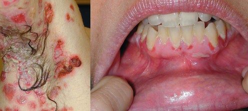
Demonstration of IgG autoantibodies on the cell surface of affected epithelium or detection of antigen-specific autoantibodies in the blood. Pemphigus vulgaris is characterized by progressively evolving fragile blisters and erosions. Oral involvement essentially ‘always’ occurs and is the major point to differentiate pemphigus vulgaris from pemphigus foliaceus. Histologic changes include suprabasilar acantholysis and cell surface-bound IgG. Circulating autoantibodies are specific for desmoglein 3 alone when lesions are restricted to the mouth and for both desmogleins 3 and 1 when cutaneous lesions are present in addition to oral lesions. Definition of the specificity of desmoglein autoantibodies can be reliably gained by enzyme-linked immunosorbent assay (ELISA).

In pemphigus foliaceus, mucosal lesions ‘never’ occur. This is the major clinical feature differentiating pemphigus foliaceus from pemphigus vulgaris. The cutaneous lesions are superficial scaling erosions. Immunopathologic studies reveal subcorneal acantholysis and tissue-bound and circulating antidesmoglein 1 antibodies.

Paraneoplastic pemphigus occurs in the context of the several lymphoproliferative disorders: non-Hodgkin lymphoma, chronic lymphocytic leukemia, Castleman disease, thymoma, and retroperitoneal sarcoma. Intractable mucositis with lichenoid erosions is the most constant clinical finding. Polymorphous cutaneous involvement with lesions that resemble erythema multiforme, pemphigus, pemphigoid, or lichenoid eruptions is observed. Histologic study shows suprabasilar acantholysis or interface/lichenoid changes. The key diagnostic finding is the presence of antibodies against desmogleins 3 and 1 and additional autoantibodies against epithelial plakin proteins, such as desmoplakin, envoplakin, and periplakin, which may be identified by immunoblotting or immunoprecipitation techniques. They may also be inferred with immunofluorescence techniques showing their reactivity with murine bladder epithelium, although the lichenoid variant of paraneoplastic pemphigus has been occasionally reported with the complete absence of the characteristic autoantibodies.

First-line therapies

Pemphigus vulgaris and foliaceus
imageSystemic corticosteroids B
imageRituximab D

image

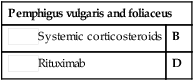
The first-line therapy for all forms of pemphigus is systemic corticosteroids. Corticosteroids work relatively quickly and are relatively safe when used at appropriate doses for limited periods of time. In the past, regimens of rapidly accelerating doses of corticosteroids were administered, but they were found to be associated with unacceptably high morbidity and mortality risks. Initial treatment should start at 1 mg/kg daily (lean body weight). A good clinical response, defined as a resolution of the majority of existing lesions and absence of newly developing lesions, should be evident within two to 3 months. The dose should then be reduced to 40 mg daily and subsequently tapered over 6 to 9 months, ideally to a maintenance dose of 5 mg every other day. Tapering can be accomplished by reduction of the prednisone by an average of 10 mg/month initially, and 5 mg/month later. There are some advantages to beginning an alternate dose regimen at 40 mg daily, so that monthly reductions would ideally be 40/20 mg alternate days, 40/0 mg, 30/0 mg, 20/0 mg, 15/0 mg, and 10/0 mg, and then 5/0 mg alternate days for maintenance.

The use of a second-line therapy is certainly indicated if significant corticosteroid side effects develop or are expected to develop during the ideal prednisone taper, if the disease does not improve sufficiently to allow continuous tapering, or the disease flares. With the introduction of better tolerated drugs such as mycophenolate mofetil, it is reasonable to use a second agent from the start of therapy in all patients with moderate to severe disease from the start of therapy, in anticipation of steroid-sparing benefits.

Monthly pulse corticosteroids have been suggested as a less toxic alternative to daily oral therapy, but pemphigus is very persistent, and more consistent daily or alternate day dosing is usually required to achieve suppression.

Patients must be monitored for corticosteroid-induced osteopenia by bone mineral density studies (DEXA scan) at institution of therapy and annually thereafter. Patients without a history of renal calculi may be given prophylactic supplemental calcium 1500 mg daily and vitamin D, 400–800 IU daily. In patients with osteopenia or osteoporosis, additional therapies may include hormone replacement in women (estrogen/progesterone, or raloxifen in those with a contraindication for estrogens, such as a history of breast carcinoma) or exogenous testosterone in men with low serum testosterone levels, or a bisphosphonate such as alendronate, or intranasal calcitonin.

In pemphigus vulgaris, therapy as outlined should commence in all patients once the diagnosis is confirmed. Even in cases with limited oral lesions, the disease will progress unless treated with systemic agents, and palliative therapy with topical agents or intralesional injections just delays definitive therapy. There is clinical evidence that early intervention with definitive treatment leads to a better long-term outcome. In pemphigus foliaceus, however, not every patient requires immediate treatment. Some patients have a very limited and smoldering disease, and can therefore benefit from some palliative treatment, such as topical corticosteroids. Paraneoplastic pemphigus is usually relentlessly progressive, justifying the immediate institution of systemic corticosteroids and a second-line treatment. If a patient has an associated benign lymphoproliferative disorder, such as thymoma, hyaline vascular Castleman disease, or sarcoma, complete surgical removal should be attempted to prolong disease remission.

A single cycle of rituximab for the treatment of severe pemphigus.

Joly P, Mouquet H, Roujeau JC, D’Incan M, Gilbert D, Jacquot S, et al. N Engl J Med 2007; 357: 545–53.

Twenty-one patients with pemphigus that was refractory to corticosteroids (at least two relapses despite prednisone treatment with doses higher than 20 mg/day; corticosteroid-dependent disease) or who had severe contraindications to corticosteroids were treated with four weekly infusions of rituximab, 375 mg/m2 body surface area. Eighteen showed complete remission at 3 months.

Depletion of CD20+ B cells by the use of anti-CD20 monoclonal antibody is emerging as a potentially powerful tool in many autoimmune diseases and is approved for use in rheumatoid arthritis. CD20 is not expressed on pre-B cells or plasma cells, so it is not profoundly immunosuppressive, and the effect only lasts 6 to 10 months, as the CD20+ cells are repopulated from stem cells. The currently approved dosing is 375 mg/m2 weekly for 4 weeks for lymphoma, or two doses of 1000 mg on days 1 and 15 for rheumatoid arthritis (RA). The original dosing regimen for lymphoma was designed to prevent tumor lysis syndrome in patients with bulky lymphomas. The authors have used the simpler, lower dose RA schedule in a small number of pemphigus vulgaris patients with equivalent effiicacy. Toxicity of rituximab is minimal, but the addition of the drug to patients already exposed to corticosteroids and immunosuppressive drugs increases the risk of infection, including one reported fatal case of Pneumocystis carinii pneumonia (PCP) in a patient treated with rituximab and cyclophosphamide. PCP prophylaxis should be considered with the use of this drug. The drug can cause rapid reduction of autoantibody levels in even severely affected cases, with excellent results and often dramatic recoveries. It is possible that a second course of rituximab has an important consolidation role for durable remission. The addition of IVIG to rituximab is reasonable in explosive acute cases; however, according to the current studies its added value in less acute cases is questionable. Rituximab is used most commonly in paraneoplastic pemphigus, where it has a dual role of treating the underlying lymphoproliferative disease as well as the associated autoimmune disease.

Second-line therapies

imageMycophenolate mofetil B
imageAzathioprine C
imageHigh-dose IVIG C

Treatment of pemphigus vulgaris and foliaceus with mycophenolate mofetil.

Mimouni D, Anhalt GJ, Cummins DL, Thorne JE, Nousari HC. Arch Dermatol 2003; 139: 739–42.

Forty-two patients were treated with mycophenolate mofetil, 1.5 g twice daily, and standard prednisone therapy. Complete clinical remission was defined as achieving no new lesions with prednisone doses less than 10 mg daily. This was achieved in 70% of patients with pemphigus vulgaris and 55% of patients with pemphigus foliaceus. Therapy was discontinued in only two cases due to adverse effects: one secondary to febrile neutropenia and one for gastrointestinal intolerance.

For pemphigus vulgaris and foliaceus, two effective antimetabolite immunosuppressive drugs are azathioprine and mycophenolate mofetil. Mycophenolate has an excellent safety profile, but it is very expensive. Azathioprine appears to be equally effective and is much cheaper, but has much more frequent toxicities. These drugs are added to the systemic corticosteroids, if the indications for their use are met. Once their beneficial effect is observed, the corticosteroids should be progressively tapered, while the second agent is used at full doses for up to 2 to 3 years to induce a durable remission. For both drugs, the doses required are greater than those needed to control other cutaneous diseases because only at high doses does one observe the required inhibition of the synthesis of autoantibodies by B cells.

Mycophenolate mofetil has approval for two dosing schedules: 1000 mg orally bid in renal transplantation, and 1500 mg orally bid in cardiac transplants. Both dosing regimens have been used in pemphigus but there is insufficient evidence to recommend one over the other. Onset of action is slow, and remissions are observed in responders after 2 to 12 months of therapy. Monitoring of complete blood count and liver enzymes should be performed monthly, but cytopenias and hepatotoxicity are rarely observed. Some lymphopenia without neutropenia is common, but has no adverse consequences and can correlate with a good clinical effect. Some nausea and diarrhea can occur, but improve with dosage reduction. Avoidance of pregnancy is mandatory, as human fetal malformations have occurred with its use.

Azathioprine in the treatment of pemphigus vulgaris. A long term follow-up.

Aberer W, Wolff-Schreiner EC, Stingl G, Wolff K. J Am Acad Dermatol 1987; 16: 527–33.

Azathioprine is given in a single daily dose of 3–4 mg/kg. At this dose, there is a risk of neutropenia, thrombocytopenia, hepatotoxicity, and severe or debilitating nausea. Monitoring should consist of complete blood count and liver enzymes, initially every 2 weeks. Patients with thiopurine methyltransferase (TPMT) deficiency cannot metabolize the drug effectively and can develop severe pancytopenia during the first 2 months of therapy. Late effects include elevation of liver enzymes and drug fever. The drug also requires 6 to 8 weeks of therapy before its effect on the disease can be judged. The drug is quite effective, but even if one screens patients for TPMT deficiency before starting treatment, the incidence of side effects is greater than with mycophenolate. It is still a useful second-line agent for those who cannot afford mycophenolate. There is also concern that exposure to this drug can increase one’s lifetime risk of leukemia or lymphoma, but the risk is very much less than that associated with the use of alkylating agents.

A comparison of oral methylprednisolone plus azathioprine or mycophenolate mofetil for the treatment of pemphigus.

Beissert S, Werfel T, Frieling U, Böhm M, Sticherling M, Stadler R, et al. Arch Dermatol 2006; 142: 1447–54.

A prospective, multicenter, randomized, non-blinded clinical trial to compare two parallel groups of patients with pemphigus treated with oral methylprednisolone plus azathioprine or oral methylprednisolone plus mycophenolate mofetil. In 13 (72%) of 18 patients with pemphigus receiving oral methylprednisolone and azathioprine, complete remission was achieved after a mean 74 days compared with 20 (95%) of 21 patients receiving oral methylprednisolone and mycophenolate mofetil in whom complete remission occurred after a mean 91 days. In six (33%) of 18 patients treated with azathioprine, severe adverse effects were documented in contrast to four (19%) of 21 patients who received mycophenolate mofetil.

Randomized controlled open-label trial of four treatment regimens for pemphigus vulgaris.

Chams-Davatchi C, Esmaili N, Daneshpazhooh M, Valikhani M, Balighi K, Hallaji Z, et al. J Am Acad Dermatol 2007; 57: 622–8.

The aim of this randomized study was to compare the efficacy and safety of four treatment regimens for new-onset pemphigus vulgaris (n=121): prednisolone alone, prednisolone plus azathioprine; prednisolone plus mycophenolate mofetil; and prednisolone plus intravenous cyclophosphamide pulse therapy. Results after 1 year were better with adjuvant treatment than with corticosteroid treatment alone. The highest efficacy was noted for azathioprine, followed by cyclophosphamide (pulse therapy), and mycophenolate mofetil. Interestingly, there were no significant differences in side effects among the groups.

Treatment of pemphigus with intravenous immunoglobulin.

Bystryn JC, Jiao D, Natow S. J Am Acad Dermatol 2002; 358: 358–63.

Both studies used IVIG in addition to treatment with prednisone and immunosuppressive therapy. IVIG produced a rapid reduction of circulating autoantibody levels, which was accompanied by significant clinical improvement in some cases.

High-dose IVIG can be used for acute control of active pemphigus. This treatment seems to accelerate the catabolism of the autoantibody and reduce circulating levels as effectively as plasmapheresis. It is generally safe and well tolerated, but has some risk. A small number of patients can develop thrombotic complications such as deep venous thrombosis or stroke. It is enormously expensive (as much as $12 000 per treatment for a 70 kg patient), and may lose its effectiveness after repeated treatment cycles. It can be given intravenously at a dose of 2 g/kg body weight, infused in divided doses over 2 to 5 days monthly.

IVIG is used frequently for acute control of severe cases. It can provide disease control for several months while slower acting drugs such as mycophenolate are used, and is safer than plasmapheresis. It can also be used effectively in combination with rituximab. The use of IVIG for extended periods to induce a remission is more controversial and requires better data.

Third-line therapies

imageCyclophosphamide B
imageCyclophosphamide plus plasmapheresis E
imageChlorambucil D

Alkylating agents such as cyclophosphamide have a profound effect on inhibition of autoantibody synthesis, and are the most effective agents for inducing a remission. They also have very significant potential toxicities, which restrict their use to third-line therapy. In countries where the high cost of rituximab limits its use, cyclophosphamide is still frequently used.

Cyclophosphamide is the preferred agent because any neutropenia associated with its use is predictable in onset, and withdrawal of the drug results in rapid recovery of neutrophils (within 1 week to 10 days). There are four ways to administer the drug:

1. Daily orally at 2.5 mg/kg. A single morning dose is followed by aggressive fluid consumption throughout the day to rinse metabolites from the bladder and prevent hemorrhagic cystitis. Weekly complete blood counts and urinalysis are required. With this use, a durable remission can be obtained after 18–24 months of therapy in almost all cases. This exposure probably increases the patient’s lifetime risk of leukemia, lymphoma, or bladder cancer by as much as 5–10% over the normal population. This risk is appreciated some 20–30 years after treatment. Such treatment can also cause sterility in young patients.

2. Monthly intravenous pulses at a dose of 750 mg/m2 body surface area. Monthly intravenous administration reduces the risk of hemorrhagic cystitis, but this intermittent use is not as effective in suppressing the disease.

3. Monthly intravenous administration with lower dose oral daily maintenance. This can also be very effective in inducing a remission.

4. Single, very high-dose immunoablative therapy. This experimental treatment is effective in many autoimmune diseases. It employs a dose of intravenous cyclophosphamide of 200 mg/kg, given over 4 days, which induces profound marrow aplasia. Upon recovery, the patients often experience a complete remission. However, unlike the experience with disorders such as aplastic anemia, where remission may last many years, most patients with pemphigus relapse within 2 years of completion of therapy, limiting its usefulness.

Synchronization of plasmapheresis and pulse cyclophosphamide therapy in pemphigus vulgaris.

Euler HH, Loffler H, Christophers E. Arch Dermatol 1987; 123: 1205–10.

Plasmapheresis is the only method by which one can rapidly reduce autoantibody levels, and is used in patients with very extensive and accelerated disease. Its use involves a total of six high-volume removals (3–3.5 L per removal, three times weekly for 2 consecutive weeks). This must be combined with the concomitant use of systemic corticosteroids and oral or pulse cyclophosphamide. If these drugs are not used, the reduction of autoantibodies removes feedback inhibition to the autoimmune B cells, and causes a rebound flare of the disease. This can be blunted only by alkylating agents due to their preferential toxicity to rapidly proliferative B cells. This causes the induction of a durable remission, though cyclophosphamide must still be used at full doses for 18–24 months to harden that remission.

Ineffectiveness of cyclosporine as an adjuvant to corticosteroids in the treatment of pemphigus.

Ioannides D, Chrysomallis F, Bystryn JC. Arch Dermatol 2000; 868: 505–6.

Thirty-three consecutive patients hospitalized with pemphigus vulgaris (n=29) or foliaceus (n=4) were randomized to receive either prednisolone or prednisolone plus cyclosporine, 5 mg/kg daily. The groups were similar in terms of disease severity, and demographics. The addition of cyclosporine produced no change in the response to treatment or the total dose of corticosteroid administered. Complications were, however, more common in those patients that received cyclosporine.

Although there are anecdotal cases reporting benefit from the use of cyclosporine, this well-performed study with an impressive number of cases is good evidence that the drug should not be used in pemphigus vulgaris and foliaceus. There is still good anecdotal evidence that cyclosporine may have a role to play in the management of paraneoplastic pemphigus, a disease with a much more complex pathophysiology.