Pediatric Heart and Heart-Lung Transplantation

Published on 25/03/2015 by admin

Filed under Pediatrics

Last modified 22/04/2025

Print this page

rate 1 star rate 2 star rate 3 star rate 4 star rate 5 star
Your rating: none, Average: 0 (0 votes)

This article have been viewed 1358 times

Chapter 437 Pediatric Heart and Heart-Lung Transplantation

437.1 Pediatric Heart Transplantation

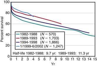
Pediatric heart transplantation is standard therapy for children with end-stage cardiomyopathy and other lesions not amenable to surgical repair. As of 2005, >6,900 heart transplants had been performed on children in the USA, with ≈375 transplants annually. Survival rates among children compare favorably with those of adults. For children transplanted in the 1980s and early 1990s, 1 yr survival has been 75-80%, whereas for those transplanted after 2000, 1 yr survival is now in the range of 90%; during the same time periods, 5 yr survival has improved from 60-65% to 75% (see imageFig. 437-1 on the Nelson Textbook of Pediatrics website at www.expertconsult.com). A growing number of children are now reaching their 15, 20, and 30 yr post-transplant anniversaries.

Recipient and Donor Selection

Potential heart transplant recipients must be free of serious noncardiac medical problems such as neurologic disease, systemic infection, severe hepatic or renal disease, or severe malnutrition. Many children with ventricular dysfunction may have pulmonary hypertension and even pulmonary vascular disease, which would preclude heart transplantation; pulmonary vascular resistance must be measured at cardiac catheterization, both at rest and in response to vasodilators. Patients with fixed elevated pulmonary vascular resistance are at higher risk for heart transplantation and may be considered candidates for either heterotopic heart transplantation (see later) or heart-lung transplantation (Chapter 437.2). A comprehensive social services evaluation is an important component of the recipient evaluation. Because of the complex post-transplantation medical regimen, the family must have a history of compliance. Detailed informed consent must be obtained.

Donor shortage is a serious problem for both adults and children. At the national registry of transplant recipients in the USA (the United Network for Organ Sharing [UNOS]), allografts are matched by ABO blood group and body weight. HLA matching is not currently performed unless the recipient has preformed antibodies against a particular HLA antigen. Recent data suggests that ABO matching is not required for young infants; the exact age under which ABO tolerance develops has not yet been determined. Contraindications to organ donation include prolonged cardiac arrest with persistent moderate to severe cardiac dysfunction, ongoing systemic illness or infection, and pre-existing severe cardiac disease. Physicians caring for a patient who may be a potential donor should always contact the organ donor coordinator at their institution, who can best judge the appropriateness of organ donation and has experience in interacting with potential donor families. A history of resuscitation alone or reparable congenital heart disease is not an automatic exclusion for donation.

The decision of when to place a patient on the transplant waiting list is based on a combination of many factors, including extremely poor ventricular function (left ventricular fractional shortening <10%; normal is 28-40%), poor exercise tolerance as determined by cardiopulmonary exercise testing (Chapter 417.5), poor response to medical anticongestive therapy, multiple hospitalizations for heart failure, arrhythmia, progressive deterioration in renal or hepatic function, early stages of pulmonary vascular disease, and poor nutritional status. In patients awaiting transplantation, those with poor left ventricular function (fractional shortening <15%) are usually started on a regimen of anticoagulation to reduce the risk of mural thrombosis and thromboembolism. Patients with cardiogenic shock unresponsive to standard pharmacologic treatment may be candidates for placement of left ventricular (LVAD) or biventricular (BiVAD) assist devices, or for extracorporeal membrane oxygenation (ECMO) support, which can stabilize hemodynamics and serve as a bridge to transplantation (Chapter 436).

Perioperative Management

In the classic operation, both donor and recipient hearts were excised so that the posterior portions of the atria containing the venae cavae and pulmonary veins are left intact. The aorta and pulmonary artery are divided above the level of the semilunar valves. The anterior portion of the donor’s atria was then connected to the remaining posterior portion of the recipient’s atria, thereby avoiding the need for delicate suturing of the venae cavae or pulmonary veins. The donor and recipient great vessels were connected via end-to-end anastomoses. This has largely been supplanted by the bicaval anastomosis, with the donor right atrium (and sinus node) left intact and the suture lines at the superior and inferior vena cavae; the left atrial connection is still performed as in the classic procedure. Heterotopic heart transplantation has been used for patients with left ventricular cardiomyopathy and elevated pulmonary vascular resistance. In this operation, the donor and recipient hearts are connected in parallel, so that the recipient right ventricle (which has hypertrophied over time due to the elevated pulmonary pressures) pumps mostly to the lungs, and the donor left ventricle pumps mostly to the body. This operation is preferable to heart-lung transplant for appropriate candidates (patients with pulmonary hypertension but without parenchymal lung disease, without evidence of right ventricular failure, and without serious congenital heart disease), as it is associated with a greater survival at all post-transplant time points.

In the immediate postoperative period, immunosuppression is most commonly achieved with a triple-drug regimen, although some centers use steroid-free regimens. The most common combinations are a calcineurin inhibitor (either cyclosporine or tacrolimus), a white blood cell enzyme inhibitor (mycophenolate mofetil or azathioprine), and prednisone. Some centers are using one of the cell-cycle inhibitors that act on the protein mTOR (target of rapamycin) instead of mycophenolate because of their potential benefit in reducing graft coronary artery disease. In many centers, induction therapy (usually an antilymphocyte preparation) is added in the 1st wk, either antithymocyte globulin (ATG), monoclonal murine antihuman T-lymphocyte antibody (OKT3), or one of the humanized anti–interleukin 2 receptor antibodies (daclizumab, basiliximab). In children who do not experience significant graft rejection, steroids can be gradually eliminated after the 1st 6-12 mo. Some centers do not use steroids as part of maintenance immunosuppression, but do use them as bolus treatment for acute rejection episodes.

Most pediatric heart transplant recipients can be extubated within the 1st 48 hr after transplantation and are out of bed within 3-4 days. These patients are often discharged within the 1st 2 wk after transplantation. In patients with pre-existing high-risk factors, postoperative recovery may be considerably prolonged. For those with preoperative pulmonary hypertension, the use of nitric oxide (NO) in the postoperative period can buy time to allow the donor right ventricle to hypertrophy in response to elevated pulmonary artery pressures. Occasionally, these patients will require right ventricular assist device (RVAD) support.

Diagnosis and Management of Acute Graft Rejection

Post-transplantation management consists of adjusting medications to maintain a balance between the risk of rejection and the side effects of over-immunosuppression. Along with infection, acute graft rejection is a leading cause of death in pediatric heart transplant recipients. The incidence of acute rejection is greatest in the 1st 3 mo after transplantation and decreases considerably thereafter. Many pediatric patients experience at least 1 episode of acute rejection in the 1st 2 yr after transplantation, although modern immunosuppressive regimens have decreased the frequency of serious rejection episodes. Because the symptoms of rejection can mimic many routine pediatric illnesses (pneumonia, gastroenteritis), it is mandatory that the transplant center be notified whenever a heart transplant recipient is seen in the pediatrician’s office or emergency room for any acute illness.

Clinical manifestations of acute rejection may include fatigue, fluid retention, fever, diaphoresis, abdominal symptoms, and a gallop rhythm. The electrocardiogram may show reduced voltage, atrial or ventricular arrhythmias, or heart block but is usually nondiagnostic. Roentgenographic examination may show an enlarged heart, effusions, or pulmonary edema, but usually only in the more advanced stages of rejection. Most rejection episodes occur without any detectable clinical symptoms. On echocardiography, indices of systolic left ventricular function may be decreased; however, these usually do not deteriorate until rejection is at least moderately severe. Techniques to evaluate wall thickening and left ventricular diastolic function, which appeared to be promising, have not fulfilled their promise as predictors of early rejection. Most transplant centers do not rely on echocardiography alone in rejection surveillance.

Myocardial biopsy is the most reliable method of monitoring patients for rejection. Biopsy specimens are taken from the right ventricular side of the interventricular septum and can be harvested relatively safely, even in small infants. In older children, myocardial biopsies may be performed as often as every 1-4 wk during the 1st 3-6 mo after transplantation. The frequency is then reduced to 2 or 4 biopsies per year unless the patient has an episode of rejection. In infants, surveillance biopsies are usually performed less often and may be as infrequent as once or twice per year. Children may have clinically unsuspected rejection episodes even 5-10 yr after transplantation; most pediatric transplant centers continue routine surveillance biopsies, albeit at less frequent intervals (every 6-12 mo).

Criteria for grading cardiac rejection are based on a system developed by the International Society for Heart and Lung Transplantation (ISHLT); these criteria take into account the degree of cellular infiltration and whether myocyte necrosis is present. ISHLT rejection grade 1R (former grades 1A, 1B, and 2) is usually mild enough that it is often not treated with bolus steroids, and many of these episodes resolve spontaneously. A repeat biopsy specimen is usually obtained within several weeks. For patients with ISHLT grade 2R (formerly grade 3A) rejection, treatment is instituted with either intravenous methylprednisolone or a “bump and taper” of oral prednisone. Asymptomatic patients >3 mo post-transplant and with normal echocardiograms are often treated as outpatients. Patients with grade 3R (formerly grades 3B or 4), or anyone with hemodynamic instability, are admitted to the hospital for intravenous steroid and potentially more aggressive anti-rejection therapy. For rejection episodes resistant to steroid therapy, additional therapeutic regimens include a repeat course of an antilymphocyte preparation (daclizumab, OKT3, antithymocyte globulin), methotrexate, or total lymphoid irradiation. Patients with repeated episodes of rejection may also benefit from being switched from cyclosporine to tacrolimus (or vice versa). Refractory rejection is not considered a good indication for retransplantation due to the relatively poor outcomes compared with other indications for retransplantation.

Gene expression profiling of peripheral blood mononuclear cells has been validated in adults as a highly sensitive, and moderately selective, method of rejection surveillance. Studies to corroborate these results in children are in progress. Progress has also been made in genetic profiling as a means to determine which patients are most at risk for rejection. Children who have single nucleotide polymorphisms (SNPs) leading to greater activity of inflammatory cytokines or decreased activity of regulatory cytokines are at increased risk of rejection. This type of profiling may be useful in designing patient-specific immunosuppressive regimens in the future.

Some rejection episodes are not associated with a cellular infiltrate on biopsy. These cases of acellular or humoral rejection are mediated by circulating antibodies and can be detected by immunostaining of the biopsy specimen for the complement component C4d and other immune markers. Humoral rejection is less responsive to standard therapies for acute cellular rejection (e.g. bolus steroids) and has been treated with plasmapheresis, intravenous immunoglobulin (IVIG) and the anti-CD20 monoclonal antibody rituximab.

Complications of Immunosuppression

Infection

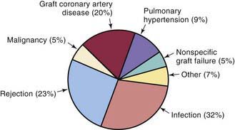
Infection is 1 of the 2 leading causes of death in pediatric transplant patients (Fig. 437-2). The incidence of infection is greatest in the 1st 3 mo after transplantation when immunosuppressive doses are highest. Viral infections are the most common, especially cytomegalovirus, which accounts for as many as 25% of infectious episodes. Cytomegalovirus infection may occur as a primary infection in patients without previous exposure to the virus or as a reactivation. Severe cytomegalovirus infection can be disseminated or associated with pneumonitis or gastroenteritis and may provoke an episode of acute graft rejection or graft coronary disease. Most centers use intravenous ganciclovir or cytomegalovirus immune globulin (CytoGam), or both, as prophylaxis in any patient receiving a heart from a donor who is positive for cytomegalovirus or in any recipient who has serologic evidence of previous cytomegalovirus disease. Polymerase chain reaction (PCR) enhances the ability to diagnose cytomegalovirus infection and to monitor the efficacy of therapy serially. Oral preparations of ganciclovir with improved absorption profiles are available for chronic therapy and have largely replaced intravenous preparations for prophylaxis.

Most normal childhood viral illnesses are well tolerated and do not usually require special treatment. Otitis media and routine upper respiratory tract infections can be treated in the outpatient setting, although fever or symptoms that last beyond the usual course require further investigation. Gastroenteritis, especially with vomiting, can result in markedly reduced absorption of immunosuppressive medications and provoke a rejection episode. In this setting, drug levels should be closely monitored and the use of intravenous medications considered. Gastroenteritis can also be a sign of rejection, so a high index of suspicion must always be maintained. Varicella is another childhood illness of some concern for immunosuppressed patients. If a heart transplant recipient acquires clinical varicella infection, treatment with intravenous acyclovir usually attenuates the illness.

Bacterial infections are the next most frequent, with the lung being the most common site of infection (35%), followed by the blood, the urinary tract, and, less commonly, the sternotomy site. Other sources of post-transplantation infection include fungi (14%) and protozoa (6%). Many centers use nystatin mouthwash to decrease fungal colonization and trimethoprim/sulfamethoxazole (Bactrim, Septra) during the time a patient is on steroids as prophylaxis to prevent Pneumocystis carinii infection.

Chronic Rejection

Graft coronary artery disease (GCAD) is a manifestation of chronic graft rejection that occurs in ≈20% of children 5 yr after transplant. The cause is still unclear, although it is thought to be a form of immunologically mediated vessel injury (chronic rejection). Hypercholesterolemia and hyperglycemia increase the risk of this disease. Unlike native coronary atherosclerosis, GCAD is a diffuse process with a high degree of distal vessel involvement. Because the transplanted heart has been denervated, patients may not experience symptoms such as angina pectoris during ischemic episodes, and the initial manifestation may be cardiovascular collapse or sudden death. Most centers perform coronary angiography annually to screen for coronary abnormalities; some also perform coronary intravascular ultrasound (IVUS) on adolescents. Standard coronary artery bypass procedures are usually not helpful because of the diffuse nature of the process, although transcatheter stenting can sometimes be effective for isolated lesions. For severe cases, repeat heart transplantation has been the only effective treatment. Thus, prevention has been the focus of most current research. The cell cycle inhibitors sirolimus and everolimus have been shown to decrease coronary arterial intimal thickening in adult transplant patients. Other drugs that have been shown to reduce the risk of graft coronary artery disease include the calcium channel blockers (such as diltiazem) and the cholesterol-lowering HMG-CoA (3-hydroxy-3-methyl-coenzyme A) reductase inhibitors (such as pravastatin or atorvastatin). The combined use of these two classes of drugs has recently been shown to reduce the incidence of GCAD in children to less than 10%.

Rehabilitation

Despite the potential risks of immunosuppression, the prospect for rehabilitation in pediatric heart transplant recipients is excellent. More than 95% of pediatric heart transplant recipients have no functional limitations in their daily lives. The majority of patients do not require rehospitalization for transplant-related problems.

Pediatric heart transplant recipients can attend daycare or school and participate in non-collision competitive sports and other age-appropriate activities. Standardized measurements of ventricular function are close to normal. Because the transplanted heart is denervated, the increase in heart rate and cardiac output during exercise is slower in transplant recipients, and maximal heart rate and cardiac output responses are mildly attenuated. These subtle abnormalities are rarely noticeable by the patient.

Growth of the transplanted heart is excellent, although a mild degree of ventricular and septal hypertrophy is commonly seen, even years after transplantation. The sites of atrial and great vessel anastomoses usually grow without the development of obstruction. In neonates who undergo transplantation for hypoplastic left heart syndrome, however, juxtaductal aortic coarctation may recur.

As assessed by standardized psychologic testing, the psychologic adjustment to heart transplantation in children is usually good. A serious problem with noncompliance often occurs once patients reach adolescence, and life-threatening rejection may result. Early intervention by social workers, counselors, and psychologists may be able to reduce this risk.

Bibliography

Almond CS, Thiagarajan RR, Piercey GE, et al. Waiting list mortality among children listed for heart transplantation in the United States. Circulation. 2009;119:717-727.

Bernstein D, Naftel D, Chin C, et al. Pediatric Heart Transplant Study. Outcome of listing for cardiac transplantation for failed Fontan: a multi-institutional study. Circulation. 2006;114:273-280.

Boucek MM, Aurora P, Edwards LB, et al. Registry of the International Society for Heart and Lung Transplantation: tenth official pediatric heart transplantation report—2007. J Heart Lung Transplant. 2007;26:796-807.

Boucek MM, Mashburn C, Dunn SM, et al. Pediatric heart transplantation after declaration of cardiocirculatory death. N Engl J Med. 2008;359:709-714.

Canter CE, Shaddy RE, Bernstein D, et al. Indications for heart transplantation in pediatric heart disease. Circulation. 2007;115:658-676.

Chin C, Lukito SS, Shek J, et al. Prevention of pediatric graft coronary artery disease: atorvastatin. Pediatr Transplant. 2008;12:442-446.

Deng MC, Eisen HJ, Mehra MR, et al. Noninvasive discrimination of rejection in cardiac allograft recipients using gene expression profiling. Am J Transplant. 2006;6:150-160.

Fan X, Ang A, Pollock-Barziv SM, et al. Donor-specific B-cell tolerance after ABO-incompatible infant heart transplantation. Nat Med. 2004;10:1227-1233.

Girnita DM, Brooks MM, Webber SA, et al. Genetic polymorphisms impact the risk of acute rejection in pediatric heart transplantation: a multi-institutional study. Transplantation. 2008;85:1632-1639.

Kulikowska A, Boslaugh SE, Huddleston CB, et al. Infectious, malignant, and autoimmune complications in pediatric heart transplant recipients. J Pediatr. 2008;152:671-677.

Pollock-BarZiv SM, Anthony SJ, Niedra R, et al. Quality of life and function following cardiac transplantation in adolescents. Transplant Proc. 2003;35:2468-2470.

Ringewald JM, Gidding SS, Crawford SE, et al. Nonadherence is associated with late rejection in pediatric heart transplant recipients. J Pediatr. 2001;139:75-78.

Rosenthal DN, Chin C, Nishimura K, et al. Identifying cardiac transplant rejection in children: diagnostic utility of echocardiography, right heart catheterization and endomyocardial biopsy data. J Heart Lung Transplant. 2004;23:323-329.

Ross M, Kouretas P, Gamberg P, et al. Ten- and 20-year survivors of pediatric orthotopic heart transplantation. J Heart Lung Transplant. 2006;25:261-270.

Russo LM, Webber SA. Pediatric heart transplantation: immunosuppression and its complications. Curr Opin Cardiol. 2004;19:104-109.

Tsang V, Yacoub M, Sridharan S, et al. Late donor cardiectomy after paediatric heterotopic cardiac transplantation. Lancet. 2009;374:387-392.

Webber SA. Immunology of pediatric heart transplantation: a clinical update. Semin Thorac Cardiovasc Surg Pediatr Card Surg Annu. 2001;4:158-184.

Webber SA, Naftel DC, Fricker FJ, et al. Lymphoproliferative disorders after pediatric heart transplantation: a multi-institutional study. Lancet. 2006;367:233-239.

West LJ, Pollock-Barziv SM, Dipchand AI, et al. ABO-incompatible heart transplantation in infants. N Engl J Med. 2001;344:793-800.

437.2 Heart-Lung and Lung Transplantation

More than 700 heart-lung and lung (single or double) transplants have been performed in children in the USA, with ≈60 procedures performed annually. Primary indications for heart-lung transplantation include cystic fibrosis, primary pulmonary hypertension, complex congenital heart disease with pulmonary hypoplasia or Eisenmenger syndrome, congenital lung abnormalities, and end-stage parenchymal lung disease (bronchopulmonary dysplasia, chronic lung disease, and interstitial fibrosis). Many of these patients with normal hearts may also be candidates for single- or double-lung transplantation if right ventricular function is preserved. In some patients with Eisenmenger physiology, double-lung transplantation can be performed in combination with repair of intracardiac defects. Patients with cystic fibrosis are not candidates for single-lung grafts because of the risk of infection from the diseased contralateral lung. Patients are selected according to many of the same criteria as for heart transplant recipients (Chapter 437.1).

Post-transplant immunosuppression is usually achieved with a triple-drug regimen, similar to that used for heart transplantation. Unlike patients with isolated heart transplants, few patients with lung transplants can be weaned totally off steroids. Prophylaxis against P. carinii infection is achieved with trimethoprim-sulfamethoxazole or aerosolized pentamidine. Ganciclovir and cytomegalovirus immune globulin prophylaxis are used as in heart transplant recipients (Chapter 437.1).

Pulmonary rejection is common in heart-lung transplant recipients, whereas heart rejection is encountered less often than in patients with isolated heart transplants. Symptoms of lung rejection may include fever and fatigue, although many episodes are minimally symptomatic. Surveillance for rejection is performed by monitoring pulmonary function (forced vital capacity; forced expiratory volume in 1 sec [FEV1]; forced expiratory flow, midexpiratory phase [FEF25-75%]), systemic arterial oxygen tension, and chest roentgenograms and by serial transbronchial biopsy. Because of technical limitations, biopsies are not usually performed in infants, who are monitored by clinical criteria alone.

Actuarial survival rates after heart-lung or lung transplantation in children are currently 75% at 1 yr and 50% at 5 yr; improved patient selection and postoperative management are continually improving these survival statistics. Graft failure and infection are the leading cause of early death, whereas a form of chronic rejection known as bronchiolitis obliterans (OB) accounts for nearly 50% of late mortality. Other causes of early morbidity and mortality include tracheal complications, pulmonary venous obstruction, donor lung dysfunction, bleeding, and acute rejection. Additional late complications include the development of airway stenosis, accelerated GCAD (though less common than in isolated heart transplantation), and other side effects of chronic immunosuppression.

Postoperative indices of cardiopulmonary function and exercise capacity show significant improvement. More than 95% of patients are without activity limitations at 2 yr follow-up. Problems of donor availability are even more severe with lung transplantation than with isolated heart transplantation. Living related lung transplantation, in which a lobe from a parent is transplanted into a child, can partially alleviate this problem.