Published on 19/03/2015 by admin
Filed under Dermatology
Last modified 22/04/2025
This article have been viewed 3013 times
Alex Milligan, Rosie Davis and Graham A. Johnston
Evidence Levels: A Double-blind study B Clinical trial ≥ 20 subjects C Clinical trial < 20 subjects D Series ≥ 5 subjects E Anecdotal case reports
The diagnosis parapsoriasis, even as an umbrella term, continues to cause diagnostic difficulties and there is still debate as to whether the variants described in this chapter are in fact precursors of cutaneous T-cell lymphoma. This chapter covers the entities small plaque parapsoriasis (SPP: chronic superficial scaly dermatitis; persistent superficial dermatitis; digitate dermatosis; xanthoerythroderma perstans) and large plaque parapsoriasis (LPP: parakeratosis variegata; retiform parapsoriasis; atrophic parapsoriasis; poikilodermatous parapsoriasis). Confusingly, the term parapsoriasis en plaque has been used for either SPP or LPP.
Other conditions sometimes grouped under the banner of parapsoriasis are pityriasis lichenoides et varioliformis acuta, pityriasis lichenoides chronica, and lymphomatoid papulosis, all of which are the subjects of separate chapters.
The diagnosis of parapsoriasis is made on clinical grounds, with histology supporting the clinical impression, especially when early cutaneous T-cell lymphoma is in the differential diagnosis. Patches of LPP are larger than 5 cm in diameter, and often 10 cm or larger, distinguishing them from SPP, which is characterized by lesions smaller than 5 cm.
If malignancy is considered in the differential diagnosis, T-cell receptor gene rearrangement studies are more likely to demonstrate monoclonality in cutaneous T-cell lymphoma, though monoclonality is not entirely sensitive or specific for the latter. Repeat studies may be warranted if progression to cutaneous T-cell lymphoma is suspected.
Although some advocate non-aggressive therapies, e.g., topical corticosteroids, for parapsoriasis, the potential for progression to cutaneous lymphoma in patients with LPP justifies the use of psoralen with UVA (PUVA). Sunlight, broadband UVB, and narrowband UVB have been used successfully as well, particularly for SPP.
The diagnosis is principally made on clinical findings
Histology is non-specific
TCR gene rearrangement studies
Immunohistochemistry cannot differentiate between the two forms.
Assessment of TCR-beta clonality in a diverse group of cutaneous T-cell infiltrates.
Plaza JA, Morrison C, Magro CM. J Cutan Pathol 2008; 35: 358–65.
Monoclonality is a reliable characteristic of CTCL, polyclonality being very infrequent. However, the authors warn that the various cutaneous lymphoid dyscrasias, including pityriasis lichenoides chronica, could manifest restricted molecular profiles in the context of an oligoclonal process or frank monoclonality.
Clonal T cell receptor gamma-chain gene rearrangement by PCR-based GeneScan analysis in the skin and blood of patients with parapsoriasis and early-stage mycosis fungoides.
Klemke CD, Dippel E, Dembinski A, Pönitz N, Assaf C, Hummel M, et al. J Pathol 2002; 197: 348–54.
Although studies have shown T-cell clonality in both skin and peripheral blood, monoclonality is neither easily demonstrable nor thought to be a prerequisite for diagnosis.
The role of immunohistochemical analysis in the diagnosis of parapsoriasis.
Bordignon M, Belloni-Fortina A, Pigozzi B, Saponeri A, Alaibac M. Acta Histochem 2011; 113: 92–5.
Immunohistochemical techniques cannot distinguish between large and small cell parapsoriasis.
SPP consists of fixed, small scaly erythematous plaques which are asymptomatic or only mildly itchy and occur mainly on the trunk. The lesions sometimes appear to run in finger-like lines parallel to the ribs (hence the name ‘digitate dermatosis’). SPP runs a chronic, indolent, and benign course.
Treatments with emollients, topical tar, and topical corticosteroid are cited in books, and appear effective in clinical practice. There have, however, been no studies or case reports to back this up, and these treatments are therefore unreferenced.
Narrowband UVB phototherapy for small plaque parapsoriasis.
Aydogan K, Karadogan SK, Tunali S, Adim SB, Ozcelik T. J Eur Acad Dermatol Venereol 2006; 20: 573–7.
Forty-five patients were treated with narrowband UVB therapy three to four times weekly. There was a complete response in 33 patients with a mean cumulative dose of 14.3 J/cm2 after a mean number of 29 exposures. There was a partial response in 12 of the 45. Relapses occurred in six patients within a mean of 7.5 months.
Treatment of small plaque parapsoriasis with narrow-band (311 nm) ultraviolet B: a retrospective study.
Herzinger T, Degitz K, Plewig G, Rocken M. Clin Exp Dermatol 2005; 30: 379–81.
Sixteen patients had complete remission after a mean number of 32.8 exposures and a mean total dose of 35.4 J/cm2. Side effects were rare and mild. Relapse occurred after an average of 29 weeks.
Narrowband (311-nm) UV-B therapy for small plaque parapsoriasis and early-stage mycosis fungoides.
Hofer A, Cerroni L, Kerl H, Wolf P. Arch Dermatol 1999; 35: 1377–80.
Fourteen patients with SPP were treated with narrowband UVB, three to four times weekly for 5 to 10 weeks. Complete response was achieved after an average of 20 exposures. All patients then relapsed after an average of 6 months, and topical corticosteroid therapy was effective at producing a second clearance in an unspecified number of patients.
Treatment of parapsoriasis and mycosis fungoides: the role of psoralen and long-wave ultraviolet light A (PUVA).
Powell FC, Spiegel GT, Muller SA. Mayo Clin Proc 1984; 59: 538–46.
Seven patients with SPP had complete clearance with as few as 15 treatments (84 J/cm2) of standard PUVA. Three patients experienced some recurrence at follow-up (mean of 13 months), and one of these was then successfully treated with topical corticosteroid.
UVA1 cold-light phototherapy has also been reported to be beneficial in SPP.
Topical nitrogen mustard therapy in patients with mycosis or parapsoriasis.
Lindahl LM, Fenger-Gron M, Iversen L. J Eur Acad Dermatol Venereol 2012.
Seventy-one patients with parapsoriasis (no information on numbers of LPP and SPP) between 1991 and 2009 were treated with mechlorethamine hydrochloride. 20 mg was dissolved in 40 mL water and applied daily to affected areas for a 14-day induction period. Maintenance was two treatments every 4 to 8 weeks until clear, insufficient response or side effects. The ‘overall’ response was 90%, with a ‘complete’ response of 41%. Relapse (no time given) was 62%. Main side effect was contact dermatitis which caused withdrawal of treatment in 14% of patients.
Topical carmustine (BCNU) for mycosis fungoides and related disorders: a 10-year experience.
Zackheim HS, Epstein EH Jr, McNutt NS, Grekin DA, Crain WR. J Am Acad Dermatol 1983; 9: 363–74.
One patient with SPP was treated with carmustine as a subgroup of the study. A variety of treatment regimens were used and not individually specified. Complete response was achieved at follow-up, but the duration is not specified.
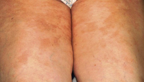
Like SPP, the trunk is mainly affected but the lesions are larger, atrophic, and even poikilodermatous, with a red or yellow-orange color.
Skin biopsy
The diagnosis is suggested clinically. Histology can vary from a mild dermatitis to epidermal atrophy, lichenoid changes at the dermoepidermal junction, and a band-like lymphocytic infiltrate in the papillary dermis.
Progression to T-cell lymphoma can occur. T-cell clonality can be demonstrated in some patients.
Large plaque parapsoriasis: clinical and genotypic correlations.
Simon M, Flaig MJ, Kind P, Sander CA, Kaudewitz P. J Cutan Pathol 2000; 27: 57–60.
TCR gene rearrangement status was assessed in 12 patients. Six of the 12 showed a clonal T-cell population, one of whom developed cutaneous T-cell lymphoma after a follow-up of 8 years. The other five patients showed no such progression after follow-up of 2 to 21 years. The authors conclude that TCR gene rearrangement status has no prognostic significance and does not allow distinction between LPP and early mycosis fungoides.
The nosology of parapsoriasis.
Lambert WC, Everett MA. J Am Acad Dermatol 1981; 5: 373–95.
Of 129 cases of LPP, 11% developed mycosis fungoides over a follow-up period ranging from 1 to 64 years.
Parapsoriasis and mycosis fungoides: the Northwestern University experience, 1970 to 1985.
Lazar AP, Caro WA, Roenigk HH, Pinski KS. J Am Acad Dermatol 1989; 21: 919–23.
Of 89 patients with LPP, 30% developed mycosis fungoides. The follow-up period was not specified.
Photochemotherapy in cutaneous T cell lymphoma and parapsoriasis en plaque. Long-term follow-up in forty-three patients.
Rosenbaum MM, Roenigk HH Jr, Caro WA, Esker A. J Am Acad Dermatol 1985; 13: 613–22.
Seven patients with LPP were included as part of the above study. They were treated with oral psoralens and UVA (PUVA). A complete response was achieved in all seven, though the total dosages of PUVA are not stated. Average follow-up for all 43 patients was 38.4 months (range 4–67 months), and during that time relapse was observed in five out of the seven.
Because cutaneous T-cell lymphoma is in the differential diagnosis of LPP, and PUVA is effective for both conditions, this therapy is useful in patients in whom it is difficult to distinguish between the two conditions.
Treatment of a case of mycosis fungoides and one of parapsoriasis en plaque with topical PUVA using a monofunctional furocoumarin derivative, 4,6,4′-trimethylangelicin.
Morita A, Takashima A, Nagai M, Dall’Acqua F. J Dermatol 1990; 17: 545–9.
A single patient with LPP was treated with 0.1% TMA topical lotion (a monofunctional psoralen), followed 2 hours later with UVA light. Clearance was achieved after nine treatments with 24 cm2. A control area did not have the topical TMA applied to it and clearance was not obtained even after 20 treatments.
See details of this study in the section above on SPP.
Evaluation of a one-hour exposure time to mechlorethamine in patients undergoing topical treatment.
Foulc P, Evrard V, Dalac S, Guillot B, Delaunay M, Verret JL, et al. Br J Dermatol 2002; 147: 926–30.
Three patients with LPP were included in this study. One patient stopped treatment because of the side effects and two of the three resulted in complete remission. The mechlorethamine regimen, however, varied between the four centers included in the study and is not specified for the individual patient.
The incidence of squamous cell carcinoma of the skin is dramatically increased in patients treated with topical nitrogen mustard and PUVA.
Successful treatment of parapsoriasis en plaques with 2,4- dinitrochlorobenzene.
Mandrea E. Arch Dermatol 1971; 103: 560–1.
A single patient with LPP was treated topically with twice-daily 1% 2,4-dinitrochlorobenzene in equal parts with olive oil and propylene glycol. The patient developed a painful, erythematous reaction and the treatment was stopped; 18 months later the skin remained clear.
Excimer-laser (308 nm) treatment of large plaque parapsoriasis and long-term follow-up.
Gebert S, Raulin C, Ockenfels HM, Gundogan C, Greve B. Eur J Dermatol 2006; 16: 198–9.
A letter describing long-term benefit with a 308 nm laser.
Treatment of Skin Disease Comprehensive Therapeutic Strategies 4e
WhatsApp us


 Emollients, tar, topical corticosteroids
 PUVA
PUVA Narrowband UVB
 Topical nitrogen mustard

 PUVA
PUVA PUVA with 4,6,4′-trimethylangelicin
 Topical nitrogen mustard
 Topical 2,4-dinitrochlorobenzene
 Excimer laser