Paediatric cardiopulmonary resuscitation

Published on 27/02/2015 by admin

Filed under Anesthesiology

Last modified 22/04/2025

Print this page

rate 1 star rate 2 star rate 3 star rate 4 star rate 5 star
Your rating: none, Average: 0 (0 votes)

This article have been viewed 1433 times

Chapter 106 Paediatric cardiopulmonary resuscitation

This chapter concerns basic and advanced cardiopulmonary resuscitation (CPR) for infants and children (Figure 106.1). The essentials of resuscitation of the ‘newly-born’ (at birth) infant are also provided (Figure 106.2). The recommendations are based on guidelines published by several authoritative resuscitation organisations13 which are in turn derived from an extensive evaluation of the science of resuscitation conducted by the International Liaison Committee on Resuscitation.4

image

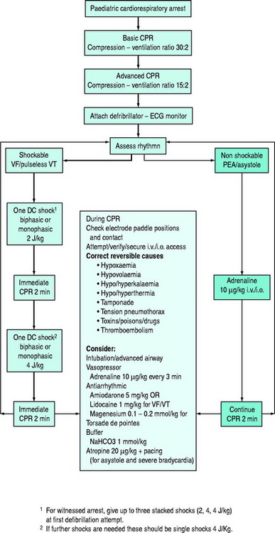
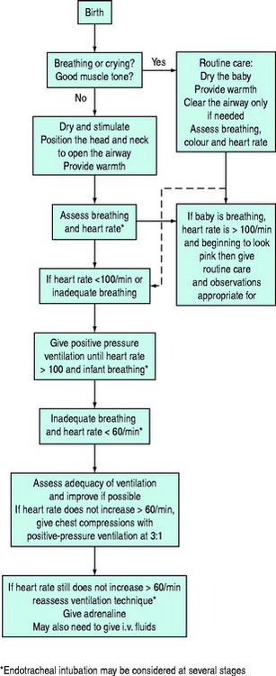
Figure 106.2 Newly-born infant resuscitation.

(Reproduced with permission from the Australian Resuscitation Council, Melbourne.)

This chapter is intended primarily for use by medical and nursing personnel in hospital (health carers). To add ability to knowledge, it is advisable to undertake a specialised paediatric cardiopulmonary resuscitation course, such as the Advanced Paediatric Life Support (APLS) or Paediatric Advanced Life Support (PALS) courses. This chapter should be regarded as an addendum to Chapters 17 (Adult cardiopulmonary resuscitation), 96 (The critically ill child) and 104 (Equipment for paediatric intensive care).

Distinctions within the term paediatric are based on combinations of physiology, physical size and age. Some aspects of CPR are different for the ‘newly-born’, infant, small (younger) child and large (older) child. ‘Newly-born’ refers to the infant at birth or within several hours of birth. ‘Infant’ refers to an infant outside the ‘newly-born’ period up to the age of 12 months. Other terms, such as newborn or neonate, do not enable that distinction. ‘Small/young child’ refers to a child of preschool and early primary school from the age of 1–8 years. ‘Large/older child’ refers to a child of late primary school from the age of 9 up to 14 years. Children older than 14 years may be treated as adults but they do not have the same propensity for ventricular fibrillation as do adults.

BASIC LIFE SUPPORT

EXTERNAL CARDIAC COMPRESSION (ECC)

Different techniques are used for infants and children of different sizes but in all patients the depth of compression is one-third the depth of their chest. For newly-born infants and infants, two techniques are in common use. In the ‘two-finger technique’, the middle and forefinger are used. This technique is taught to lay-persons and is also the preferred technique by a single health care rescuer. With the ‘two-thumb technique’, the hands encircle the thorax, approaching the chest from either above or below, and the thumbs are placed either opposite, alongside or atop one another. With this technique, the rescuer must take care to avoid restriction of the patient’s chest during inflation. With premature newly-borns and small infants, the rescuer’s encircling fingers may reach and stabilise the vertebral column, without limiting chest inflation. With both techniques, the sternum is compressed above the xiphoid or about one finger breadth below the internipple line.

Either a single hand or both hands may be used for infants and children as determined by the relationship between the size of the patient’s chest and the hands of the rescuer. For young children, ECC can be performed with the heel of one hand. For older children, a bimanual technique as per adults may be used. In all ages, the ‘centre of the chest’ – which corresponds to the lower sternum – is compressed. Approximately 50% of each cycle should be compression.

RATES OF COMPRESSION AND RATIO OF COMPRESSION TO VENTILATION

In hospitals where there are usually two (or more) rescuers, the ratio of compressions to ventilation for infants and children should be 15:2. After every 15 compressions, a pause should allow delivery of two ventilations whenever expired air resuscitation or any type of mask ventilation is given. ECC can be given during the second exhalation. The aim should be to achieve about five cycles per minute, i.e. about 75 compressions and 10 breaths. If circulation returns, but respiration remains inadequate, the number of ventilations should be higher but care should be taken to avoid hypocarbia which results in cerebral ischaemia. A health care rescuer, when alone, may use the lay-person ratio of 30:2, aiming to achieve about five cycles in 2 minutes, i.e. about 75 compressions and 5 breaths per minute.

For infants and children, compressions should be delivered at a rate of 100/min, i.e. one compression every 0.6 seconds or approximately two per second. This does not mean that 100 actual compressions are given each minute. When ventilation is interposed between compressions, the actual compressions delivered will be less than 100 each minute.

If the airway has been secured, e.g. by intubation, strict coordination of compression and ventilation is not crucial: ventilation can be given against resistance imposed by chest compression. In this case about 100 compressions/minute will be achieved but ventilation should be limited to about 10–12 per minute and wherever possible guided by arterial blood gas analysis.

For newly-born infants the total number of recommended ‘events’ per minute is 120, with the aim of achieving 90 compressions and 30 inflations each minute, i.e. in a ratio of 3:1.

ADVANCED LIFE SUPPORT

As soon as practicable, mechanical ventilation with added oxygen should be commenced with either a bag-valve-mask or via endotracheal intubation. Although intubation is preferred (see below), valuable time should not be wasted in numerous unsuccessful attempts. Initial effective bag-valve-mask ventilation is a necessary prerequisite for successful paediatric CPR. Bags of appropriate sizes should be available for infants, small children and large children. A bag of ∼500 ml volume should be available for newly-born infants. Insertion of an oropharyngeal (Guedel) airway may be necessary to facilitate bag-valve-mask ventilation. Access to the circulation and display of the electrocardiograph should also be achieved as soon as possible. Thereafter treatment should be guided by the cardiac rhythm. Underlying causes of CPA should be sought and treated.

AIRWAY MANAGEMENT

If attending personnel are skilled, the trachea should be intubated as soon as practicable. This establishes and maintains an airway, facilitates mechanical ventilation with 100% oxygen, minimises the risk of pulmonary aspiration, enables suctioning of the trachea and provides a route for the administration of selected drugs. If difficulty is experienced at initial intubation, oxygenation should be established with bag-valve-mask ventilation before a reattempt at intubation. Initial intubation should be via the oral route, not via the nasal route.

Intubation via the oral route:

On the other hand, a tube placed nasally:

A nasogastric tube should be inserted after intubation to relieve possible gaseous distension of the stomach sustained during bag-valve-mask ventilation.

Correct placement of the endotracheal tube in the trachea must be confirmed. In the hurried conditions of emergency intubation at cardiopulmonary arrest, it is not difficult to mistakenly intubate the oesophagus or to intubate a bronchus. There is no substitute for:

It is recommended that correct placement of the endotracheal tube be confirmed by capnography or colorimetric CO2 detection after initial placement with the realisation that CO2 excretion can only occur with effective pulmonary blood flow. This implies that CO2 detection cannot be expected unless spontaneous cardiac output returns or unless external cardiac compression is effective. Oxygenation should be confirmed with use of a pulse oximeter or measurement of arterial gas tension.

Tube size

Tubes larger and smaller should be available. The correct size should allow a small leak on application of moderate pressure but also enable adequate pulmonary inflation.

The tube is inserted a specific depth to avoid accidental extubation or endobronchial intubation. Assessment of the depth of insertion at laryngoscopy is not reliable because this is performed with the neck extended. When the laryngoscope is removed the head assumes a position of neutrality or flexion and the tube depth increases.

The appropriate depth of insertion measured from the centre of the lips for an oral tube is:

An alternative formula for oral tube depth of insertion is: depth (cm) = size (mm) ×3. After 1 year the depth is given by the formula: depth (cm) = age (years)/2 + 12. The appropriate depth of insertion for a nasal tube in this age group is: depth (cm) = age (years)/2 + 15. On a chest X-ray taken with the head in neutral position, the tip of the tube should be at the interclavicular line.

A laryngeal mask airway (LMA) may be used to establish an airway in the setting of CPA. However, its role in provision of mechanical ventilation remains uncertain. Like bag-valve-mask ventilation, it does not protect the airway from aspiration. Although insertion of an LMA is easier to learn than endotracheal intubation, training should not replace mastery of bag-valve-mask ventilation. An LMA is not suitable for long-term use or during transport when endotracheal intubation is preferred.

INTRAOSSEOUS INJECTION

If intravenous (i.v.) access cannot be achieved rapidly, say within 60 seconds, intraosseous (i.o.) access should be obtained. This route has been used for patients of all ages and provides rapid, safe and reliable access to the circulation. It serves as an adequate route for any parenteral drug and fluid administration.

A metal needle with a trocar (e.g. disposable intraosseous needle; Cook, Illinois, USA) is preferable although a short lumbar puncture type of needle with an inner trocar may suffice. Although many sites have been used for bone marrow injection, the easiest to identify is the anteromedial surface of the upper or lower tibia, especially in children less than 6 years. The site of the former is a few centimetres below the anterior tuberosity and the latter a few centimetres above the medial malleolus. The handle of the device needle is held in the palm of the hand while the fingers grip the shaft about a centimetre from the tip. It is inserted perpendicular to the bone surface and a rotary action is used to traverse the cortex. Sudden loss of resistance signifies entry to bone marrow. Correct positioning of the needle is confirmed by aspiration of bone marrow (which may be used for biochemical and haematological purposes) but that is not always possible. Bone injection guns (Wais Med Ltd), which fire a needle a preset distance according to the size of the patient, enable easy i.o. injection for infants, children and adults.

Volume expanders need to be given by syringe to achieve rapid restoration of circulating volume and rapid access of drugs to the central circulation. This is best achieved using a 10-ml syringe with a three-way tap in the i.v. tubing so that the i.o. needle does not become dislodged inadvertently.

Care should be exercised to avoid complications, particularly cutaneous extravasation, compartment syndrome of the leg and osteomyelitis. Contraindications include local trauma and infection.

DC SHOCK

Defibrillators should have paediatric paddles of cross-sectional area 12–20 cm2 for use in children <10 kg body weight. For others, adult-sized paddles (50–80 cm2) are satisfactory provided the paddles do not contact each other. Selectable energy levels should enable delivery of doses 0.5–4 J/kg. The closest level to the dose should be selected. One paddle is placed over the mid-axilla opposite the xiphoid or nipple, the other to the right of the upper sternum below the clavicle. Conductive gel (confined to the area beneath the paddles) or gel pads and firm pressure are needed to deliver optimum energy to the heart without causing skin burns. The doses for monophasic and biphasic shock are the same.

In the treatment of refractory arrhythmias, equipment failure should be excluded. An anteroposterior position of the paddles (one over the cardiac apex, one over the left scapula) may be efficacious in refractory arrhythmia. Dextrocardia may be present with congenital heart disease and the position of the paddles should be altered accordingly.

Monophasic and biphasic automated external defibrillators (AEDs) with attenuated energy doses (approximately 50 J or less) may be used for children 1–8 years of age (approximately 10–25 kg). Adult energy doses of 150–200 J may be used in children older than 8 years of age or 25 kg body weight. AEDs are not recommended for use in infants since they cannot reliably distinguish ventricular fibrillation from tachycardia and hence pose a risk of harmful DC shock.

Rescuers should be cognisant of the risk of inadvertent shock to themselves and colleagues during operation of defibrillators. They should ensure that they have no physical contact with the patient directly or indirectly at the time of electrical discharge. Paddles should be charged only when already placed on the chest of the patient. If the need to give DC shock abates while the paddles are charged, they should be disarmed before replacing them in their cradles.

ASYSTOLE AND BRADYCARDIA

Asystole and hypotensive bradycardia (<60 beats/min) should be treated with adrenaline 10 μg/kg i.v. or i.o. If these routes are not available, adrenaline 100 μg/kg should be administered via endotracheal tube (ETT).

Unresponsive asystole should be treated with similar doses of adrenaline (10 μg/kg i.v., i.o.; 100 μg/kg ETT) every 3–5 minutes. Higher doses, up to 200 μg/kg i.v. or i.o., may be indicated in special circumstances (e.g. β-blocker toxicity) but should not be used routinely as they have not altered outcome and predispose to complications (postarrest myocardial dysfunction, hypertension, tachycardia). In newly-born infants, the initial bolus dose is 100–300 μg/kg.

Recurrent bradycardia or asystole may require an infusion of adrenaline at 0.05–3 μg/kg per min: doses <0.3 μg/kg per min are predominantly β-adrenergic; doses >0.3 μg/kg per min are predominantly α-adrenergic. Infuse into a secure large vein. If sinus rhythm cannot be restored, sodium bicarbonate 1 mmol/kg i.v. or i.o. may be helpful but do not allow mixing with adrenaline since catecholamines are inactivated in alkaline solution.

Sinus bradycardia, sinus arrest with slow junctional or idioventricular rhythm, and atrioventricular block are the most common preterminal arrhythmias in paediatric practice. Untreated, bradycardia progresses to asystole.

Bradycardia caused by vagal stimulation should be managed with cessation of the stimulus and/or atropine 20 μg/kg i.v. or i.o. (minimum dose 100 μg) but persistent vagal-mediated bradycardia should be treated with adrenaline 10 μg/kg i.v. or i.o.

If facilities are available, bradycardia may be treated with pacing (oesophageal, transcutaneous, transvenous, epicardial) if sinus node dysfunction or heart block exists, but it is not helpful in asystole.

VENTRICULAR FIBRILLATION AND PULSELESS VENTRICULAR TACHYCARDIA

The initial dose for ventricular fibrillation (VF) or pulseless ventricular tachycardia (VT) is a single unsynchronised shock at 2 J/kg followed by immediate resumption of ECC. If VF or VT persists, subsequent single shocks of 4 J/kg are given, each separated by 2 minutes of ECC.

The witnessed (monitored) onset of VF or pulseless VT should be treated with unsynchronised DC shock initially at 2 J/kg, progressing to 4 J/kg in a stack (salvo) of three shocks. Rescuers should maintain the paddles on the chest and be prepared to deliver a series of three shocks in quick succession, pausing only to verify the rhythm. A precordial thump may be given before DC shock but its efficacy has not been proven.

Failure to revert to sinus rhythm should be treated with adrenaline 10 μg/kg i.v. or i.o. or 100 μg/kg ETT and another shock at 4 J/kg after an interval of ECC. Persistent (refractory) or recurrent VF or VT may also be treated with antiarrhythmics (amiodarone, lidocaine, magnesium) interspersed with single DC shocks. Irrespective of other drug therapy, adrenaline should be administered every 3–5 minutes. The dose of lidocaine is 1 mg/kg i.v. or i.o. bolus followed by an infusion if successful at 20–50 μg/kg per min. The dose of amiodarone is 5 mg/kg i.v. or i.o. as a bolus which may be repeated to maximum of 15 mg/kg. Magnesium, 25–50 mg/kg (0.1–0.2 mmol/kg) is indicated for polymorphic VT (torsade de pointes). The alternative to adrenaline as a vasopressor, vasopressin, has not been adequately investigated for use during CPR for children.

SUPRAVENTRICULAR TACHYCARDIA

Supraventricular tachycardia (SVT) is the most common spontaneous arrhythmia in childhood and infancy. It may cause life-threatening hypotension. It is usually re-entrant with a rate of 220–300/min in infants, less in children. The QRS complex is usually narrow (<0.08 seconds) making it difficult sometimes to discern from sinus tachycardia (ST). However, whereas ST is a part of other features of illness, SVT is a singular entity, and whereas the rate in ST is variable with activity or stimulation, it is uniform in SVT. In both rhythms, a P wave may be discernible. SVT with aberrant conduction (wide QRS) may resemble VT.

If haemodynamically stable (adequate perfusion and blood pressure), initial treatment of SVT should be vagal stimulation. For infants and young children, application to the face of a plastic bag filled with iced-water is often effective. Older children may be treated with carotid sinus massage or perform a Valsalva (e.g. blowing through a narrow straw). If unsuccessful, give adenosine 100 μg/kg i.v. by rapid bolus (maximum dose 6 mg), doubling once to 200 μg/kg. If unsuccessful, give synchronised DC shock (cardioversion) commencing with 0.5 J/kg, progressing if unsuccessful to 2 J/kg under sedation/anaesthesia. If SVT is accompanied by haemodynamic instability (i.e. hypotension), proceed to cardioversion (synchronised 0.5–2 J/kg) immediately although vagal stimulation or adenosine (i.v. or i.o.) may be used provided they do not delay cardioversion. Verapamil should not be used to treat SVT in infants and should be avoided in children because it induces hypotension and cardiac depression.

POST RESUSCITATION CARE

Supportive therapy should be provided until there is recovery of function of vital organs. This may require provision of oxygen therapy, mechanical ventilation, inotropic/vasopressor infusion, renal support, parenteral nutrition and other therapy for several days or longer. Recovery of infants and children is usually slow because cardiorespiratory arrest is often secondary to prolonged global ischaemia or hypoxaemia which implies that other organs sustain damage before cardiorespiratory arrest.

Particular care should be taken to ensure adequate cerebral perfusion with well-oxygenated blood. Hyperventilation is not useful for this purpose. Therapeutic hypothermia post resuscitation (33–36 °C) up to 3 days may improve neurological outcome. Inadvertently hypothermic patients, provided temperature is above 33 °C, should not be actively warmed but hyperthermia should be aggressively treated. During deliberate hypothermia shivering should be prevented with sedation and/or neuromuscular blockade, seizures should be actively sought and treated with anticonvulsant and the cause of the CPA should be investigated and treated appropriately, e.g. sepsis or drug overdose.

Complications of CPR should be sought, especially if secondary deterioration occurs. A chest X-ray should be obtained to check the position of the endotracheal tube, to exclude pneumothorax, lung collapse, contusion or aspiration, and to check the cardiac silhouette although an echocardiographic examination is preferable to specifically check contractility and to exclude a pericardial effusion. Measurement of haemoglobin, pH, gas tensions, electrolytes and glucose is important.