[level-membership-for-dermatology-category]27
Other Vesiculobullous Diseases
There are a number of disorders that can present with vesicles and bullae, including exaggerated insect bite reactions (see Chapter 20), autoimmune and inherited blistering diseases (see Chapters 23–26), porphyrias (see Chapter 41), the Stevens–Johnson syndrome–toxic epidermal necrolysis spectrum (see Chapter 16), and phototoxicity, from sunburn to phototoxic drug reactions (e.g. due to doxycycline). This chapter examines a miscellaneous group of disorders, several of which favor the lower extremities, whereas Chapter 28 reviews vesiculobullous diseases in newborns and infants.
Friction Blisters
Edema Bullae (Edema Blisters)
• The most common location is the distal lower extremities, often in the setting of an acute exacerbation of chronic edema in an elderly patient; in patients with anasarca and those who are bedridden, the distribution can be more widespread (Fig. 27.1).
Bullosis Diabeticorum (Diabetic Bullae)
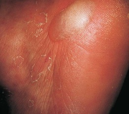
• In patients with diabetes mellitus, tense bland bullae arise rather suddenly within normal-appearing skin (Fig. 27.2); the diameter varies from 0.5 to several centimeters; the blister fluid is sterile and clear, but may be more viscous than that of friction or edema blisters.
Delayed Postburn/Postgraft Blisters
Coma Bullae (Coma Blisters)
• Tense cutaneous vesicles and bullae can appear within 2 or 3 days of prolonged pressure secondary to immobilization (Figs. 27.3 and 27.4); there is often preceding blanchable erythema.
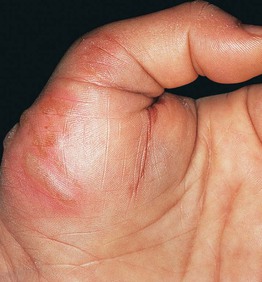
Fig. 27.3 Coma bullae. Blisters developed in an area of pressure in a previously comatose patient. Courtesy, José M. Mascaró Jr., MD.
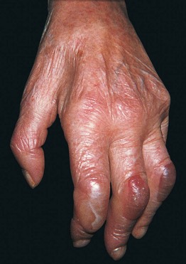
Fig. 27.4 Neurologic blisters. Tense blisters on the dorsal aspect of the fingers on the hemiplegic side of a patient with a previous cerebrovascular accident. Courtesy, José M. Mascaró Jr., MD.
• The prolonged pressure may occur in the setting of a coma, whose cause can vary from drug-induced to metabolic (e.g. hepatic encephalopathy); immobility can also result from neurologic disorders, especially cerebral vascular accidents.
For further information see Ch. 33. From Dermatology, Third Edition.
[/level-membership-for-dermatology-category][not-level-membership-for-dermatology-category]27
Other Vesiculobullous Diseases
There are a number of disorders that can present with vesicles and bullae, including exaggerated insect bite reactions (see Chapter 20), autoimmune and inherited blistering diseases (see Chapters 23–26), porphyrias (see Chapter 41), the Stevens–Johnson syndrome–toxic epidermal necrolysis spectrum (see Chapter 16), and phototoxicity, from sunburn to phototoxic drug reactions (e.g. due to doxycycline). This chapter examines a miscellaneous group of disorders, several of which favor the lower extremities, whereas Chapter 28 reviews vesiculobullous diseases in newborns and infants.
Friction Blisters
Edema Bullae (Edema Blisters)
• The most common location is the distal lower extremities, often in the setting of an acute exacerbation of chronic edema in an elderly patient; in patients with anasarca and those who are bedridden, the distribution can be more widespread (Fig. 27.1).
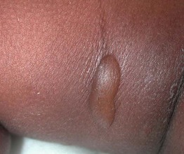
Fig. 27.1
[/not-level-membership-for-dermatology-category]
