Other Neuromuscular Transmission Disorders

Published on 03/03/2015 by admin

Filed under Neurology

Last modified 22/04/2025

Print this page

rate 1 star rate 2 star rate 3 star rate 4 star rate 5 star
Your rating: none, Average: 0 (0 votes)

This article have been viewed 2528 times

74 Other Neuromuscular Transmission Disorders

Clinical Vignette

A 58-year-old woman presented with a 3-month history of progressive fatigue and lower extremity weakness, especially noticeable when she was walking up and down stairs or arising from a chair. Lately she had noted mildly slurred speech, transient double vision with eyelid droop, and dry mouth. Her past medical history was unremarkable. She had a 60-pack-year history of cigarette smoking. Examination was remarkable for mild weakness of orbicularis oculi, neck flexor, deltoid, triceps, and hip flexor and hip extensor muscles bilaterally. She had difficulty arising from a chair. Muscle stretch reflexes were absent on routine testing but elicitable after 10 seconds of isometric exercise.

Serum creatine kinase and acetylcholine (ACh) receptor antibody study results were normal. Nerve conduction studies (NCS) demonstrated low compound muscle action potentials (CMAPs) on most motor nerve conduction studies. A 15% decrement of CMAP amplitude was observed with 3-Hz repetitive motor nerve stimulation of the ulnar nerve. After 10 seconds of exercise, the ulnar CMAP amplitude facilitated approximately 300%, which is typical of a presynaptic neuromuscular transmission disorder. She was diagnosed with Lambert–Eaton myasthenic syndrome (LEMS). Serum voltage-gated P/Q calcium channel (VGCC) antibodies were later found to be elevated, consistent with LEMS.

A chest radiograph showed a right hilar mass; its biopsy revealed small cell lung cancer (SCLC). The patient received chemotherapy, as well as 3,4-diaminopyridine for symptomatic treatment of LEMS, resulting in some modest improvement in strength. She died 16 months later from complications of SCLC.

Lambert–Eaton Myasthenic Syndrome

Although rare, LEMS is the most frequently occurring presynaptic neuromuscular transmission disorder in adults. In approximately 50% of cases, LEMS is associated with cancer, especially SCLC. When LEMS is not associated with cancer, a primary autoimmune etiology is suspected. The paraneoplastic and nonparaneoplastic forms of LEMS share an autoimmune pathophysiologic mechanism. VGCC antibodies are detected in more than 90% of patients with either paraneoplastic or nonparaneoplastic LEMS. Antibodies to SOX1 have been identified in the majority of paraneoplastic LEMS patients but are not found in patients with nonparaneoplastic LEMS, and thus may prove valuable in evaluating for the presence or absence of cancer in a patient with LEMS. LEMS associated with lung cancer usually presents in past or present smokers, and clinical manifestations of LEMS often begin months to years before diagnosis of the malignancy. Therefore, a timely LEMS diagnosis may expedite the lung cancer diagnosis and influence treatment and prognosis. LEMS unassociated with cancer usually occurs in younger nonsmokers, especially women.

The vignette in this chapter illustrates cardinal LEMS symptoms and signs: proximal extremity weakness, reduced or absent muscle stretch reflexes, and complaints of fatigue and dry mouth, with milder symptoms of ocular and oropharyngeal weakness. Because of the patient’s history of cigarette smoking, paraneoplastic LEMS was the prime consideration. The muscle stretch reflexes demonstrated improvement after 10 seconds of exercise, indicative of a presynaptic neuromuscular junction transmission disorder, and this corresponded to the NCS of postexercise facilitation of the CMAP amplitude.

LEMS often provides an early diagnosis of SCLC because the immune response producing LEMS is believed to begin early in tumor evolution. However, by the time LEMS was diagnosed in the preceding vignette, the SCLC had metastasized and prognosis was poor.

Etiology and Pathophysiology

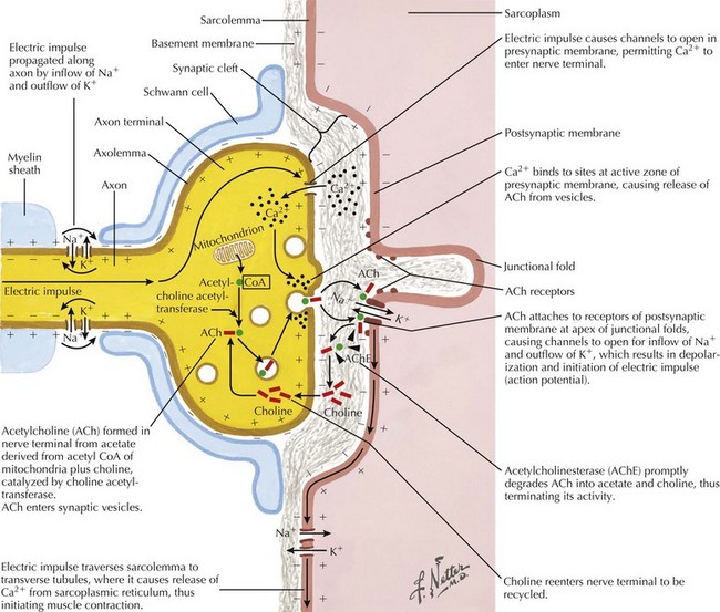
Under normal conditions, neuronal depolarization opens VGCCs on the presynaptic axon terminal membrane, resulting in calcium influx within the nerve terminal. Intracellular calcium binds to calmodulin and mobilizes acetylcholine (ACh) vesicles that are released into the synaptic cleft (Fig. 74-1). The VGCCs are the primary site of immunopathology in LEMS. Divalent IgG autoantibodies cross-link the calcium channels, disrupting their function, resulting in inadequate release of presynaptic ACh vesicles from motor and autonomic cholinergic nerve terminals. The release of fewer ACh quanta at the neuromuscular junction decreases the probability of reaching the “all or none” depolarization threshold of a muscle fiber and thus the likelihood of the muscle fiber action potential (MFAP) generation. It is this drop out of a significant number of MFAP generation that results in low CMAPs on routine NCS.

The VGCCs expressed on SCLC cells and other neoplasms provide the presumed antigenic stimulus for antibody production to VGCC in the paraneoplastic form of LEMS. The precise antigenic stimulus in the nonparaneoplastic varieties of LEMS remains to be identified. Muscle weakness, fatigue, and autonomic symptoms result because the VGCC antibodies reduce the ACh release at motor and autonomic nerve terminals, impairing synaptic transmission.

Diagnostic Approach

Appropriate NCS testing is crucial in the diagnosis of LEMS (Fig. 74-3). Most patients with LEMS have low-amplitude CMAPs on routine nerve conduction studies. The identification of low baseline CMAP amplitudes should prompt postexercise facilitation testing. This is performed by having the patient isometrically contract the muscle from which the CMAP is being recorded (e.g., abductor digiti quinti minimi) for 10 seconds followed by nerve stimulation and measurement of the CMAP. In patients without LEMS (or another presynaptic neuromuscular junction transmission disorder), there will be no change or only modest increase (<100%) in the size of the CMAP amplitude. The differentiation between a presynaptic and a postsynaptic neuromuscular junction transmission disorder is that brief (10–15 seconds) voluntary exercise, or rarely, if necessary, high-frequency (20–50 Hz) repetitive motor nerve stimulation, leads to marked (>100%) facilitation of the baseline CMAP in patients with LEMS (presynaptic) but not those with myasthenia gravis (MG) (postsynaptic). In addition to testing for postexercise facilitation, slow (e.g., 2- or 3-Hz) repetitive motor nerve stimulation demonstrating decrement of CMAP amplitude of more than 10% can further support the diagnosis of a neuromuscular transmission disorder. Many electromyography (EMG) laboratories routinely combine the slow repetitive motor nerve testing with the 10-second exercise testing in order to search simultaneously for both the postexercise facilitation (of the first CMAP following brief exercise) and the decrement (of the subsequent CMAPs during slow repetitive nerve studies) seen in postsynaptic disorders.

Serum VGCC antibody testing is also very helpful in diagnosing LEMS. Abnormally high titers of VGCC antibodies are found in approximately 90% of patients with LEMS. High titers of VGCC are also found in 20–40% of patients with SCLC who do not have LEMS. Therefore, a positive VGCC antibody test result does not necessarily diagnose LEMS; the typical clinical and EMG findings are needed to support the diagnosis.

Approximately 50–70% of patients with LEMS have an associated cancer (usually SCLC); therefore, a search for malignancy must be initiated in each patient diagnosed with LEMS. Antibodies to SOX1 have recently been identified in the majority of patients with paraneoplastic LEMS but not in patients with LEMS unassociated with cancer, and thus serological testing for SOX1 may become a valuable aid in searching for cancer in patients who present with LEMS. Imaging for cancer (e.g., chest computed tomographic [CT] scan, chest magnetic resonance imaging scan, and positron emission tomographic [PET] scan) is indicated even if the initial chest radiograph is normal. If the CT is negative, pulmonary cytologic studies, including sputum analysis and bronchial washings, may be valuable to diagnose occult lung tumors, particularly in patients who have a smoking history. Follow-up chest imaging for cancer surveillance is indicated for at least 4 years after the diagnosis of LEMS, in patients with a smoking history.

Differential Diagnosis

The combination of symptoms of weakness and dry mouth sometimes mimics the hyperventilation syndrome, hysteria, malingering, or depression. These conditions should be in the differential early in the evaluation but an appropriate history-taking and examination, combined with appropriate NCS/EMG testing, should allow the physician to easily distinguish a psychological etiology from a neurologic cause. The neurologic disorders that most often mimic LEMS are MG, myopathy, and a chronic polyneuropathy. Unlike LEMS, MG typically has a preponderance of ocular or bulbar symptoms—diplopia, ptosis, dysarthria, and dysphagia—early in its course, whereas in LEMS, oculobulbar symptoms are less commonly the presenting symptoms and usually remain mild. Prominent autonomic symptoms, such as dry mouth and erectile dysfunction, are characteristic of patients with LEMS but are often overlooked by patients and/or clinicians. Other rare presynaptic neuromuscular transmission disorders (e.g., botulism and magnesium intoxication) usually present acutely. Patients with inflammatory myopathies also have predominant proximal limb and neck weakness. However, these patients do not demonstrate facilitation of strength immediately after testing of each muscle. The muscle stretch reflexes are usually preserved in patients with myopathy. Furthermore, patients with myopathy typically do not complain of autonomic and sensory symptoms; serum CK is usually elevated, which would be unexpected in LEMS. Chronic inflammatory demyelinating polyradiculoneuropathy (CIDP), with its insidious onset of proximal weakness and areflexia, also enters the LEMS differential diagnosis. Nerve conduction studies in CIDP demonstrate features of acquired demyelination (e.g., slowed conduction velocities, conduction block, temporal dispersion), findings never seen in LEMS. Moreover, patients with CIDP usually have prominent sensory nerve symptoms and signs, whereas in LEMS sensory symptoms are typically mild.

Treatment and Prognosis

Chemotherapy, radiation therapy, and surgery are the primary treatment modalities for LEMS-associated lung cancer. Cancer treatment often leads to symptomatic improvement in LEMS, presumably by removing the antigenic stimulus and thus downgrading the immune response. Symptomatic treatment of LEMS aims to improve neuromuscular transmission. The anticholinesterase medication pyridostigmine (Mestinon) can improve neuromuscular transmission by inhibiting breakdown of Ach at the neuromuscular junction. In contrast to MG, pyridostigmine alone is not very effective for LEMS but it is often worth trying because it is a relatively benign treatment option. Common and dose-limiting side effects are abdominal cramps and diarrhea.

3,4-Diaminopyridine (3,4-DAP) promotes ACh release from the presynaptic portion of the neuromuscular junction by prolonging the VGCC open time. It is available for use primarily in Europe but in the United States requires local institutional review board approval and a concomitant submission to the FDA for compassionate use. Caution is advised because central nervous system irritability, manifested by seizures, is a major adverse effect. Anticholinesterases, such as pyridostigmine, potentiate the effects of 3,4-DAP and thus may be particularly beneficial when combined with 3,4-DAP.

Immunomodulation therapy is also sometimes used to inhibit the immune system’s response directed at the neuromuscular junction. Unfortunately, for LEMS patients with cancer, immunomodulation may also inhibit the response of the host’s immune system to the cancer, potentially worsening the cancer. Corticosteroids (e.g., prednisone), intravenous immunoglobulin, and plasmapheresis are some of the immunotherapies that may be beneficial to LEMS patients with profound weakness. These treatments are more commonly prescribed to nonparaneoplastic LEMS patients.

Several medications may exacerbate LEMS symptoms and should be used cautiously in LEMS; these include cardiac drugs, that is, adrenergic and calcium channel–blocking agents, and antiarrhythmic agents, such as procainamide and quinidine. Aminoglycoside antibiotics, magnesium citrate cathartics, quinine, and lithium may also worsen the neuromuscular transmission defect, increasing weakness. Anesthesiologists must be aware of the diagnosis of LEMS in their patient and use medications that do not prolong postoperative respiratory depression. On rare occasions, postanesthesia respiratory failure is the initial manifestation of LEMS.

Prognosis depends on whether LEMS is associated with malignancy and on the stage of malignancy. Most patients with LEMS and SCLC have a median survival of a few years. However, early detection improves prognosis, and consequently it is important to diagnose LEMS quickly and to search aggressively for cancer. In contrast, the prognosis in patients with primary autoimmune LEMS without SCLC is good; some patients live for more than 20 years after diagnosis. These patients with nonparaneoplastic LEMS often respond positively to immunomodulation and symptomatic therapy.

Botulism

Botulism is a rare, naturally occurring disease that is caused by release of toxin from C. botulinum. The toxins of C. botulinum exert their action on the presynaptic neuromuscular junction by blocking the release of ACh. Infantile botulism is more common than adult botulism. Early recognition of infantile and adult botulism can lead to expedited treatment.

Clinical Presentation
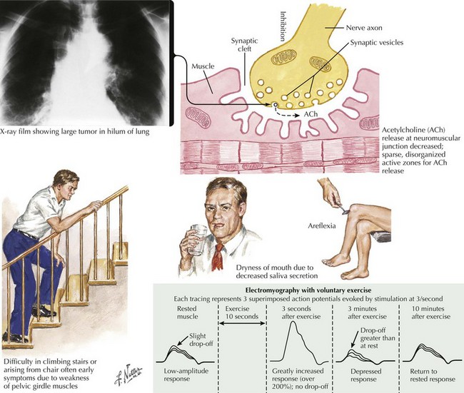
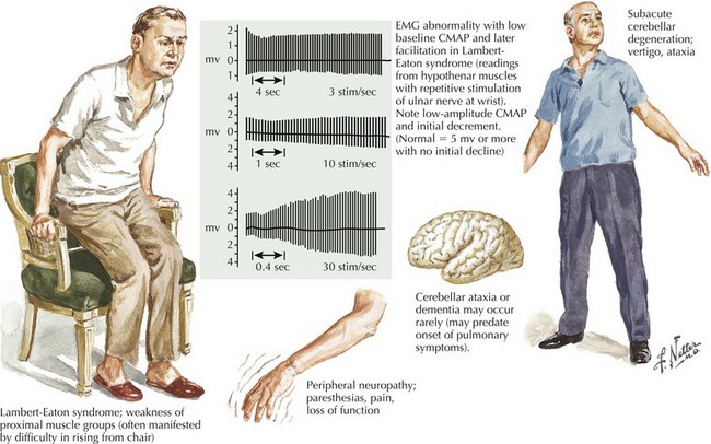
, Diagnostic Approach, and Differential Diagnosis

The presentation of botulism is distinctive with symmetrical cranial motor nerve palsies followed by descending flaccid paralysis, including the respiratory muscles. Extraocular muscle weakness (cranial nerves 3, 4, and 6) causes diplopia. Bilateral ptosis is common. Pupillary response is often impaired, resulting in blurry vision. Facial and oropharyngeal weakness, causing dysphagia and dysarthria, are prominent. Limb and axial weakness often develop; diaphragmatic and accessory breathing muscle weakness can cause respiratory compromise. Muscle stretch reflexes are lost. Autonomic nervous system dysfunction, including dry mouth, constipation, and postural hypotension, also occur. Cognition and the sensory system are unaffected.

The differential diagnosis includes MG, Guillain–Barré syndrome (GBS), LEMS, stroke syndromes, and tick paralysis. The very rapid progression of relatively symmetric cranial motor neuropathies followed by axial and limb weakness and dysautonomias (pupillary abnormalities, dry mouth, and constipation) are atypical for MG. On NCS/EMG testing, the presence of, or absence of, significant postexercise or posttetanic facilitation also helps to differentiate a presynaptic from postsynaptic neuromuscular junction transmission disorder. Furthermore, infants are unlikely to develop autoimmune MG and adults are unlikely to develop botulism, so age of onset is an important consideration. Demonstration of C. botulinum in a patient’s stool sample or in cultures of a wound strongly favors botulism, whereas antibodies in serum for the acetylcholine receptor support a diagnosis of MG. GBS, particularly the Miller Fisher variant of GBS, can mimic botulism. Age of onset can again be helpful as GBS is uncommon in infants. Cerebrospinal fluid (CSF) protein is elevated in most cases of GBS, including most cases of Miller Fisher syndrome, whereas CSF protein is normal in botulism. NCS/EMG is often very helpful in detecting evidence of demyelination in GBS and the characteristic electrodiagnostic features of a presynaptic disorder of neuromuscular transmission in botulism (as discussed above in the LEMS section). Tick paralysis can essentially be excluded by a meticulous skin examination, including the scalp; in cases of North American tick paralysis, tick removal leads to a rapid recovery. However in Australia, the inciting tick’s toxin has an effect sometimes lasting a few months.

Additional Resources

Burns TM, Russell JA, Lachance DH, Jones HR. Oculobulbar involvement is typical with Lambert-Eaton Myasthenic Syndrome. Ann Neurol. 2003;53:270-273.

Gutmann L, Phillips LH, Gutmann L. Trends in the association of Lambert-Eaton myasthenic syndrome with carcinoma. Neurology. 1992;42:848-850.

Lambert EH, Eaton LM, Rooke ED. Defect of neuromuscular conduction associated with malignant neoplasms. Am J Physiol. 1956;187:612-613.

Lambert EH, Rooke ED. Myasthenic state and lung cancer. In: Brain WR, Norris FH, editors. The Remote Effects of Cancer on the Nervous System. New York, NY: Grune and Stratton; 1965:67-80.

Lennon VA, Kryzer TJ, Griesmann GE, et al. Calcium-channel antibodies in the Lambert-Eaton syndrome and other paraneoplastic syndromes. N Engl J Med. 1995;332:1467-1474.

McEvoy KM, Windebank AJ, Daube JR, Low PA. 3,4-Diaminopyridine in the treatment of Lambert-Eaton myasthenic syndrome. N Engl J Med. 1989;321:1567-1571.

O’Neil JH, Murray NMF, Newsom-Davis J. The Lambert Eaton myasthenic syndrome: a review of 50 cases. Brain. 1988;111:577-596.

Sabater L, Titulaer M, Saiz A, Verschuuren J, Güre AO, Graus F. SOX1 antibodies are markers of paraneoplastic Lambert-Eaton myasthenic syndrome. Neurology. 2008;70:924-928.

Sanders DB. Lambert-Eaton myasthenic syndrome: clinical diagnosis, immune-mediated mechanisms, and update on therapies. Ann Neurol. 1995;37(suppl 1):S63-S73.

Sobel J. Botulism. Clinical Infectious Diseases. 2005;41:1167-1173.

Tim RW, Sanders DB. Repetitive nerve stimulation studies in the Lambert-Eaton myasthenic syndrome. Muscle Nerve. 1994;17:995-1001.

Tseng-Ong L, Mitchell WG. Infant botulism: 20 years’ experience at a single institution. J Child Neurol. 2007;22:1333-1337.