Other neonatal emergencies: Tracheoesophageal fistula and omphalocele

Published on 07/02/2015 by admin

Filed under Anesthesiology

Last modified 22/04/2025

Print this page

rate 1 star rate 2 star rate 3 star rate 4 star rate 5 star
Your rating: none, Average: 0 (0 votes)

This article have been viewed 1739 times

Other neonatal emergencies: Tracheoesophageal fistula and omphalocele

Robert J. Friedhoff, MD

Tracheoesophageal fistula

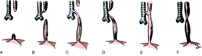
Tracheoesophageal fistula (TEF) occurs as the result of failure of the tracheal bud to develop normally from the primitive foregut. TEF occurs in several forms (Figure 203-1); type C—esophageal atresia (more correctly agenesis because the proximal part of the primitive foregut develops primarily into a trachea rather than an esophagus) with a distal TEF—is the most common form (accounts for 90% of all TEFs). Maternal polyhydramnios may indicate the presence of the lesion before birth. Diagnosis is suspected at birth when the neonate has excessive drooling, cyanotic episodes, or coughing relieved by suctioning or the clinician is unable to pass a soft catheter into the infant’s stomach. TEF can be confirmed by radiography by showing a curled catheter in the upper esophageal pouch with an air bubble in the stomach. Contrast medium is unnecessary and contraindicated because the neonate may aspirate the medium. Associated conditions include prematurity (20%-25%), congenital heart disease (20%-25%), and other midline defects.

Intraoperative management

Induction techniques for repair of a TEF include the use of inhalation agents with a rapid sequence or awake intubation. The use of N2O, which will add to gastric distention, should be avoided. Care must be taken to avoid intubating the fistula. A tracheal tube with the Murphy eye facing anteriorly should be inserted into the right main bronchus while listening for unilateral breath sounds; the tracheal tube is then pulled back until bilateral breath sounds are heard. Some clinicians prefer to cut the distal end of the tracheal tube, eliminating the Murphy eye. Use of a cuffed tracheal tube to both ventilate and occlude the fistula has been reported. Placement of a Fogarty catheter to identify and occlude the fistula using a pediatric bronchoscope can be attempted. Spontaneous ventilation, to avoid gastric distention until the fistula has been ligated, should be performed, and then controlled ventilation can be used.

Thoracoscopic repair of the fistula will avoid a thoracotomy and its sequelae. Use of a Fogarty catheter placed in the right main bronchus with the aid of a bronchoscope will facilitate one-lung ventilation. An alternative technique is to manipulate the tracheal tube to the left main bronchus with a bronchoscope. Surgical insufflation of CO2 (5-6 mm Hg) into the right hemithorax will cause right lung collapse and possible hypercapnia and hypoxemia. Time to extubation and discharge from the neonatal intensive care unit are decreased with thoracoscopic repair.

A precordial stethoscope should be placed under the dependent lung. Routine intraoperative monitors and an arterial catheter are typically used. Regional anesthesia can be added as an adjuvant.

Omphalocele

Anesthetic management for omphalocele and gastroschisis are essentially the same, but knowledge of the associated anomalies will influence anesthetic decisions. Omphalocele and gastroschisis are congenital defects of the anterior abdominal wall, permitting external herniation of abdominal viscera. Gastroschisis is not midline (usually occurs on the right), has a normally situated umbilical cord (not covered with a hernia sac), and is rarely associated with other congenital anomalies but is associated with an increased incidence of prematurity.

Omphalocele has a 75% incidence of other congenital defects, including cardiac anomalies (ventricular septal defects most common), trisomy 21, and Beckwith-Wiedemann syndrome (omphalocele, organomegaly, macroglossia, and hypoglycemia). Epigastric omphaloceles are associated with cardiac and lung anomalies. Hypogastric omphaloceles are associated with exstrophy of the bladder and other genitourinary anomalies.