Published on 07/02/2015 by admin
Filed under Anesthesiology
Last modified 22/04/2025
This article have been viewed 1901 times
C. Thomas Wass, MD
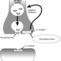
Nonhemolytic transfusion reactions (NHTRs) often occur in patients receiving blood product transfusions. Fever is the most common NHTR, with a median frequency of 4%. Fever, defined as an increase in body temperature of 1°C or more during or within several hours of transfusion, usually persists for less than 12 h, is most often associated with transfusion of cellular components (e.g., red blood cells, platelets, and granulocytes), but has also been observed with transfusion of noncellular components (e.g., fresh frozen plasma or cryoprecipitate). Although the etiology has yet to be fully elucidated, it is hypothesized that recipient alloimmunization (i.e., antibody production in response to a previous transfusion or pregnancy) toward donor white blood cells or platelets triggers release of leukocyte-derived or platelet-derived pyrogenic cytokines (e.g., IL-1β, IL-6, IL-8, TNF-α, CD40L) that increase the hypothalamic thermoregulatory set point. Alternatively, fever may occur in response to direct transfusion of pyrogenic cytokines or other inflammatory mediators that accumulate during storage of blood products such that the greater the interval between collection and transfusion, the higher the frequency of febrile NHTR. However, prestorage leukocyte reduction (e.g., using leukocyte filtration techniques) mitigates transfusion-related fever.
Should the patient develop a fever while receiving a transfusion, the transfusion must be discontinued or slowed. Bacterial contamination (diagnosed via Gram stain and cultures) and hemolytic transfusion reaction (diagnosed via repeat crossmatch and direct Coombs test, which detects antibody to transfused donor red blood cells) should be ruled out. Antipyretic drugs (e.g., acetaminophen) may be used prophylactically or to treat febrile NHTRs; however, these medications may not prevent associated symptoms (e.g., chills, rigor, soreness at the transfusion site, headache, nausea, myalgia, chest tightness).
Mild allergic reactions are the second most common NHTR, occurring with a frequency of 0.5%. Signs and symptoms are usually mild and include urticarial rash and generalized pruritus as a result of IgE-mediated histamine release from degranulated mast cells and basophils in response to foreign substances (e.g., transfused plasma proteins) found in any plasma-containing blood products (especially platelets and fresh frozen plasma). Patients who do not show signs of having an anaphylactic reaction should be treated symptomatically with diphenhydramine, and the transfusion may be continued.
Anaphylaxis, which represents the most severe form of NHTR, occurs in 1 in 20,000 to 1 in 50,000 transfusions. Patients experiencing these reactions typically have hereditary IgA deficiency, which is relatively common (1 in 700 persons). During exposure to “foreign” IgA from a previous transfusion or pregnancy, patients become alloimmunized (i.e., recipients develop IgE directed against donor IgA). IgE elicits an immune response by binding to Fc receptors on the surface of mast cells and basophils, resulting in degranulation and release of vasoactive mediators (e.g., histamine, leukotrienes, and prostaglandins). Transfusion of any plasma-containing blood product may result in an anaphylactic response. Signs, symptoms, and treatment do not differ from those of other anaphylactic reactions.
The diagnosis of an anaphylactic transfusion reaction requires quantitative confirmation of IgA deficiency and the presence of anti-IgA in recipient plasma. Levels of serum β-tryptase, a marker for mast cell degranulation, may be measured. However, these laboratory studies are often time consuming and may not be readily available. Thus, once a diagnosis of anaphylactic transfusion reaction is suspected, the transfusion should be stopped immediately. If blood transfusion must be continued, IgA-deficient blood products (e.g., blood from donors known to be IgA deficient or washed or deglycerolized red blood cells) should be used.
Both mild allergic and IgA anaphylactic reactions usually begin within 45 min after blood transfusion is started but may be delayed for as long as 1 to 3 h. Shorter onset times tend to be associated with more severe reactions.
Pulmonary edema following blood transfusion is often attributed to intravascular volume overload that overwhelms myocardial Frank-Starling forces (i.e., cardiogenic pulmonary edema). In contrast, transfusion-related acute lung injury (TRALI) is a noncardiogenic form of pulmonary edema that is difficult to distinguish from acute respiratory distress syndrome or other causes of acute lung injury. TRALI, a diagnosis of exclusion, usually occurs within 1 to 6 h of blood product transfusion and is characterized by acute respiratory distress, radiograph evidence of bilateral pulmonary edema, severe hypoxemia (PaO2/FIO2 < 300 mm Hg), and no evidence of a cardiogenic cause. TRALI is likely underdiagnosed and underreported; however, it is estimated to occur in 1 in 5000 patients who receive a transfusion.
The pathogenesis of TRALI is incompletely understood but is likely multifactorial. In 65% to 90% of patients who develop TRALI, white blood cell (including class I and II HLA or neutrophil-specific) antibodies that bind recipient white blood cell antigens can be identified in donor plasma. When white blood cell antibodies are not present in the donor’s serum, another explanation for the development of TRALI may be the two-hit theory—an initial insult (e.g., infection, surgery, or trauma) attracts and “primes” neutrophils that adhere to pulmonary vascular endothelium. A subsequent “activating stimulus” (e.g., transfusion of plasma containing biologically active mediators) causes these marginated neutrophils to release oxidases, O2 free-radical species, and proteases, resulting in endothelial damage and extravasation of intravascular fluid into lung parenchyma.
Transfusion of any blood product containing plasma can cause TRALI. Interestingly, the vast majority of implicated donors are multiparous women who have been alloimmunized to paternal HLA antigens (reported to occur in up to 25% women with more than three pregnancies). Thus, some centers restrict the use of fresh frozen plasma donated by multiparous women. Treatment is supportive, and depending on the severity of TRALI, the patient may require tracheal intubation, oxygenation, and mechanical ventilation.
With a mortality rate approaching 10%, TRALI is the leading cause of transfusion-related death in the United States. However, most patients with TRALI improve clinically, physiologically, and radiographically within 48 to 96 h.
Blood transfusion can significantly improve (in a dose-dependent manner) allograft survival following renal transplantation, yet it worsens tumor recurrence and mortality rate following resection of many cancers (e.g., breast, colorectal, gastric, head and neck, hepatocellular, lung, prostate, renal, soft tissue sarcoma) when compared with patients who do not receive transfusions or individuals who receive leukocyte-reduced blood transfusions. In either case, alterations in patient outcome have been attributed to transfusion-mediated immunomodulation, referred to as a “tolerogenic effect.” Such an effect may be due to upregulation of humoral immunity (i.e., B-cell function and antibody production), down regulation of cell-mediated immunity (i.e., T-cell function), or both.
Despite improved renal allograft survival in transfused transplant recipients, routine perioperative blood transfusion is not indicated because of the effectiveness and safety of immunosuppressant drugs (e.g., cyclosporine) and concerns about transfusion-related infection.
Fausts Anesthesiology Review Expert Consult 4e
WhatsApp us