141 Non-Snake Reptile Bites
 Key Points
• Alligator and crocodile bites may inflict significant internal injury and should be managed as major trauma.
• Gila monster bites may leave teeth in the wound that are not visible by radiography.
• Patients with a Gila monster bite should be observed for 8 hours after the bite because of the risk for systemic toxicity.
• The Komodo dragon has joined the Gila monster and the beaded lizard as the only known venomous lizards of medical significance; its bites are known to cause hypotension and coagulopathy, in addition to infections.
• Green iguanas and snapping turtles may carry Salmonella.
• Delayed wound closure and prophylactic antibiotics should be considered in all patients with reptile bites.
Epidemiology
Crocodilians account for more human injuries and fatalities worldwide than any other non-snake reptile. Three of the largest species are known for unprovoked attacks on humans: the American alligator (Alligator mississippiensis), the saltwater crocodile (Crocodylus porosus), and the Nile crocodile (Crocodylus niloticus). From 1928 through 2008, 567 reports of adverse encounters with American alligators resulted in 24 deaths in the United States. Most injuries were due to a single bite.1,2 In Australia, 80% of unprovoked attacks by the saltwater crocodile involved people who were either swimming or wading in water. The Nile crocodile of Africa, though smaller and less territorial, probably accounts for more human fatalities than all other 23 species of crocodilians combined.3
Most Gila monster bites are nonaccidental and occur as a result of handling the reptile. Accidental bites have become exceedingly rare. No deaths from Gila monster bites have been reported in the United States in the past 50 years.4,5 The Komodo dragon is now known to have a venom apparatus similar to that of the Gila monster, and deaths have been reported as a result of their bites.
Crocodilians
Pathophysiology
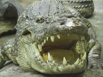
Crocodilians survived the Ice Age and are the dominant predators in many of the world’s tropical waterways. The American alligator may be found in the southeastern United States, from North Carolina to Louisiana. The saltwater crocodile is found in Southeast Asia and Australia, with males reaching lengths of up to 23 feet (7 m). Victims of crocodilian attacks sustain trauma from a combination of penetrating, blunt, and sheer force. The saltwater crocodile can generate 2000 psi when it bites. The sheer magnitude of the jaw muscle force sustained with extensive penetrating injuries devitalizes large areas of tissue, thereby making such injuries slow to heal and susceptible to infection. Bites must be considered to be heavily contaminated with multiple bacteria, including Aeromonas hydrophila, Pseudomonas, and Proteus (Fig. 141.1).
Presenting Signs and Symptoms
Crocodilian bites are characterized by punctures and tears. Their teeth are conical and not designed for chewing but for grasping their prey. Among survivors, the extremities were the most commonly injured site, with less than 10% sustaining torso trauma.3,6 Crocodilians may also roll their entire body (known as the “death roll”) to disorient and drown the victim, as well as to tear pieces from the victim’s body.3 The force of the massive jaws may lead to extensive internal injury, but even when bite wounds are not present, the force of the animal’s movement—or even blunt trauma from its tail—may also inflict significant internal trauma. Initial survivors of severe attacks may exhibit hypotension from massive hemorrhage, in addition to respiratory distress from submersion injury.
Differential Diagnosis and Medical Decision Making
 Priority Actions
Reptile Bite Wounds
Treat all victims of crocodilian bites as major trauma patients with the potential for massive internal injuries.
Extremity bites should be evaluated for underlying vascular, tendon, nerve, or bone injury.
Copiously irrigate and débride all wounds, especially crocodilian and Komodo dragon bites.
Facial wounds and others with significant cosmetic concern should be closed if the patient is seen early after the bite and does not show signs of infection.
Consider a 5-day course of prophylactic antibiotics with close outpatient follow-up to monitor for signs of infection.
Gila Monster and Beaded Lizard
Pathophysiology
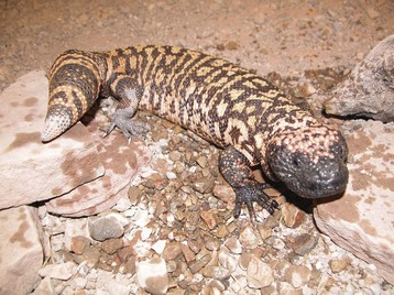
The Gila monster (Heloderma suspectum) and the closely related beaded lizard (Heloderma horridum) are native to the southwestern United States and Mexico but may be found throughout the world in zoos and as illegal pets. They are characterized by black beadlike scales mixed with bandlike patterns of yellow, white, or pink scales (Fig. 141.2). These reptiles typically range in length from 9 to 32 inches (22 to 81 cm); the beaded lizard is the larger of the two species.
Unlike pit vipers, which inject their venom through fangs, Heloderma species have a very rudimentary venom delivery mechanism. The animal has individual vertical grooves in its lancet-shaped teeth to deliver venom by capillary-type action from anterior mandibular venom glands. The lizards tend to stay attached to their victims by using mastication to augment the delivery of venom.5,6 Their venom is similar in some respects to rattlesnake venom and can stimulate the release of vasoactive kinin, which leads to hypotension.5
Presenting Signs and Symptoms
Patients may arrive at the emergency department (ED) with the lizard still attached. Bite wounds are classically indurated and erythematous. Significant edema may develop at the site of the wound. Patients often describe excruciating pain associated with the bites. In envenomated patients, weakness, hypotension, and tachycardia are commonly seen as a result of systemic effects of the reptile’s venom.4,5
Differential Diagnosis and Medical Decision Making
 Priority Actions
Envenomation by a Gila Monster or Beaded Lizard
Observe all patients for 8 hours to watch for the development of signs of envenomation.
Initiate fluid therapy for hypotension and tachycardia.
Initiate vasopressor therapy for hypotension and tachycardia unresponsive to fluids.
Use adequate opiate analgesia to control pain.
Admit patients with systemic symptoms to the hospital for observation and resolution of symptoms.
Treatment
Envenomation may cause hypotension, tachycardia, and generalized weakness. These symptoms generally respond well to intravenous crystalloid administration. Refractory hypotension may require treatment with vasopressors such as dopamine. No antivenom is commercially available for Gila monster or beaded lizard envenomation.4,6
Tips and Tricks
Animal Removal
Apply a flame under the animal’s mandible.
Immerse the animal in cold water.
Use a large Kelly clamp or handheld cast spreader to manually disengage the animal’s mouth from the victim.
Place a thin rod across the animal’s mouth between the bite site and the jaw and push back on the jaw.
Avoid pulling the animal off, if possible; this may cause further tissue damage and detach teeth into the wound.
The most important aspect of evaluation of the wound is physical examination for evidence of vascular or tendon injury and local wound exploration for retained teeth from the animal. The pain typically requires large amounts of opiate analgesics; the few patients who have experienced both rattlesnake and Gila monster bites report much greater pain associated with a Gila monster bite. The pain generally peaks between 15 and 45 minutes following the bite and may last for days.4,7
Komodo Dragon
Pathophysiology
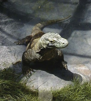
Native to the islands of Indonesia, the Komodo dragon (Varanus komodoensis) is the largest lizard in the world; it can reach lengths greater than 10 feet (3 m) and weigh as much as 300 lb (136 kg) (Fig. 141.3). These lizards can move as fast as 13 mph (20 km/hr) over short distances and take down prey as large as water buffalo. Komodo dragons’ teeth are sharklike, with posterior serrations that create deep open wounds to facilitate envenomation. Its venom is known to cause coagulopathy, increased vascular permeability, and vasodilation.8 Victims bleed profusely from large wounds. hypotension and shock develop rapidly and lead to death. If the initial attack does not kill the victim, infections from multiple pathogenic bacteria in the Komodo’s saliva can lead to sepsis and death.9
Presenting Signs and Symptoms
Most evaluations of Komodo dragon bites in the United States would expectedly come from exposure at zoos or zoologic parks. These bites typically occur on the extremities and produce puncture wounds and lacerations of the skin, along with the potential for local tendon, bone, and neurovascular damage. Bleeding from the wounds may be significant because of envenomation and subsequent coagulopathy. patients who delay medical evaluation of their wounds will most likely show signs of local and/or systemic bacterial infection. Komodo dragon attacks in the wild may result in multiple large lacerations and areas of tissue loss, not unlike victims of shark attacks. The combination of extensive tissue loss and venom toxicity can lead to hypotension, exsanguination, and sudden death.8
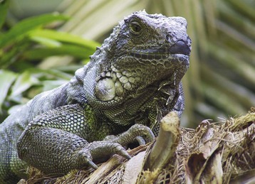
Green Iguana
Pathophysiology
The green iguana (Iguana iguana) is native to Central and South America. It is the most common lizard sold as a pet in the United States (Fig. 141.4). Feral iguana populations can now be found in Florida, Hawaii, and southern Texas. Iguanas are usually docile but can cause injuries with their teeth, claws, and tail. They can also be a source of Salmonella infection.10 No specific venom is associated with iguana bites.
Presenting Signs and Symptoms
Most trauma from iguana bites is superficial soft tissue injury, although tendon injuries have been reported.11 The majority of bites (80%) occur on the upper extremities, particularly the fingers, with 19% occurring on the face.10 As with any bite, patients may have infectious complications, particularly those with delayed care or immunosuppression.
Differential Diagnosis and Medical Decision Making
Extensive diagnostic testing is not generally necessary in cases of iguana bites. Blood and local wound cultures may be useful for patients with established wound infections and systemic signs and symptoms. During physical examination the physician should look for underlying tendon and neurovascular injury. Associated bone damage is rare with iguana bites.11
Snapping Turtle
Presenting Signs and Symptoms
 Facts and Formulas
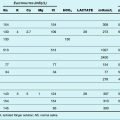
Antibiotic Therapy
Crocodilian—multiple pathogens: Broad-spectrum coverage, such as with amoxicillin-clavulanate (Augmentin)
Gila monster—multiple pathogens: Broad-spectrum antibiotic coverage, such as with amoxicillin-clavulanate (Augmentin)
Komodo dragon—multiple pathogens, including Escherichia coli and Staphylococcus spp.: Consider hospitalization for intravenous broad-spectrum antibiotics, such as ampicillin-sulbactam (Unasyn), because of the high risk for infection
Green iguana—may carry Salmonella spp.: Salmonella coverage, such as with ciprofloxacin
Snapping turtle—may carry Salmonella spp.: Salmonella coverage, such as with ciprofloxacin
Fry BJ, Wroe S, Teeuwisse W, et al. A central role for venom in predation by Varanus komodoensis (Komodo dragon) and the extinct giant Varanus (Megalania) priscus. Proc Natl Acad Sci U S A. 2009;106:8969–8974.
Gruen RL. Crocodile attacks in Australia: challenges in injury prevention and trauma care. World J Surg. 2009;33:1554–1561.
Hooker KR, Caravati EM, Hartsell SC. Gila monster envenomation. Ann Emerg Med. 1994;24:731–735.
1 Harding BE, Wolf BC. Alligator attacks in southwest Florida. J Forensic Sci. 2006;51:674–677.
2 Langley RL. Adverse encounters with alligators in the United States: an update. Wilderness Environ Med. 2010;21:156–163.
3 Caldicott DG, Croser D, Manolis C, et al. Crocodile attack in Australia: an analysis of its incidence and review of the pathology and management of crocodilian attacks in general. Wilderness Environ Med. 2005;16:143–159.
4 Hooker KR, Caravati EM, Hartsell SC. Gila monster envenomation. Ann Emerg Med. 1994;24:731–735.
5 Mebs D. Clinical toxicology of Helodermatidae lizard bites. In: Meier J, White J. Handbook of clinical toxicology of animal venoms and poisons. Boca Raton, Fla: CRC Press, 1995.
6 Gruen RL. Crocodile attacks in Australia: challenges in injury prevention and trauma care. World J Surg. 2009;33:1554–1561.
7 Miller MF. Gila monster envenomation. Ann Emerg Med. 1995;25:720.
8 Fry BJ, Wroe S, Teeuwisse W, et al. A central role for venom in predation by Varanus komodoensis (Komodo dragon) and the extinct giant Varanus (Megalania) priscus. Proc Natl Acad Sci U S A. 2009;106:8969–8974.
9 Montgomery JM, Gillespie D, Sastrawan P, et al. Aerobic salivary bacteria in wild and captive Komodo dragons. J Wildl Dis. 2002;38:545–551.
10 Forrester MB. Iguana bites reported to Texas poison centers. Am J Emerg Med. 2010;28:817–819.
11 Merin DS, Bush SP. Severe hand injury following a green iguana bite. Wilderness Environ Med. 2000;11:225–226.