Published on 19/03/2015 by admin
Filed under Dermatology
Last modified 22/04/2025
This article have been viewed 2329 times
Stuart R. Lessin and Clifford S. Perlis
Evidence Levels: A Double-blind study B Clinical trial ≥ 20 subjects C Clinical trial < 20 subjects D Series ≥ 5 subjects E Anecdotal case reports
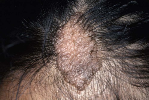
Nevus sebaceus, first described by Jadassohn in 1895, is a term for a congenital hamartoma of the epidermis and adnexal structures typically involving the scalp and face. It presents at birth or appears in early childhood as a pink, orange to yellow waxy plaque with a granular pitted surface that is often hairless. The size and configuration can be variable. During puberty, the lesion thickens and becomes verrucous as glands enlarge within the dermis. Development of cutaneous and adenexal neoplasms has been reported in 10–20% of lesions after puberty and in adulthood. These neoplasms are most commonly benign. Surgical treatment addresses cosmetic issues, as well as prophylaxis or treatment of neoplasms.
For most lesions, clinical examination is sufficient to establish the diagnosis. A skin biopsy can confirm the clinical impression when indicated. Most neoplastic growths that arise in a nevus sebaceous do not develop until after the age of 16 years; the overwhelming majority of cases are benign. These include syringocystadenoma papilliferum, trichoblastoma, tricholemmoma, sebaceoma, nevocellular nevus, and seborrheic keratosis. The most common malignancy that develops within a nevus sebaceous is a basal cell carcinoma, but the absolute incidence is very rare. Isolated case reports describe rare patients who have developed sebaceous carcinoma, squamous cell carcinoma, tricholemmal carcinoma or microcystic adnexal carcinoma within nevus sebaceus.
In 2000, a case series established that trichoblastoma was the most common neoplasm arising in nevus sebaceus. With this evidence of minimal risk of malignant neoplastic transformation, prophylactic surgery is not justified. Conservative management with clinical observation and biopsy of any lesions suspicious for malignancy appears to be most prudent.
Cribier B, Scrivener Y, Grosshans E. J Am Acad Dermatol 2000; 42(2 Pt 1): 263–8.
This is a retrospective case series of 596 cases demonstrating a 1.7% occurrence of benign tumors in childhood. Most tumors arising in nevus sebaceus occurred in adults over 40 years; in these adults, 2.1% of the neoplasms were basal cell carcinomas. The authors conclude that prophylactic surgery in children is of uncertain benefit.
Santibanez-Gallerani A, Marshall D, Duarte AM, Melnick SJ, Thaller S. J Craniofac Surg 2003; 14: 658–60.
This is a retrospective case series of 757 cases of children aged 16 years or younger. No cases of basal cell cancer were found. The authors questioned the need for prophylactic excision in children.
Jaqueti G, Requena L, Sánchez Yus E. Am J Dermatopathol 2000; 22: 108–18.
Retrospective case series of 155 cases failed to reveal any cases of basal cell carcinoma. Trichoblastoma was the most common (7.7%) basaloid tumor. It appears that histological misinterpretation of trichoblastomas as basal cell carcinomas may have been responsible for the erroneous reporting of an increased risk of basal cell carcinoma development within nevus sebaceus. The authors concluded that early prophylactic surgery seems inappropriate.
Rosen H, Schmidt B, Lam HP, Meara JG, Labow BI. Pediatr Dermatol 2009; 26: 676–81.
A retrospective, single-institution chart review evaluated 651 distinct nevus sebaceus lesions among 631 patients and 690 excisions. Twenty-one intralesional diagnoses were found in 18 patients including five (0.8%) basal cell carcinomas (mean age 12.5 years, range 9.7–17.4 years) and seven (1.1%) syringocystadenoma papilliferum (mean age 8.8 years, range 1.7–16.9 years).
Moody MN, Landau JM, Goldberg LH. Pediatr Dermatol 2012; 29: 15–23.
A comprehensive review of reported nevus sebaceus outcomes and considerations for prophylactic excision is provided. It is recommended that all cases of nevus sebaceus be evaluated individually with a thorough medical history, physical examination and review of therapeutic options in order to determine if prophylactic excision is warranted. Nevus sebaceus in cosmetically apparent regions in healthy children is recognized as a reasonable indication for consideration of prophylactic excision.
Skin biopsy
A skin biopsy may be performed to confirm a clinical diagnosis. Additionally, clinical changes suggestive of neoplastic transformation should be investigated by skin biopsy.
Based on recent case series and histological analyses that demonstrate the incidence of basal carcinoma is rare, particularly in children, clinical observation should be considered as the first option for any intervention. Prophylactic surgical excision is not warranted based on current data. Elective surgical excision may be considered if lesions become symptomatic or impact appearance. In instances of biopsy-confirmed neoplastic transformation, management should be dictated by the histology of the specific tumor.
Barkham MC, White N, Brundler MA, Richard B, Moss C. J Plast Reconstr Aesthet Surg 2007; 60: 1269–70.
Retrospective case series of 63 cases: 37 seen by plastic surgeons and 26 seen by dermatologists. Plastic surgeons excised 28 of 37 cases and dermatologists excised four of 26 cases. No malignant changes were seen in the 32 excisions; only one apocrine adenoma was found. The authors concluded that prophylactic surgery is not warranted.
A variety of destructive methods have been described for treatment of nevus sebaceus and provide an alternative to surgical excision; however, they may not be as effective in removal of deeper structures.
Dierickx CC, Goldenhersh M, Dwyer P, Stratigos A, Mihm M, Anderson RR. Arch Dermatol 1999; 135: 637–40.
This is a case report utilizing photodynamic therapy with topical δ-aminolevulinic acid for elective treatment of a large nevus sebaceus of the face in a patient with cosmetic concerns who did not wish to undergo surgical excision. An excellent cosmetic result was obtained, though post-treatment skin biopsy revealed residual sebaceous tissue in the reticular dermis, underlying the normal appearing epidermis, and a widened, fibrotic dermis.
Ashinoff R. Pediatr Dermatol 1993; 10: 189–91.
This is a case report of the use of a CO2 laser to electively treat a nevus sebaceus on the nose in a 10-year-old boy. CO2 laser vaporization provided partial and superficial destruction with a good palliative appearance-enhancing result. The treatment did not eliminate the need for continued clinical surveillance.
In S-I, Lee JY, Kim YC. Eur J Dermatol 2010; 20: 590–2.
A total of 12 patients were treated with topical 20% aminolevulinic acid or methyl aminolevulinate after CO2 laser ablation. Lesions were irradiated with a light emitting diodes device. Treatment was repeated at 1- to 4-week intervals to each patient. All 12 patients responded with three (25%) mild (25–50% improvement), seven (58%) moderate (51–75% improvement), and two (17%) marked (>75%) improvement. Two patients showed partial recurrences after completion of treatments. There were no significant side effects.
Treatment of Skin Disease Comprehensive Therapeutic Strategies 4e
WhatsApp us


 Observation
 Surgical excision
 Curettage and cautery
 Cryotherapy
 Laser resurfacing
 Photodynamic therapy