Neural tumors

Published on 08/03/2015 by admin

Filed under Dermatology

Last modified 22/04/2025

Print this page

rate 1 star rate 2 star rate 3 star rate 4 star rate 5 star
Your rating: none, Average: 5 (1 votes)

This article have been viewed 2654 times

Chapter 22

Neural tumors

Neurofibroma

Neurofibromas may occur as solitary lesions. Multiple widespread neurofibromas characterize neurofibromatosis (von Recklinghausen’s disease) in which they are seen in association with café-au-lait macules, axillary freckling, and pigmented hamartomas of the iris (Lisch nodules).

Diffuse neurofibroma

Differential Diagnosis

Dermatofibrosarcoma protuberans has a similar growth pattern and can be myxoid, bearing a considerable resemblance to diffuse neurofibroma. Both may be CD34 positive, but S100 staining of the neurofibroma can distinguish the two. Spindle cell lipoma can appear similar, but occurs as an encapsulated nodule.

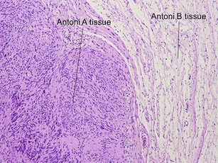
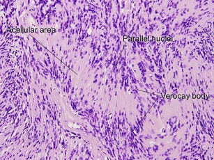
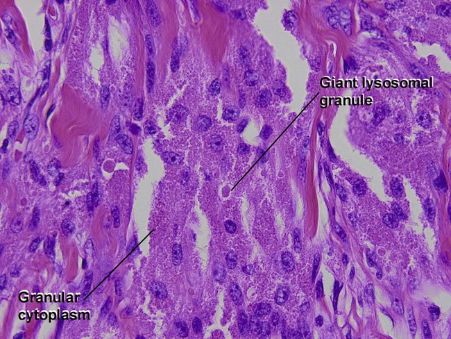
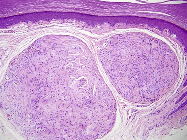
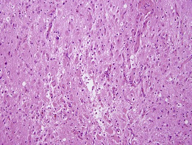
Schwannoma (neurilemmoma)

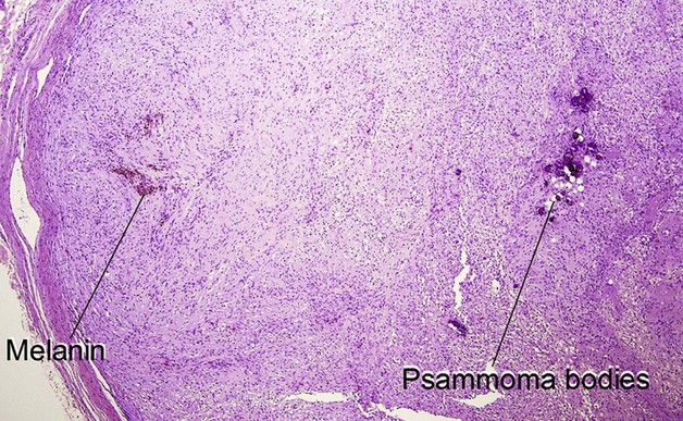
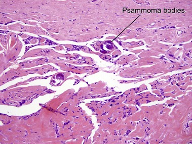
Psammomatous melanotic schwannoma is a variant with psammoma bodies and melanin. It is associated with Carney’s complex (myxomas, spotty pigmentation, and endocrinopathy).
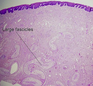
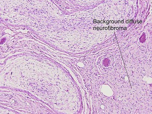
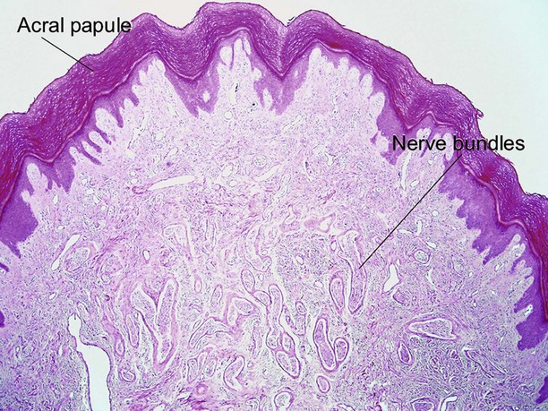
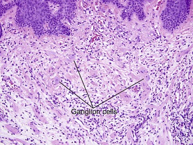
Neuromas

Neuromas are nerve sheath tumors with a roughly 1 : 1 ratio of axons to Schwann cells.
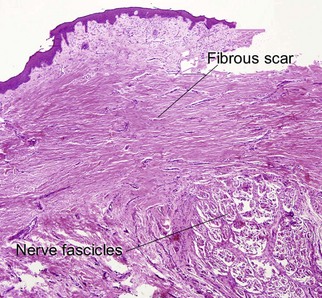
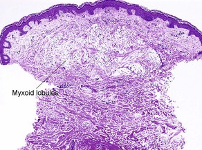
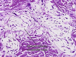
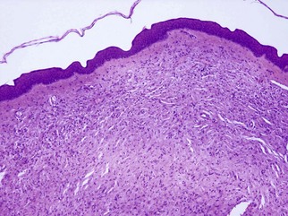
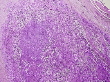
Palisaded encapsulated neuroma

Palisaded encapsulated neuromas are solitary painless papules that occur most commonly on the lower central face. The name is somewhat of a misnomer, as there is usually no palisading and only an inconspicuous capsule. Histologically, they resemble the mucosal neuromas of multiple endocrine neoplasia syndrome type 2b, an autosomal-dominant syndrome caused by a mutation of the RET proto-oncogene. In addition to the multiple mucosal neuromas, the syndrome is characterized by a marfanoid habitus, medullary carcinoma of the thyroid, pheochromocytoma, and hyperparathyroidism.

Differential Diagnosis

Unlike schwannomas, palisaded encapsulated neuromas are superficial tumors with adjacent sebaceous glands and small vellus follicles typical of facial skin. Schwannomas lack the fascicles with clefts seen in palisaded encapsulated neuromas.

Supernumerary digit (rudimentary polydactyly)

Supernumerary digit presents as a congenital papule, typically located at the base of the fifth digit along the ulnar border.

Differential Diagnosis

Acquired digital fibrokeratoma is also an acral papule; however, there are no nerve bundles and large stellate factor XIIIa-positive dendrocytes may be present. There is often longitudinal streaking of collagen.

Merkel cell carcinoma (primary neuroendocrine carcinoma of the skin, trabecular carcinoma)

Membrane-bound dense core granules on electron microscopy are characteristic. Metastatic small (oat) cell carcinoma of the lung also consists of small blue cells, but CK20 is typically negative and thyroid transcription factor (TTF-1) is positive. Also in the differential diagnosis of small blue cell tumors is lymphoma, which can be distinguished by positivity for hematopoietic markers. Melanoma is S100 positive. Neuroblastoma often demonstrates elongated, angulated “carrot-shaped” blue cells and may form rosettes.

Differential Diagnosis

The microscopic differential diagnosis for “small blue cell” tumors can be remembered by the mnemonic “LEMONS”:

Neurothekeoma

Neurothekeomas are benign neoplasms that are divided into myxoid, mixed, and cellular types. The myxoid type is strongly S100 positive, whereas those on the cellular end of the spectrum are composed of undifferentiated cells with partial features of Schwann cells, smooth muscle cells, myofibroblasts, and fibroblasts.

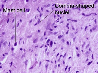
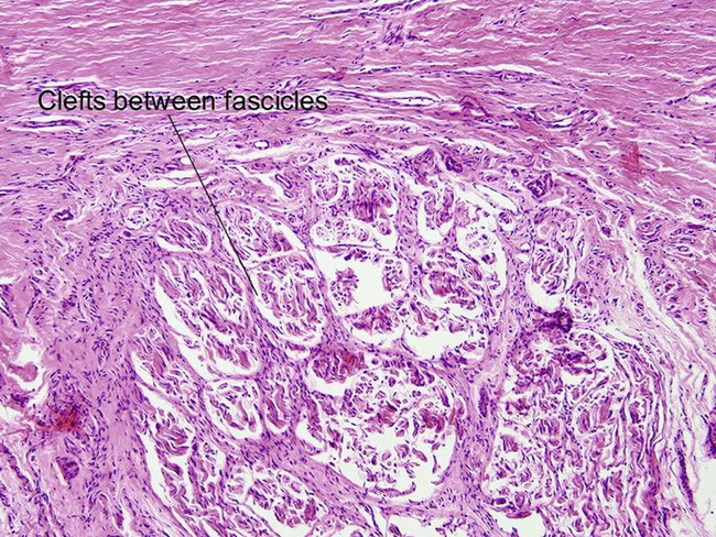
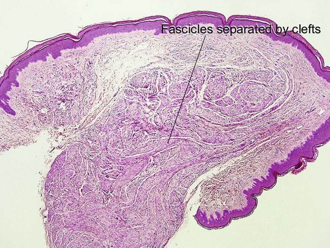
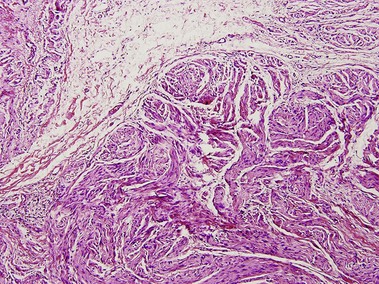
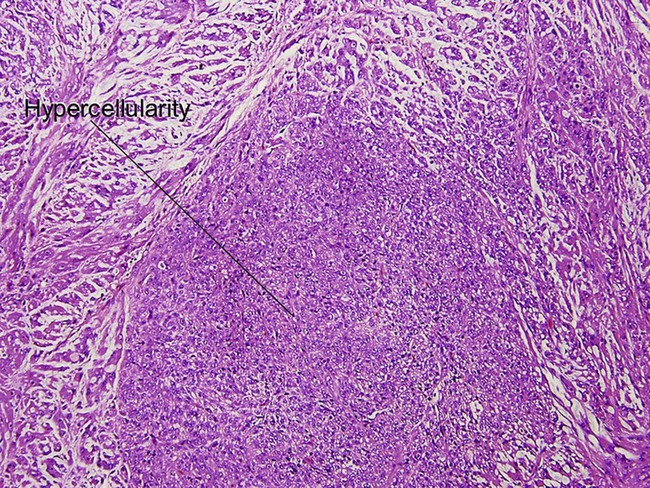
Cellular neurothekeoma

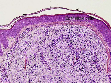
The cellular variant is negative for S100 and desmin, but sometimes positive for smooth muscle actin, neuron-specific enolase, and factor XIIIa. It is frequently positive for NK1/C-3, S100A6, and PGP9.5 with antigen retrieval. S100A6 also stains histiocytic tumors, including atypical fibroxanthoma, as well as Spitz nevi.

Differential Diagnosis

Histologically, cellular neurothekeomas may be confused with melanocytic lesions such as dermal Spitz nevi. S100 positivity strongly favors the melanocytic lesion. The variable reactivity for factor XIIIa in cellular neurothekeoma can result in confusion with epithelioid fibrous histiocytoma. S100A6 positivity can lead to confusion with spindled atypical fibroxanthoma. Of the two, atypical fibroxanthoma is less likely to demonstrate fascicles of tumor cells. Epithelioid pilar leiomyomas may also be considered in the differential diagnosis due to the variable smooth muscle actin positivity in cellular neurothekeomas. However, the absence of desmin in cellular neurothekeoma aids in the distinction.

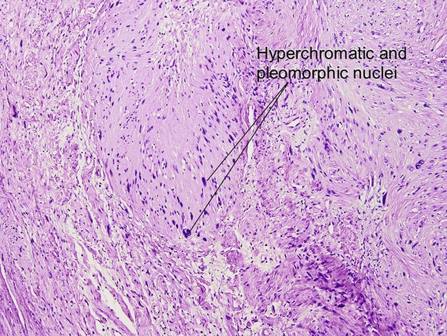
Malignant peripheral nerve sheath tumor (MPNST) (neurofibrosarcoma, malignant schwannoma)

Foci of divergent differentiation may be present, including osseous, chondroid, and rhabdoid foci as well as foci of adenocarcinoma or angiosarcoma. Malignant peripheral nerve sheath tumor with focal rhabdomyosarcoma is known as malignant Triton tumor. S100 staining is usually focal and weak in MPNSTs but diffusely positive in its mimicker, desmoplastic melanoma.

Further reading

Argenyi, ZB. Cutaneous neural heterotopias and related tumors relevant for the dermatopathologist. Semin Diagn Pathol. 1996; 13(1):60–71.

Argenyi, ZB, Balogh, K, Abraham, AA. Degenerative (“ancient”) changes in benign cutaneous schwannoma. A light microscopic, histochemical and immunohistochemical study. J Cutan Pathol. 1993; 20(2):148–153.

Argenyi, ZB, Kutzner, H, Seaba, MM. Ultrastructural spectrum of cutaneous nerve sheath myxoma/cellular neurothekeoma. J Cutan Pathol. 1995; 22(2):137–145.

Calonje, E, Wilson-Jones, E, Smith, NP, et al. Cellular “neurothekeoma”: an epithelioid variant of pilar leiomyoma? Morphological and immunohistochemical analysis of a series. Histopathology. 1992; 20(5):397–404.

Carney, JA, Stratakis, CA. Epithelioid blue nevus and psammomatous melanotic schwannoma: the unusual pigmented skin tumors of the Carney complex. Semin Diagn Pathol. 1998; 15(3):216–224.

Chambers, PW, Schwinn, CP. Chordoma. A clinicopathologic study of metastasis. Am J Clin Pathol. 1979; 72(5):765–776.

Dewit, L, Albus-Lutter, CE, de Jong, AS, et al. Malignant schwannoma with a rhabdomyoblastic component, a so-called triton tumor. A clinicopathologic study. Cancer. 1986; 58(6):1350–1356.

Ducatman, BS, Scheithauer, BW. Malignant peripheral nerve sheath tumors with divergent differentiation. Cancer. 1984; 54(6):1049–1057.

Kluwe, L, Friedrich, RE, Mautner, VF. Allelic loss of the NF1 gene in NF1-associated plexiform neurofibromas. Cancer Genet Cytogenet. 1999; 113(1):65–69.

Macarenco, RS, Ellinger, F, Oliveira, AM. Perineurioma: a distinctive and underrecognized peripheral nerve sheath neoplasm. Arch Pathol Lab Med. 2007; 131(4):625–636.

Miedema, JR, Zedek, D. Cutaneous meningioma. Arch Pathol Lab Med. 2012; 136(2):208–211.

Wallace, CA, Hallman, JR, Sangueza, OP. Primary cutaneous ganglioneuroma: a report of two cases and literature review. Am J Dermatopathol. 2003; 25(3):239–242.

Wang, AR, May, D, Bourne, P, et al. PGP9. 5: a marker for cellular neurothekeoma. Am J Surg Pathol. 1999; 23(11):1401–1407.