Panniculitis
Lobular panniculitis
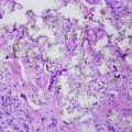
Lupus panniculitis (lupus profundus)
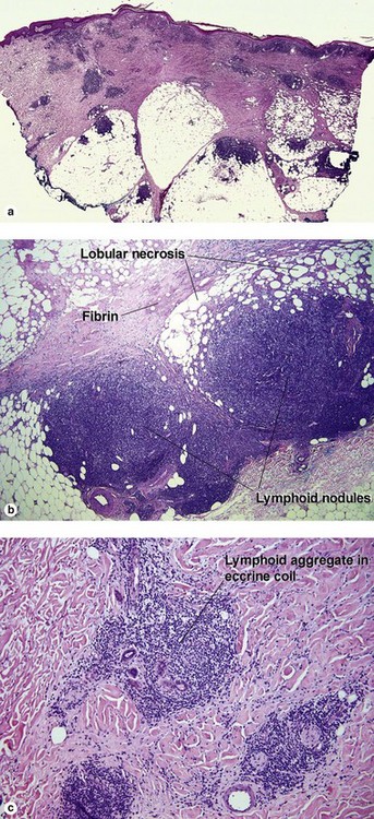
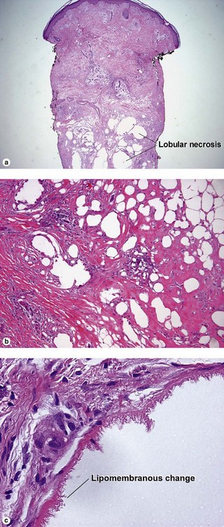
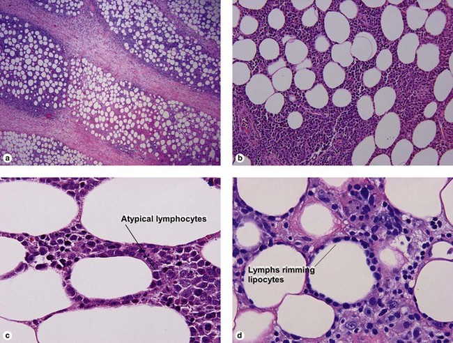
Although connective tissue disease can produce less specific patterns of granulomatous panniculitis, the features noted above are highly characteristic of lupus panniculitis. Plasma cells are common in the nodular lymphoid foci. Overlying features of lupus erythematosus may be present. The overlying dermis may be sclerotic. Lipomembranous changes may be present (eosinophilic “frost on the windowpane” or “ferning” pattern at the edge of the lipid vacuole).
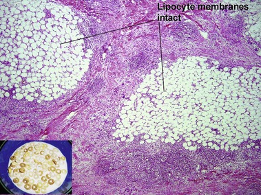
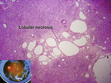
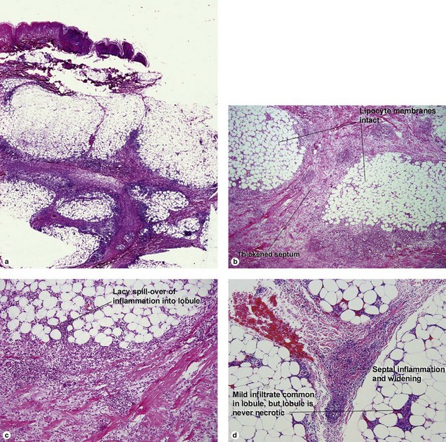
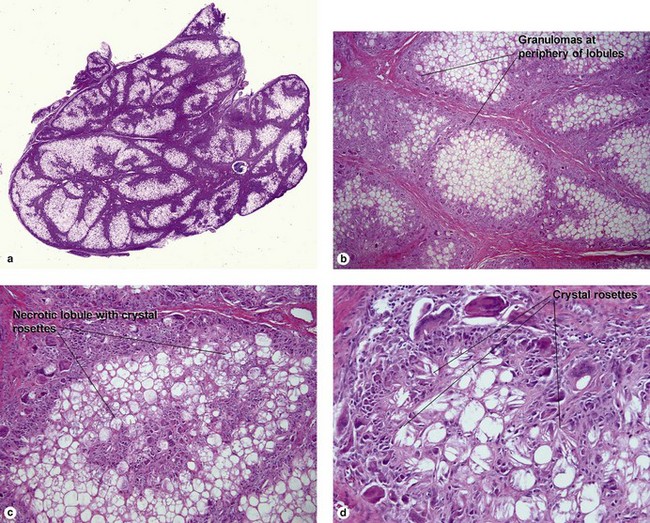
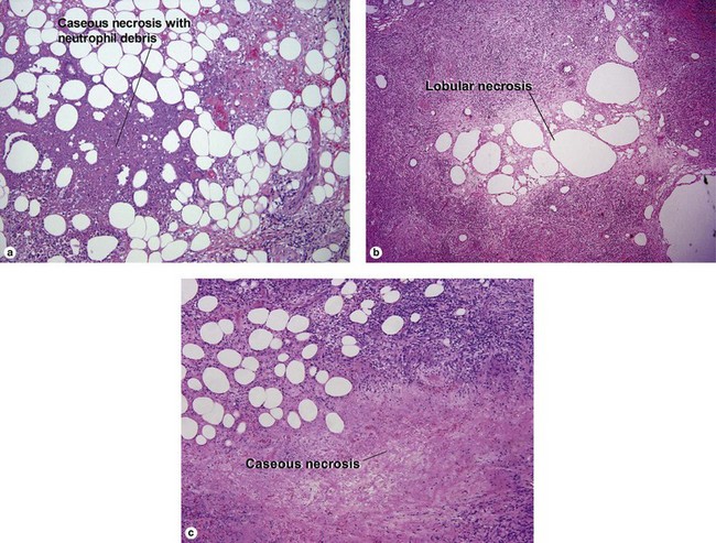
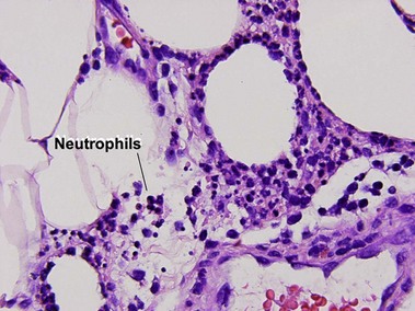
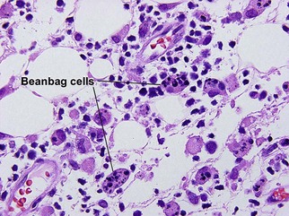
Pancreatic panniculitis
Pancreatic panniculitis may occur in the setting of pancreatitis or pancreatic neoplasm.
Geraminejad, P, DeBloom, JR, 2nd., Walling, HW, et al. Alpha-1-antitrypsin associated panniculitis: the MS variant. J Am Acad Dermatol. 2004; 51(4):645–655.
Gonzalez, EG, Selvi, E, Lorenzini, S, et al. Subcutaneous panniculitis-like T-cell lymphoma misdiagnosed as lupus erythematosus panniculitis. Clin Rheumatol. 2006; 26(2):244–246.
Heymann, WR. Panniculitis. J Am Acad Dermatol. 2005; 52(4):683–685.
Ma, L, Bandarchi, B, Glusac, EJ. Fatal subcutaneous panniculitis-like T-cell lymphoma with interface change and dermal mucin, a dead ringer for lupus erythematosus. J Cutan Pathol. 2005; 32(5):360–365.
Massone, C, Kodama, K, Salmhofer, W, et al. Lupus erythematosus panniculitis (lupus profundus): clinical, histopathological, and molecular analysis of nine cases. J Cutan Pathol. 2005; 32(6):396–404.
McBean, J, Sable, A, Maude, J, et al. Alpha1-antitrypsin deficiency panniculitis. Cutis. 2003; 71(3):205–209.
Phelps, RG, Shoji, T. Update on panniculitis. Mt Sinai J Med. 2001; 68(4–5):262–267.
Requena, L, Sánchez Yus, E. Panniculitis. Part II. Mostly lobular panniculitis. J Am Acad Dermatol. 2001; 45(3):325–361.
Schneider, JW, Jordaan, HF. The histopathologic spectrum of erythema induratum of Bazin. Am J Dermatopathol. 1997; 19(4):323–333.
Sutra-Loubet, C, Carlotti, A, Guillemette, J, et al. Neutrophilic panniculitis. J Am Acad Dermatol. 2004; 50(2):280–285.