Nerve Block at the Ankle

Published on 07/02/2015 by admin

Filed under Anesthesiology

Last modified 22/04/2025

Print this page

rate 1 star rate 2 star rate 3 star rate 4 star rate 5 star
Your rating: none, Average: 0 (0 votes)

This article have been viewed 2918 times

Nerve Block at the Ankle

Douglas A. Dubbink, MD

Anesthesia distal to the ankle is accomplished by interrupting the five major nerves that innervate the foot: the tibial nerve and the deep peroneal nerve, which supply the deep structures of the foot, and the superficial peroneal nerve, the sural nerve, and the saphenous nerve, which supply sensory innervation to the skin. The ankle block is a relatively easy block to learn if the anatomy is well understood. Ankle blocks can be effective for nearly any surgical procedure of the foot. Major complications are rare; however, prolonged paresthesias have been reported. No epinephrine-containing local anesthetic agents should be used when performing an ankle block.

Technique

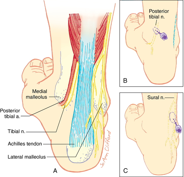
The tibial nerve supplies the sole and plantar portions of the toes up to the nails. It lies behind the posterior tibial artery anteromedial to the Achilles tendon and deep to the flexor retinaculum, which must be penetrated for a successful block (Figure 130-1).

The block is started by injecting a small amount of local anesthetic agent medial to the Achilles tendon at the level of the upper border of the medial malleolus. A 3-cm to 5-cm, 22G or 25G needle is directed at right angles to the tibia. The needle tip is slowly advanced until a paresthesia is elicited (a nerve stimulator can be used) or bone is contacted. At this point, 5 to 7 mL of local anesthetic agent is injected near the posterior aspect of the tibia, with an equal volume of local anesthetic injected during withdrawal of the needle to the skin surface if a paresthesia is not elicited.

The sural nerve is a superficial nerve that provides cutaneous sensation to the lower posterolateral ankle, lateral foot, and fifth toe. Five to 10 mL of local anesthetic agent is administered by infiltrating the solution posterior to the lateral malleolus to the Achilles tendon at the level of the upper border of the lateral malleolus.

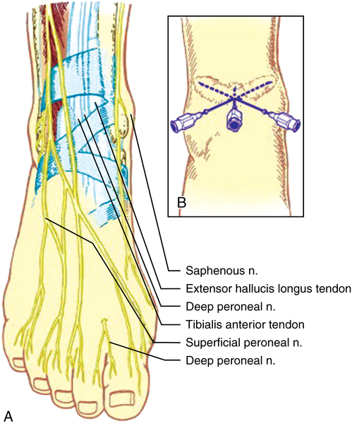
The deep peroneal, superficial peroneal, and saphenous nerves can all be blocked using a single injection site. The deep peroneal nerve courses midway between the malleoli before assuming a position between the anterior tibial tendon and the extensor hallucis longus tendon beneath the extensor retinaculum at the dorsum of the foot. It innervates the short extensors of the toes and provides skin sensation to the interdigital cleft between the great and second toes. With the patient dorsiflexing the foot, the tendons of the anterior tibial and extensor hallucis longus muscles can be readily identified at a level just above a line connecting the malleoli. The pulsation of the anterior tibial (dorsalis pedis) artery will often be felt. The nerve is lateral to the artery and deep to the extensor retinaculum. A 25G, 3-cm to 5-cm needle is inserted perpendicular to skin, as depicted in Figure 130-2. A loss of resistance will often be felt during passage through the extensor retinaculum, at which time between 3 mL and 5 mL of local anesthetic agent is injected.

The superficial peroneal nerve supplies cutaneous sensation to the dorsum of the foot and toes (except between great and second toes). Blockade of this nerve can be achieved by injecting local anesthetic agent subcutaneously laterally from the site of injection of the deep peroneal nerve toward the superior aspect of the lateral malleolus using 5 mL to 10 mL of solution.

The saphenous nerve is anterior to the medial malleolus near the long saphenous vein supplying cutaneous innervation to the anteromedial side of the lower leg and medial foot midway to the toes. The saphenous nerve is blocked with 3 mL to 5 mL of local anesthetic agent injected subcutaneously medially from the site of injection of the deep peroneal nerve toward the saphenous vein.