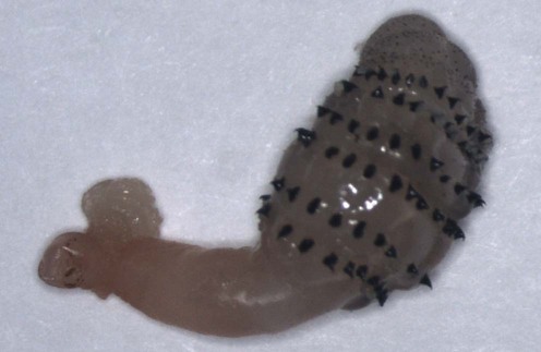
Myiasis


 Detailed travel history
 Morphologic identification of the parasite
First-line therapies
 Local anesthesia with or without surgical incision
 Squeezing the skin surrounding the furuncle with fingers or wooden spatulas
 Vaseline® therapy
 Bacon therapy
 Pork fat therapy
 Hair gel
 Occlusion with chimo (paste-like smokeless tobacco)
 Small wad of cotton soaked in sap of the Thevetia ahouai (Apocynaceae) tree
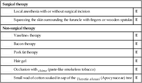
Surgical therapy
Second-line therapies
 Systemic ivermectin
 Topical ivermectin
 Chloroform/ether
 Ethanol spray
 Oil of betel leaf
 Mineral turpentine
Larvicidal effects of mineral turpentine, low aromatic white spirits, aqueous extracts of Cassia alata, and aqueous extracts, ethanolic extracts and essential oil of betel leaf (Piper betle) on Chrysomya megacephala.
Kumarasinghe SP, Karunaweera ND, Ihalamulla RL, Arambewela LS. Int J Dermatol 2002; 41: 877–80.

