Mycobacterial (atypical) skin infections

Published on 19/03/2015 by admin

Filed under Dermatology

Last modified 22/04/2025

Print this page

rate 1 star rate 2 star rate 3 star rate 4 star rate 5 star
Your rating: none, Average: 0 (0 votes)

This article have been viewed 2262 times

Mycobacterial (atypical) skin infections

Ure Eke and John Berth-Jones

Evidence Levels:  A Double-blind study  B Clinical trial ≥ 20 subjects  C Clinical trial < 20 subjects  D Series ≥ 5 subjects  E Anecdotal case reports

image

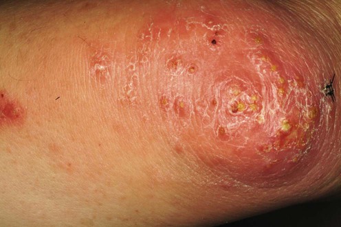
Fish tank (swimming pool) granuloma

Fish tank granuloma is an infection of the skin caused by Mycobacterium marinum, causing plaques and nodules, commonly on the upper extremities, which may spread in a sporotrichoid fashion after an incubation period of 2 to 6 weeks. The most common sources of infection are tropical fish aquariums, and swimming pools. The infection is commonly limited to the skin, but tenosynovitis, osteomyelitis, and arthritis have been reported with deeper infections. Rarely, disseminated infection may occur, especially in the immunocompromised. There are reports of the development of cutaneous M. marinum infection whilst on infliximab and adalimumab although paradoxically, infliximab has been found to be a useful adjunctive therapy in the management of M. marinum infection.

Management strategy

No controlled trials have been conducted, probably owing to the paucity of cases. Successful treatment is reported with antibiotics given singly or in combination. The mean duration of treatment in various reports ranges from 6 to 20 weeks. No statistical difference in efficacy between treatments has been demonstrated. Lesions can often be effectively treated by simple excision, but occasionally this seems to result in a prolonged infection. Heat treatment of the infected area may have an adjunctive role. Although spontaneous resolution may occur within three years, treatment is required for rapid recovery from infection and the prevention of dissemination. The second-line treatments described below have been so designated as there is relatively little published information about them. The third-line treatments are probably best regarded as adjunctive.

Specific investigations

Histology shows non-caseating granulomas, but a complete absence of epithelioid cells and multinucleate giant cells is not unusual in acute lesions. Suppurative, tuberculoid and palisading patterns of granulomas have been described. Dermal small vessel proliferation with mixed inflammation may be a good indicator of cutaneous atypical mycobacterial infections. Ziehl–Nielsen staining is positive for acid-fast bacilli in 30% of biopsies. Culture of the biopsy specimen at 30–33°C yields pigmented colonies of M. marinum. PCR can provide rapid and sensitive detection of mycobacterial DNA in formalin-fixed, paraffin-embedded specimens. In vitro sensitivity studies have not been uniformly predictive of clinical response to the antibiotics. Although they do not have a routine role in directing initial treatment, they may be useful in resistant cases.

First-line therapies

image Minocycline 100–200 mg once daily for 6–12 weeks C
image Doxycycline 100 mg twice daily for 3 to 4 months C
image Clarithromycin 500 mg once or twice daily for 3 to 4 months C
image Rifampin 600 mg and ethambutol 1.2 g daily for 3 to 6 months D
image Co-trimoxazole 2–3 tablets twice daily for 6 weeks D
image Clarithromycin 250 mg twice daily and ethambutol 800 mg once daily for 2 to 6 months D

Epidemiological, clinical, and therapeutic pattern of Mycobacterium marinum infection: a retrospective series of 35 cases from southern France.

Eberst E, Dereure O, Guillot B, Trento C, Terru D, van de Perre P, Godreuil S. J Am Acad Dermatol 2012; 66: e15–16.

In this retrospective series, the treatment of 35 consecutive cases of M. marinum was reviewed. Thirty-four of 35 (97%) had complete clearance of skin lesions with one course of oral antimicrobial therapy given over a period of four to 24 weeks (median 12.4 weeks). Single antibiotic therapy was given successfully in 25 patients. Fourteen of these received minocycline 200 mg od, five patients received doxycycline 200 mg od and six patients received clarithromycin 500 mg od. Nine patients had dual antibiotic therapy using a combination of clarithromycin with doxycycline, minocycline, rifampicin or ofloxacin.

Second-line therapies

image Ciprofloxacin 500 mg + clarithromycin 250 mg twice daily for 4 months E
image Rifabutin 600 mg + clarithromycin 500 mg twice daily + ciprofloxacin 500 mg twice daily for 4 months E
image Azithromycin 500 mg three times a week for 2 months E

Third-line therapies

image Simple excision E
image Curettage and electrodesiccation E
image Incision and drainage E
image Heat therapy by gloves, hot water or heated armlet E
image Photodynamic therapy E
image Cryotherapy E
image Adjunctive anti-TNF-α inhibitors E

Mycobacterium ulcerans

Mycobacterium ulcerans (Buruli ulcer) is the third most common mycobacterial infection after leprosy and tuberculosis in immunocompetent hosts. It occurs most commonly in wetlands of tropical and subtropical countries (Africa and Australia). Children under 15 years of age are most commonly affected. After inoculation into the skin, M. ulcerans proliferates and produces a toxin, mycolactone, which enters the cells and causes necrosis of the dermis, panniculus, and deep fascia. Initially firm, painless nodules, papules, or plaques are seen. As the necrosis spreads, overlying ulceration develops over 1 or 2 months. The ulcers have a characteristic undermined edge and necrotic base. Although indolent in the majority, the ulcers can grow rapidly to more than 15 cm, with resultant extensive scarring and deformity. Up to 13% of patients may develop systemic involvement.

Management strategy

There are anecdotal reports of spontaneous resolution of cutaneous M. ulcerans infection, but early treatment is recommended to limit disease progression and prevent contractures. The WHO recommends combination chemotherapy with oral rifampicin and intramuscular streptomycin as first-line treatment. Adjunctive surgical excision may be required for necrotic or unhealed skin tissue. Antimicrobial therapy may reduce the extent of surgical excision. Recent combined therapy options contain oral preparations which are easier to administer and show similar efficacy. There can be paradoxical clinical deterioration peaking around week 8 of antimicrobial therapy, before subsequent improvement. This response does not indicate treatment failure and may be the result of temporary immunostimulation from antimicrobial treatment. Adequate wound care and the use of bednets offer protection against M. ulcerans infections in endemic areas. Independent risk factors for relapse after surgery alone include incomplete excision, age under 16 years, leg edema and plaque-like lesions before ulceration.

Specific investigations

Smears from the necrotic base of ulcers often reveal clumps of acid-fast bacilli on Ziehl–Nielsen staining. Appropriately selected biopsy specimens that include the necrotic base and the undermined edge of lesions with subcutaneous tissue are nearly always diagnostic. Histology reveals necrosis of the deep dermis and panniculitis. Inflammatory cells are few, presumably owing to the immunosuppressive activity of the toxin. With healing, there is a granulomatous response and the ulcerated area is eventually replaced by a depressed scar which may result in functional disability. M. ulcerans can be cultured from exudates or tissue fragments, but visible growth often requires 6 to 8 weeks incubation at 33°C. PCR is quicker and has the highest sensitivity compared with microscopy, culture, and histopathology. For PCR, a 3 mm punch biopsy is the best sample for non-ulcerative lesions whilst a swab is best for ulcerative lesions.

First-line therapies

image Oral rifampin daily at 10 mg/kg and streptomycin intramuscularly at 15 mg/kg daily for 4 to 12 weeks B
image Wide surgical excision alone B
image Wide surgical excision and rifampin 10 mg/kg daily and ciprofloxacin (250–500 mg twice daily) for 3 to 6 months B
image Rifampicin 10 mg/kg/day and clarithromycin 12–15 mg/kg od for 2 to 6 months ± adjunctive surgery B
image Rifampicin 10 mg/kg/day and moxifloxacin 400 mg od for 3 to 6 months and adjunctive surgery C
image Ciprofloxacin 500 mg bd and clarithromycin 500 mg bd for 3 to 6 months and adjunctive surgery C
image Rifampicin 10 mg/kg/day and ciprofloxacin 500 mg bd for 3 to 6 months C
image Local heat (40°C) C

Clinical efficacy of combination of rifampin and streptomycin for treatment of Mycobacterium ulcerans disease.

Sarfo FS, Phillips R, Asiedu K, Ampadu E, Bobi N, Adentwe E, et al. Antimicrob Agents Chemother 2010; 54: 3678–85.

A study of 160 patients with confirmed M. ulcerans infection treated with oral rifampicin 10 mg/kg/day and intramuscular streptomycin 15 mg/kg daily for 8 weeks. Ninety-five percent (152 patients) with early and late forms of disease responded to therapy without surgery. In eight cases, surgical excision followed by split skin graft repair was carried out at week 8 due to progressive lesions or non-healing lesions. Complete healing was achieved between 2 and 48 weeks. There were no recurrences in 158 (98%) of cases. Treatment was generally well tolerated. Two patients changed from streptomycin to oral moxifloxacin 400 mg od due to nausea, vomiting, and dizziness.

Effect of a control project on clinical profiles and outcomes in Buruli ulcer: a before/after study in Bas-Congo, Democratic Republic of Congo.

Phanzu DM, Suykerbuyk P, Imposo DB, Lukanu PN, Minuku JB, Lehman LF, et al. PLoS Negl Trop Dis 2011; 5: e1402.

A prospective observational study in Congo comparing clinical outcomes of patients admitted with M. ulcerans ulcers. Between 2004 and 2007, 64 patients were treated with surgery alone resulting in healing in 48 patients; 15 patients were left with disability; 12 patents died due to sepsis. Between 2005 and 2007, 107/190 patients were treated with rifampicin and streptomycin. The majority of these patients were also treated with surgery. In this group, healing occurred in 176 of 190 patients, 37 were left with functional disability and death occurred in 6 patients. Sepsis, malnutrition and anemia, and postsurgical shock were reported causes of fatality.

Mycobacterium kansasii

This organism most commonly causes pulmonary disease; skin lesions are rare., Most reported cases have occurred in immunocompromised subjects. The gross morphology of such lesions varies greatly and can be verrucous, nodular, pustular, ulcerated, or sporotrichoid. Cutaneous lesions may progress slowly or run an acute course.

First-line therapies

image Combination of antituberculous drugs for 6 to 18 months D
image Antituberculous drugs with intramuscular kanamycin 500 mg three times a week for 3 months E
image Minocycline 100–200 mg daily for 16 weeks E
image Erythromycin 2 g daily for 6 months E

Rapidly growing mycobacteria

Mycobacterium fortuitum, Mycobacterium chelonae, and Mycobacterium abscessus are environmental pathogens also known as rapidly growing mycobacteria (RGM), so termed because colonies are visible to the naked eye within 7 days of culture. Cutaneous lesions due to these pathogens usually occur after surgery, percutaneous catheter insertion, or accidental inoculation. Dark red nodules develop, with abscess formation and clear fluid drainage. Disseminated disease occurs most commonly in immunosuppressed hosts.

Management strategy

RGM should be individually identified and in vitro sensitivity tests performed before treatment. M. fortuitum and M. chelonae are resistant to most antituberculous drugs except kanamycin and amikacin. M. fortuitum may also be susceptible to cefoxitin, ciprofloxacin, and imipenem. M. abscessus is usually sensitive to amikacin, cefoxitin, and clarithromycin. Tobramycin and clarithromycin are more effective than amikacin in the treatment of M. chelonae. The wide variability in antibiotic sensitivity means that each case must be considered individually. For persistent non-healing cutaneous lesions, wide excisional surgery with delayed closure or skin grafting can be undertaken. The duration of therapy can vary from 6 weeks to 7 months and is dictated by clinical and microbiological response.

First-line therapies

Mycobacterium chelonae

image Clarithromycin 500 mg bd for 3 to 8 months ± surgery D
image Azithromycin 250 mg od for at least 6 months E
image Clarithromycin as part of dual or triple therapy with ciprofloxacin, tobramycin, and tigecycline E

Clinical management of rapidly growing mycobacterial cutaneous infections in patients after mesotherapy.

Regnier S, Cambau E, Meningaud JP, Guihot A, Deforges L, Carbonne A, Bricaire F, Caumes E. Clin Infect Dis 2009; 49: 1358–64.

Sixteen patients were diagnosed with cutaneous infection due to RGM following an outbreak after mesotherapy in France. M. chelonae was identified in 11 patients and M. frederiksbergense in two patients. Patients were treated with drug regimens containing clarithromycin. Six received triple therapy initially and eight received dual therapy. Fourteen received clarithromycin 1–2 g/day, six received intravenous tobramycin 3 mg/kg/day; 14 received ciprofloxacin 500 mg bd; and six received intravenous tigecycline initially at 100 mg followed by 50 mg/day. Patients were treated for a mean duration of 14 weeks (range 1–24 weeks) with time to healing estimated at 6.2 months (range 1–15 months). Success was noted in all but one patient.

Mycobacterium fortuitum

image Clarithromycin 500 mg bd and levofloxacin 500 mg bd for 3 to 6 months B
image Clarithromycin 250–500 mg bd ± IM amikacin 250 mg three times a week for 3 to 6 months C
image Ciprofloxacin 500 mg bd for 3 to 6 months C
image Ciprofloxacin 500 mg bd, clarithromycin 500 mg bd and amoxicillin–clavulanic acid 500 mg bd for 6 weeks E
image Co-trimoxazole 800/160 mg bd, clarithromycin 500 mg bd, and amoxicillin–clavulanic acid 500 mg bd for 6 weeks E
image Levofloxacin 300 mg od E
image Trimethoprim D

Mycobacterium abscessus

image Clarithromycin 1 g/day for adults and 0.5 g/day for children for 3 to 6 months + adjunctive surgery B
image Clarithromycin 250 mg bd + moxifloxacin 400 mg od for 4 to 5 months B
image Clarithromycin 250–500 mg bd ± IM amikacin 250 mg three times a week for 3 to 6 months B
image Multiple antibiotic therapy + adjunctive interferon-γ for refractory lesions E

Report on an outbreak of postinjection abscesses due to Mycobacterium abscessus, including management with surgery and clarithromycin therapy and comparison of strains by random amplified polymorphic DNA polymerase chain reaction.

Villanueva A, Calderon RV, Vargas BA, Ruiz F, Aguero S, Zhang Y, Brown BA, Wallace RJ Jr. Clin Infect Dis 1997; 24: 1147–53.

A cohort of 350 patients were clinically diagnosed with M. abscessus infections following an outbreak in a physician’s office in Columbia (205 cases culture-confirmed). Patients received either surgery alone or clarithromycin alone or combination of drugs and surgery. Of 35 patients treated with clarithromycin alone, eight patients were cured (23%). Of 22 patients treated with surgical resection alone, seven were cured (32%). Of 148 patients treated with combined surgery and clarithromycin, 140 were cured (95%). Clarithromycin was given at a dose of 1 g/day in adults and 0.5 g/day in children for 3 to 6 months.

Share this: