Published on 19/03/2015 by admin
Filed under Dermatology
Last modified 22/04/2025
This article have been viewed 2457 times
Mahreen Ameen and Wanda Sonia Robles
Evidence Levels: A Double-blind study B Clinical trial ≥ 20 subjects C Clinical trial < 20 subjects D Series ≥ 5 subjects E Anecdotal case reports
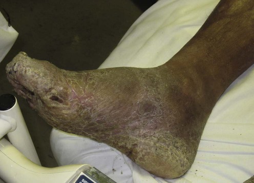
Mycetomas (Madura foot) are endemic in the tropics and sub-tropics. They are chronic, granulomatous, subcutaneous infections caused by either actinomycetes bacteria or eumycetes fungi, giving rising to actinomycetomas and eumycetomas, respectively. The infectious agents are saprophytes existing in soil or plants, and infection usually results from traumatic inoculation into the skin. Consequently, the disease most commonly affects agriculturalists and those who are bare-foot. The disease is characterized by abscesses, draining sinuses and discharging grains, and it slowly progresses with risks of bone and visceral involvement. The discharging grains represent aggregates of fungal hyphae or bacterial filaments. Actinomycetomas are produced by agents of the genera Nocardia, Actinomadura, Streptomyces and Nocardiopsis. Nocardia is the commonest agent, particularly in the Americas, but Streptomyces somaliensis is more common in Sudan and the Middle East. Eumycetomas are caused by a large number of fungi: Madurella mycetomatis is of particular significance as it is the most prevalent causative agent in regions of India and Africa.
Treatment of mycetomas is generally difficult, and management varies from a very conservative approach to chemotherapy and surgery. Effective chemotherapy is available for actinomycetomas. However, eumycetomas are more refractory to drug therapy.
Eumycetomas may sometimes be managed conservatively as they are usually indolent and seldom life threatening. Treatment is then symptomatic with relief of pain and applications of dressings to affected areas, particularly the sinuses. Any secondary bacterial infection requires treatment. More active management consists of long courses of antifungals, 18–24 months or even longer, together with aggressive surgical excision and debulking. Antifungal therapy is initiated before surgery and continued afterwards to reduce the risk of recurrence. Small lesions have a more favorable prognosis as they are more easily excised completely. Advanced disease with bony involvement characteristically shows poor therapy response, and often requires surgical amputation. Of all the antifungal drugs, azoles have been most commonly used. Fluconazole has been found to be ineffective but ketoconazole and itraconazole have both demonstrated efficacy, particularly at high doses (200–400 mg daily for ketoconazole and 300–400 mg daily for itraconazole). Itraconazole is preferred as it is better tolerated for longer periods of time and is thought to demonstrate greater efficacy than ketoconazole, although there are no published studies comparing their efficacy.
There are reports of the high tolerability and efficacy of the newer broad-spectrum triazoles such as voriconazole and posaconazole against Madurella species and Scedosporium apiospermum infection. This is supported by in vitro susceptibility testing. However, their high costs are prohibitive for use in most endemic regions. Griseofulvin appears to be ineffective. Amphotericin has shown variable responses in the few cases that it has been used in. High-dose terbinafine (500 mg twice daily) has demonstrated limited clinical efficacy that correlates with in vitro susceptibility testing which has shown only moderate efficacy of terbinafine against some black grain mycetomas. In vitro studies have not demonstrated any efficacy of echinocandins against Madurella mycetomatis.
Actinomycetomas are usually amenable to antibiotic therapy, but cure rates vary widely from 60% to 90%. Combined drug therapy is preferred in order to prevent the development of drug resistance as well as to eradicate any residual infection. The duration of drug therapy depends on clinical response. Cure is defined by a lack of clinical activity, absence of grains and negative cultures. Treatment with sulfonamides and sulfonamide combinations such as trimethoprim–sulfamethoxazole (co-trimoxazole) are usually first line. Aminoglycosides, tetracyclines, rifampicin, ciprofloxacin and amoxicillin-clavulanate have also been successfully used. Parenteral amikacin and oral co-trimoxazole combination therapy is especially advocated for cases at risk of bone or visceral involvement. Actinomycetomas seldom require surgical management.
Direct microscopy
Culture
Histopathology
It is imperative to differentiate between eumycetomas, which respond poorly to chemotherapy, and actinomycetomas, which generally respond well. Furthermore, species identification is important as it has treatment and prognostic implications, some species having demonstrated higher efficacy to some chemotherapy regimens than others.
The clinical diagnosis can be confirmed by the demonstration and identification of grains, which can be obtained by direct extraction, fine needle aspiration, or deep tissue biopsy. Direct microscopy of a crushed grain in 20% potassium hydroxide gives an indication of the size and shape of the grain, which provides an initial clue to the causative agent, whether bacterial or fungal. Histopathology of a deep surgical biopsy demonstrates a granulomatous, inflammatory reaction with abscesses containing grains. Culture of grains using Sabouraud or blood agar media permits species identification. However, fungal culture can be particularly difficult, as morphological differentiation of fungi may be poor or delayed. Molecular tests have therefore been developed for species identification of several black-grained eumycetomas, including species-specific polymerase chain reaction (PCR) analysis. Serological tests such as enzyme-linked immunosorbent assay (ELISA) are employed by some centers to support diagnosis as well as to assess therapy response. Radiology and ultrasound enable assessment of disease extent and bony involvement. Helical computed tomography can provide detailed assessments of soft tissue and visceral involvement.
Fahal AH. Trans R Soc Trop Med Hyg 2004; 98: 3–11.
A review article based on the experiences of the Mycetoma Research Centre in Sudan. Combined antibiotic treatment for actinomycetomas and aggressive surgery in combination with antifungals for eumycetomas is recommended.
Ahmed AO, van Leeuwen W, Fahal A, van de Sande W, Verbrugh H, van Belkum A. Lancet Infect Dis 2004; 4: 566–74.
A good review article on developments in the clinical, epidemiological and diagnostic management of Madurella mycetomatis eumycetoma.
Welsh O, Vera-Cabrera L, Salinas-Carmona MC. Dermatol Clin 2007; 25(2): 195–202.
A comprehensive review article covering the etiopathogenesis, diagnosis and treatment options for mycetomas. The authors describe their long experience in Mexico of using parenteral amikacin therapy (15 mg/kg/day for 3 weeks) in combination with oral trimethoprim–sulfamethoxazole (800/160 mg/kg/day for 5 weeks) for the treatment of severe or refractory actinomycetomas. Up to four 5-weekly cycles can be given, and treatment is administered continuously to minimize the development of secondary resistance to amikacin. They recommend that audiometry and creatinine clearance are performed during the last 2 weeks of each cycle.
Ameen M, Arenas R. Expert Opin Pharmacother 2008; 9(12): 2077–85.
A comprehensive review of trial data supporting therapies for both eumycetomas and actinomycetomas and the potential of the newer antifungal agents in the treatment of mycetomas.
Dieng MT, Niang SO, Diop B, Ndiaye B. Bull Soc Pathol Exot Filiales 2005; 98: 18–20.
Ninety patients with actinomycetomas (A. pelletieri, n=60; A. madurae, n=25; S. somaliensis, n=5) in Senegal were treated with sulfamethoxazole monotherapy. Despite bone involvement in 55% of cases, cure was achieved after 1 year of treatment in 83% of patients. 50% of these cases were localized to the foot. Despite treatment two patients died of visceral involvement.
Bonifaz A, Ibarra G, Saul A, Paredes-Solis V, Carrasco-Gerard E, Fierro-Arias L. Pediatr Infect Dis J 2007; 26(1): 50–2.
Mycetomas are very rare in children but the clinical presentation and course is similar to that in adults. There were 13 cases of Nocardia actinomycetoma and two cases of M. mycetomatis eumycetoma. Sulfonamide combinations were advocated as first-line therapy for actinomycetomas and co-amoxiclav as second-line therapy. Itraconazole and ketoconazole were used in the management of eumycetomas.
Ramam M, Bhat R, Garg T, Sharma VK, Ray R, Singh MK, et al. Indian J Dermatol Venereol Leprol 2007; 73: 235–9.
The authors emphasise that, although parenteral therapy regimes demonstrate high efficacy, they are costly in terms of inpatient stays. They describe a reduced parenteral regime (intravenous gentamicin 80 mg twice daily together with oral co-trimoxazole 320/1600 mg twice daily for 1 month), followed by a longer phase of oral medication (doxycycline 100 mg twice daily together with co-trimoxazole at the same dose). All 21 patients demonstrated significant clinical response at the end of the parenteral phase of treatment. The oral phase needed to be continued for 3.5–16 months (mean 9.1 months) until cure, and in the majority of these patients this included a treatment period of 5 to 6 months after complete healing of the lesions to prevent any relapse.
Castro LG, Piquero-Casals J. Int J Dermatol 2008; 47: 160–3.
This article describes the treatment of 13 cases of eumycetoma (with itraconazole) and 14 cases of actinomycetoma (with co-trimoxazole). Combination drug therapy was used in more than half of the cases. There was higher efficacy in three patients with actinomycetoma treated with co-trimoxazole in combination with amikacin. However, two of these patients developed hearing loss after treatment. The authors emphasise the problems of diagnostic testing, even in secondary and tertiary health centres, and were only able to identify the etiological agent in fewer than half of their cases.
Smith EL, Kutbi S. J Am Acad Dermatol 1997; 34: 279–80.
This study from Saudi Arabia treated 25 patients with a follow-up period of 12 years. The authors recommend a combination itraconazole drug therapy together with surgical excision or debulking. Drainage of sinuses with removal of grains that can cause inflammation reduced pain and swelling.
Fahal AH, Sabaa AH. Trans R Soc Trop Med Hyg 2010; 104: 117–21.
A large retrospective study of 722 children (age range 4–17 years) with confirmed mycetoma seen at the Mycetoma Research Centre, Sudan, during a 20-year period until 2009. Diagnosis was established by cytological and ultrasound examinations of the lesions and histological examination of the surgical biopsies. Most of the patients had eumycetomas. Combined medical treatment and surgical excision was the standard treatment. Disease recurrence after surgical excision was reported in 17.9% of patients.
Fahal AH, Rahman IA, El-Hassan AM, Rahman ME, Zijlstra EE. Trans R Soc Trop Med Hyg 2011; 105: 127–32.
This prospective study of 13 patients demonstrated that pre-operative treatment with a 1-year course of itraconazole enhances lesion encapsulation that facilitates wide local excision avoiding unnecessary mutilating surgery. Itraconazole was given at 400 mg daily for 3 months followed by a reduced dose of 200 mg daily for 9 months. All patients showed a good clinical response to the 400 mg daily dose but a slower response to the 200 mg daily dose. Post-treatment surgical exploration demonstrated that all lesions were well localized and encapsulated and easily removed. There was only a single recurrence after a follow-up period of 18–36 months.
Bonifaz A, Flores P, Saul A, Carrasco-Gerard E, Ponce RM. Br J Dermatol 2007; 156 (2): 308–11.
This study advocates co-amoxiclav as rescue therapy in patients with Nocardia spp. refractory to other therapy regimes. Twenty-one patients who had previously failed on other therapy regimes were treated with oral co-amoxiclav 875/125 mg twice daily. There was clinical and microbiological cure in 15 (71%) patients after a mean treatment period of 10 months. Patients with bone and visceral involvement required longer treatment periods.
N’Diaye B, Dieng MT, Perez A, Stockmeyer M, Bakshi R. Int J Dermatol 2006; 45: 54–157.
High-dose terbinafine, 500 mg twice daily, was given to 23 patients with eumycetomas in Senegal. After 24–48 weeks of treatment mycological cure was seen in 25% patients, and a further 55% of patients demonstrated clinical improvement. Treatment was well tolerated.
This is the only study of terbinafine monotherapy for the treatment of eumycetomas perhaps because the drug is prohibitively expensive in countries endemic for the infection.
Mahgoub ES, Gumaa SA. Trans R Soc Trop Med Hyg 1984; 78: 376–9.
This trial consisted of 13 patients treated in the Sudan and Saudi Arabia. Ketoconazole was given at doses of 200–400 mg daily for three to 36 months (median 13 months). Treatment was well tolerated. Five patients were cured, and a further four improved. Response appeared to be dose-dependent. The authors recommend a 400 mg daily dose of ketoconazole, and a minimum treatment period of 12 months where there is bony involvement irrespective of clinical improvement.
Given the availability of newer antifungals, there are few recent trials evaluating the use of ketoconazole for eumycetomas. However, it is a less expensive drug than itraconazole and therefore is more commonly used in endemic regions.
Negroni R, Tobon A, Bustamante B, Shikanai-Yasuda MA, Patino H, Restrepo A. Rev Inst Med Trop Sao Paulo 2005; 47: 339–46.
In this study from Argentina, posaconazole (800 mg daily in divided doses) was given to six patients with eumycetoma (M. grisea, n = 3; M. mycetomatis, n = 2; S. apiospermum, n = 1) resistant to standard therapy. There was partial and complete clinical response in five of the six patients. Treatment was well tolerated, even after long-term administration of more than 2 years.
Ameen M, Arenas R, Vásquez del Mercado, Torres E, Zacarias R. J Am Acad Dermatol 2010; 62: 239–46.
Eight patients with severe and protracted infection (two with visceral and a further two with bone involvement) refractory to previous sulfonamide monotherapy received a 3-week course of parenteral imipenem (1.5 g daily, n=3) given as either monotherapy or in combination with amikacin (1 g daily, n=5). Treatment cycles were repeated at 6-month intervals. Oral co-trimoxazole was also given and continued between cycles. Treatment was well tolerated and four patients achieved clinical and microbiological cure after one to two cycles of treatment, the others demonstrating greater than 75% clinical improvement and negative culture results.
This study also demonstrated that sulfonamides are effective for limited disease of relatively short duration. Their partial efficacy in severe cases was the reason for continuing treatment with sulfonamides in combination with imipenem.
Lacroix C, De Kerviller E, Morel P, Derouin F, Feuilhade de Chavin M. Br J Dermatol 2005; 152: 1067–8.
Voriconazole has been reported to be effective in the management of disseminated fungal infections with Scedosporium and Fusarium sp. This is the first report of successful treatment of a eumycetoma with oral voriconazole monotherapy given for 16 months at a dose of 300 mg twice daily. The treatment was well tolerated and the patient remained disease-free 4 years after the end of treatment.
Porte L, Khatibi S, Hajj LE, Cassaing S, Berry A, Massip P, et al. Trans R Soc Trop Med Hyg 2006; 100: 891–4.
Scedosporium apiospermum mycetoma usually requires limb amputation. This case with bone involvement which had previously failed with itraconazole, fluconazole, and co-trimoxazole was given voriconazole 400 mg daily for 18 months. There was a good clinical response and magnetic resonance imaging demonstrated impressive regression of bony lesions. Treatment however was discontinued because of hepatic impairment.
This report suggests voriconazole as a promising treatment option for S. apiospermum mycetomas.
Moylett EH, Pacheco SE, Brown-Elliott BA, Perry TR, Buescher ES, Birmingham MC. Clin Infect Dis 2003; 36: 313–18.
Oral linezolid belongs to the new broad-spectrum oxazolidinones. This report describes its successful use in Nocardia infection. There was only single case of cutaneous infection with N. brasilienesis. Linezolid produced clinical cure after only 2 months of treatment at a dose of 600 mg twice daily.
Damle DK, Mahajan PM, Pradhan SN, Belgaumkar VA, Gosavi AP, Tolat SN, et al. J Drugs Dermatol 2008; 7: 853–6.
Eighteen patients with poor responses to previous therapies were treated with the standard ‘Welsh regimen’ consisting of amikacin and co-trimoxazole in combination with rifampicin. Sixteen patients who completed treatment were cured and remained in remission during a follow-up period of up to 18 months.
This study has demonstrated efficacy and tolerability of these three drugs in combination. There are also reported cases of rifampicin being added to sulfonamides to improve efficacy.
Barbaric D, Shaw PJ. Med Pediatr Oncol 2001; 37: 122–5.
A report of five cases of Scedosporium infection in immunosuppressed patients. Three of these patients died despite treatment with various combinations of amphotericin B and itraconazole. Two patients were successfully treated with liposomal amphotericin B and itraconazole.
Treatment of Skin Disease Comprehensive Therapeutic Strategies 4e
WhatsApp us


 Sulfonamides
 Aminoglycosides
 Itraconazole
 Amoxicillin–clavulanate
 Terbinafine
 Ketoconazole
 Posaconazole
 Imipenem
 Voriconazole
 Oxazolidinones
 Rifampicin
 Amphotericin B