[level-membership-for-dermatology-category]
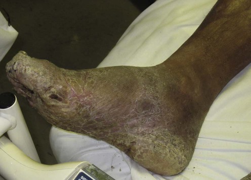
Mycetoma
eumycetoma and actinomycetoma
Specific investigations


First-line therapies
 Sulfonamides
 Aminoglycosides
 Itraconazole
Second-line therapies
 Amoxicillin–clavulanate
 Terbinafine
 Ketoconazole
 Posaconazole
 Imipenem
 Voriconazole
 Oxazolidinones
 Rifampicin
 Amphotericin B
[/level-membership-for-dermatology-category][not-level-membership-for-dermatology-category]
Mycetoma
eumycetoma and actinomycetoma
