Chapter 26 Muscle Pain and Cramps
Muscle Pain: Basic Concepts
Nociceptor Terminal Stimulation and Sensitization
Stimuli of afferent axons can be chemical and mechanical. Increased levels of glutamate in muscle correlate temporally with the appearance of pain after exercise or experimental injections of hypertonic saline. Glutamate injection into muscle in humans both produces pain and sensitizes muscle afferents to other stimuli. Some mechanosensitive unmyelinated afferent axons in muscle are stimulated by acromelic acid-A, a kainoid mushroom toxin that produces long-lasting allodynia and burning pain when ingested. The injection of acid (H+) into skeletal muscle elicits pain. Acid-sensing ion channel 3 (ASIC3) channels are expressed on sensory neurons innervating skeletal and cardiac muscle. H+ ions may produce muscle pain by activating ASIC3 channels on afferent axons. ASIC3 channels may initiate the anginal pain associated with myocardial ischemia. Lactate, an anaerobic metabolite, probably does not play a primary role in directly stimulating muscle pain. Patients with myophosphorylase deficiency do not produce lactate under ischemia yet experience pain. Lactate may potentiate the effects of H+ ions on ASIC3 channels in activating pain-related axons. Adenosine triphosphate (ATP), another metabolite, is present in increased levels in muscle interstitium during ischemic muscle contraction. Injection of ATP also elicits pain. Many peripheral nociceptors express ATP purinergic receptors, specifically P2X3, P2Y2, and P2Y3. P2X3/P2X2/3 receptor antagonists can reverse the mechanical hyperalgesia that occurs with inflammation. Individual algogenic molecules only activate subsets of peripheral nociceptors. Combinations of protons, lactate, and other molecules synergistically activate the most sensory afferents in muscle tissue (Light et al., 2008).
Sensitization of nociceptive axon terminals is reduction of the threshold for their stimulation into the innocuous range. Sensitization of nociceptor terminals can have two effects on axons: (1) an increase in the frequency of action potentials in normally active nociceptors or (2) induction of new action potentials in a population of normally silent small axons that are especially prominent in viscera. Vanilloid receptors and tetrodotoxin-resistant sodium channels can play roles in the sensitization of nerve terminals. The heat and capsaicin receptor, transient receptor potential cation channel V1 (TRPV1) can be activated in strong acidic conditions. However, the main TRPV1 activator may be endogenous ligands such as oxidized metabolites of linoleic acid, which damaged myocytes potentially release. Factors released during damage or repetitive stimulation induce a reduction in the axon terminal thresholds in muscle. Substance P induces a low-frequency discharge in afferent axons that could contribute to spontaneous pain. Leukotriene D4 may have a desensitizing effect on muscle nociceptors. The depression of muscle nociceptor activity by aspirin may reflect inhibition of the effects of prostaglandin E2. Other endogenous substances proposed to play roles in activating or sensitizing peripheral nociceptive afferents include neurotransmitters (serotonin, histamine, glutamate, nitric oxide, adrenaline), neuropeptides (substance P, neurokinin 1, bradykinin, nerve growth factor [NGF], calcitonin gene-related peptide), inflammatory mediators (prostaglandins, cytokines), and potassium ions (Mizumura, 2009). GTP cyclohydrolase 1 (GCH1), a rate-limiting enzyme in the tetrahydrobiopterin synthetic pathway, may play a role in enhancing inflammatory and neuropathic pain sensitivity. Certain haplotypes of GCH1 are associated with reduced GCH1 activity and may be protective against pain in patients experiencing pain and normal healthy controls.
Nociceptive Axons
Many of the afferent nerve fibers that transmit painful stimuli from muscle (nociceptors) have small unmyelinated (free) axon terminals (Graven-Nielsen and Mense, 2001; Julius and Basbaum, 2001). These terminal axons (nerve endings) are mainly located near blood vessels and in connective tissue but do not contact muscle fibers. Free nerve endings have a small diameter (0.5 µm) with varicosities (expansions). They contain glutamate and neuropeptides. Noxious stimuli produce graded receptor potentials in nerve endings, with the amplitude dependent on strength of the stimulus. An action potential develops if the amplitude of the receptor potential is large enough to reach threshold. Action potentials arising in nociceptor terminals induce or potentiate pain by two mechanisms. Centripetal conduction to central branches of afferent axons brings nociceptive signals directly to the CNS. Centrifugal conduction of action potentials along peripheral axon branches causes indirect effects by invading other nerve terminals and causing release of glutamate and neuropeptides into the extracellular medium. These algesic substances can stimulate or sensitize terminals on other nociceptive axons.
Aδ-class (group III) and C-class (group IV) afferent axons play important roles in the conduction of pain-inducing stimuli from muscle to the CNS (Arendt-Nielsen and Graven-Nielsen, 2008). Blockade of both Aδ- and C-class axons eliminates the ability to detect acute noxious stimuli. Selective expression of sodium channel isoforms Nav1.7, Nav1.8, and Nav1.9 occurs on axons and sensory ganglia in peripheral nerve pain pathways. Aδ-class nociceptive axons are thinly myelinated, conduct impulses at moderately slow velocities (3 to 13 m/sec), and have membrane sodium channels that are tetrodotoxin sensitive. Aδ axons are high-threshold mechanoceptors stimulated by strong local pressure and mediate rapid, acute, sharp (first-phase) muscle pain. Aδ-class axons probably mediate spontaneous pain and dysesthesias. C-class nociceptive axons are unmyelinated, conduct impulses at very slow velocities (0.6 to 1.2 m/sec), and have membrane sodium channels that are tetrodotoxin resistant. C-class axons in muscle are often polymodal, responding to a range of stimuli, but stimulus-specific axon terminals are also present. C fibers mediate somewhat delayed, diffuse, dull or burning (second-phase) pain evoked by noxious stimuli. Constituents of muscle nociceptor C axons include substance P, calcitonin gene-related peptide, and somatostatin. These constituents may place the nociceptive axons in a subgroup of C fibers that mediate hyperalgesia in response to inflammation. Aβ-class axons are large, myelinated, and conduct impulses at rapid velocities. They normally mediate innocuous stimuli, and stimulation may reduce the perception of pain. Inflammation or repetitive stimulation can sensitize Aβ axons, which then mediate mechanical allodynia in some tissues. Mediation of this “phenotypic switch” in Aβ axons may occur via up-regulation of neuropeptide Y and sprouting of terminals in the spinal cord from lamina III and IV into lamina II, with subsequent stimulation of ascending central pain pathways.
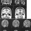
Central terminals of nociceptive axons from muscle end in lamina I in the dorsal horn of the spinal cord. Ascending central neurons with cell bodies in laminae I or II are stimulated by glutamate from the terminals of primary afferent axons and convey sensory pain modalities via a lateral nociceptive system that includes the contralateral spinothalamic tract, thalamic nuclei (Ren and Dubner, 2002), and somatosensory cortex. Some models suggest that tonic muscle pain may involve a medial set of central pathways including the ipsilateral insula and medial prefrontal regions (Thunberg et al., 2005). Transmission of affective features of pain may also involve medial pathways to the parabrachial nucleus, amygdala, thalamic intralaminar nucleus, and anterior cingulate gyrus. Interneurons and descending CNS pathways modulate afferent input, especially with chronic pain. Central sensitization to pain is associated with neurons containing substance P receptors. Glutamate acting at N-methyl-d-aspartate (NMDA) receptors is essential for the initiation of central sensitization and for the hyperexcitability of spinal cord neurons and persistent pain. Facilitation via descending CNS pathways may lead to allodynia and the maintenance of hyperalgesia. Decreased activation of inhibitory descending pathways is associated with increased opioid sensitivity and may provide a system of endogenous analgesia. There is enhanced net descending inhibition at sites of primary hyperalgesia associated with inflammation.
Pathological Conditions Producing Muscle Pain
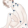
Fibromyalgia is a syndrome with diffuse chronic muscle pain and tenderness to palpation over trigger points. Central sensitization may underlie some of the symptoms of fibromyalgia and related syndromes of myofascial pain and stress-induced syndromes in which pain localizes to muscle, but without prominent physiological or morphological evidence of muscle damage (Bennett, 2005).
Clinical Features of Muscle Pain
Evaluation of Muscle Discomfort
The basis for the classification of disorders underlying muscle discomfort can be anatomy, temporal relation to exercise, muscle pathology, and the presence or absence of active muscle contraction during the discomfort (Kincaid, 1997; Pestronk, 2010). Evaluation of muscle discomfort typically begins with a history that includes the type, localization, inducing factors, and evolution of the pain; drug use; and mood disorders. The physical examination requires special attention to the localization of any tenderness or weakness. Accurate assessment of strength may be difficult in the presence of pain. The sensory examination is important because small-fiber sensory neuropathies commonly cause discomfort with apparent localization in muscle. A general examination is important to evaluate the possibility that pain may be arising from other tissues such as joints. Blood studies may include a complete blood cell count, sedimentation rate, creatine kinase (CK), aldolase, potassium, calcium, phosphate, lactate, thyroid functions, and evaluation for systemic immune disorders. Evaluate urine myoglobin in patients with a high CK and severe myalgias, especially when they relate to exercise. Electromyography (EMG) is a sensitive test for myopathy. A normal EMG can suggest that muscle pain is arising from anatomical loci other than muscle. Nerve-conduction studies may detect an underlying neuropathy, but objective documentation of small-fiber axonopathies can require quantitative sensory testing or skin biopsy with staining of distal nerve fibers. Magnetic resonance imaging, ultrasound, or radionuclide scans may reveal focal or diffuse anomalies in muscle, joints, or fascia that can be useful to guide biopsy procedures. Phosphorous magnetic resonance spectroscopy may become useful in evaluating and monitoring some metabolic myopathies, but its utility remains to be determined. Muscle ultrasound can be a useful and noninvasive method of localizing and defining types of muscle pathology. Muscle biopsy is most often useful in the presence of another abnormal test result such as a high serum lactate, aldolase, CK, or an abnormal EMG. However, important clues to treatable disorders such as fasciitis or systemic immune disorders (connective tissue pathology, perivascular inflammation or granulomas) may be present in muscle in the absence of other positive testing. Examination of both muscle and connective tissue increases the yield of muscle biopsy in syndromes with muscle discomfort. There is increased diagnostic yield from muscle biopsies if in addition to routine morphological analysis and processing, histochemical analysis includes staining for acid phosphatase, alkaline phosphatase, esterase, mitochondrial enzymes, glycolytic enzymes, C5b-9 complement, and MHC Class I. Measurement of oxidative enzyme activities can reveal causes of muscle discomfort or fatigue, even in disorders with no histopathological abnormalities. Ultrastructural examination of muscle rarely provides additional information in muscle pain syndromes.
Muscle Discomfort: Specific Causes
Muscle pain is broadly divisible into groups depending on its origin and relation to the time of muscle contraction. Myopathies may be associated with muscle pain without associated muscle contraction (myalgias) (Boxes 26.1 and 26.2). Muscle pain during muscle activity (Box 26.3; also see Box 26.2) may occur with muscle injury, myopathy, cramps, or tonic (relatively long-term) contraction. Some pain syndromes perceived as arising from muscle originate in other tissues, such as connective tissue, nerve, or bone, or have no clear morphological explanation for the pain (Box 26.4).
Box 26.1 Myopathic Pain Syndromes*
Box 26.2 Muscle Discomfort Associated with Drugs and Toxins
Box 26.3 Cramps* and Other Involuntary Muscle Contraction Syndromes
bid, Twice daily; qhs, daily at bedtime; tid, three times daily.
* Usual features: sudden involuntary painful muscle contractions (usually involve single muscles, especially gastrocnemius); local cramps in other muscles often associated with neuromuscular disease. Precipitants: muscle contraction, occasionally during sleep. Relief: passive muscle stretch, local massage.
Myopathies with Muscle Pain
Myopathies that produce muscle pain (see Box 26.1) are usually associated with weakness, a high serum CK or aldolase, or an abnormal EMG (Pestronk, 2010). Immune-mediated or inflammatory myopathies may produce muscle pain or tenderness, especially with an associated systemic connective tissue disease or pathological involvement of connective tissue (including myopathies with anti-tRNA synthetase antibodies). Pain is common in childhood dermatomyositis, immune myopathies associated with systemic disorders, eosinophilia-myalgia syndromes, focal myositis, and infections. Myopathies due to direct infections (e.g., bacterial, viral, toxoplasmosis, trichinosis) are usually painful. Metabolic myopathies, including myophosphorylase and carnitine palmitoyltransferase (CPT) II deficiencies, typically produce muscle discomfort or fatigue with exercise and less prominently at rest. As a rule, disorders of carbohydrate utilization like myophosphorylase deficiency produce pain and fatigue after short, intense exercise, whereas lipid disorders (CPT II deficiency) cause muscle discomfort with sustained exercise. Rhabdomyolysis is usually associated with muscle pain and tenderness that can persist for days after the initial event. It may occur with a defined metabolic or toxic myopathy or sporadically in the setting of unaccustomed exercise, especially in hot weather. Rhabdomyolysis may produce renal failure—a life-threatening complication. Aggressively pursue diagnosis and treatment and avoid precipitating factors. Medications, including ε-aminocaproic acid and cholesterol-lowering agents, may produce a painful myopathy with prominent muscle fiber necrosis and a very high serum CK. Cholesterol-lowering agents more commonly produce a myalgia syndrome with no defined muscle pathology but can also produce rhabdomyolysis syndromes, especially in the setting of high serum drug levels. Muscular dystrophy and mitochondrial disorders are usually painless. Occasional patients with mild Becker muscular dystrophy or mitochondrial syndromes with minimal or no weakness may experience a sense of discomfort such as myalgias, fatigue, or cramps, especially after exercise. Hereditary myopathies with occasional reports of muscle discomfort or spasms in patients (or carriers) include limb-girdle dystrophy types 1A, 1C, 2D, 2H (sarcotubular myopathy), myotonic dystrophy type 2, and some mild congenital myopathies. Several myopathies defined by specific morphological changes in muscle but whose cause is unknown commonly have myalgias or exercise-related discomfort. These syndromes include tubular aggregates with or without cylindrical spirals, focal depletion of mitochondria, internalized capillaries, and adult-onset rod myopathies.
Muscle Cramps
Muscle spasms are abnormal involuntary muscle contractions. Muscle cramps (see Box 26.3) are localized, typically uncomfortable muscle contractions or spasms (Miller and Layzer, 2005). Characteristic features include a sudden involuntary onset in a single muscle or muscle group, with durations of seconds to minutes and a palpable region of contraction. Occasionally there is distortion of posture. Fasciculations often occur before and after the cramp. Cramps usually arise during sleep or exercise and are more likely to occur when muscle contracts while in a shortened position. Pain syndromes associated with cramps include discomfort during a muscle contraction and soreness after the contraction due to muscle injury.
Cramps, especially those in the calf or foot muscles, are common in normal people of any age. They may be more common in the elderly (up to 50%), at the onset of exercise, at night, during pregnancy, and with fasciculations. These types of cramps are usually idiopathic and benign. Cramps that occur frequently in muscles other than the gastrocnemius often herald an underlying neuromuscular disorder. The presence of fasciculations with mild cramps but no weakness usually represents a benign condition (benign fasciculation syndrome). When the muscle cramps are more disabling, the condition is the cramp-fasciculation syndrome. EMG is normal except for the presence of fasciculations. Slow, repetitive nerve stimulation provokes after-discharges of motor unit action potentials. Neurogenic disorders that produce partial denervation of muscles (e.g., amyotrophic lateral sclerosis, radiculopathies, polyneuropathies) can be associated with cramps, painful muscle spasms, or muscle discomfort. Other causes of cramps (see Boxes 26.2 and 26.3) include drugs and metabolic, neuropathic, and inherited disorders.
Other Involuntary Muscle Contraction Syndromes
Diffuse muscle contraction syndromes, usually arising from the PNS or CNS (dystonias), often show widespread and continuous spontaneous motor unit fiber discharges. They may produce considerable discomfort. Causes are hereditary syndromes, CNS disorders, drugs, or toxins (see Boxes 26.2 and 26.3). Specific syndromes include stiff person syndrome, Isaac syndrome (neuromyotonia with contractions due to repetitively discharging motor units at high frequencies), Schwartz-Jampel syndrome, restless legs, and toxic disorders related to phencyclidine, amphetamine, tetanus, and strychnine. Tetany, typically associated with hypocalcemia or alkalosis, manifests with neuromyotonic-like discharges. Myokymic rippling of muscles shows recurring bursts of motor units at 30 to 80 Hz. In the postoperative period, a muscle contraction syndrome with fasciculations and myalgia often occurs after the use of succinylcholine (suxamethonium). Two diffuse contraction disorders, malignant hyperthermia and neuroleptic malignant syndromes, have not undergone a systematic evaluation by electrodiagnostic testing during episodes.
Myalgia Syndromes without Chronic Myopathy
Pain originating from muscle, often acute, may occur in the absence of a chronic myopathy. Muscle ischemia causes a squeezing pain in the affected muscles during exercise. Ischemia produces pain that develops especially rapidly (within minutes) if muscle is forced to contract at the same time; the pain subsides quickly with rest. Cramps and overuse syndromes are associated with pain during or immediately after muscle use. DOMS occurs 12 to 48 hours after exercise and lasts for hours to days. Muscle contraction or palpation exacerbates discomfort. Serum CK is often increased. DOMS is most commonly precipitated by eccentric muscle contraction (contraction during muscle stretching) or unaccustomed exercise. The discomfort after eccentric contractions may be associated with repetitive overstretching of elastic non-contractile tissues. Exercise training typically protects against DOMS. Muscle fatigue after exercise, with reduced maximum levels of voluntary contraction, may occur via a disorder separate from DOMS involving disruption of excitation/contraction systems (Iguchi and Shields, 2010).
Polymyalgia syndromes (see Box 26.4) have pain localized to muscle and other structures. The pain may produce the appearance of weakness by preventing full effort. Typical of this type of “weakness” on examination is sudden reduction in the apparent level of effort, rather than smooth movement through the range of motion expected with true muscle weakness. Polymyalgic pain is often present at rest and variably affected by movement. Serum CK and EMG are normal. No major pathological change in muscle occurs unless the discomfort produces disuse and atrophy of type II muscle fibers. Muscle biopsies may also show changes associated with systemic immune disorders, including inflammation around blood vessels or in connective tissue. Polymyalgia syndromes can have identified causes including systemic immune disease, drug toxicity, and small-fiber polyneuropathies.
Arendt-Nielsen L., Graven-Nielsen T. Muscle pain: sensory implications and interaction with motor control. Clin J Pain. 2008;24:291-298.
Bennett R. Fibromyalgia: present to future. Curr Rheumatol Rep. 2005;7:371-376.
Graven-Nielsen T., Mense S. The peripheral apparatus of muscle pain: evidence from animal and human studies. Clin J Pain. 2001;17:2-10.
Iguchi M., Shields R.K. Quadriceps low-frequency fatigue and muscle pain are contraction-type-dependent. Muscle Nerve. 2010;42:230-238.
Julius D., Basbaum A.I. Molecular mechanisms of nociception. Nature. 2001;413:203-210.
Kincaid J.C. Muscle pain, fatigue and fasciculations. Neurol Clin. 1997;15:697-709.
Light A.R., Hughen R.W., Zhang J., et al. Dorsal root ganglion neurons innervating skeletal muscle respond to physiological combinations of protons, ATP, and lactate mediated by ASIC, P2X, TRPV1. J Neurophysiol. 2008;100:1184-1201.
Miller T.M., Layzer R.B. Muscle cramps. Muscle Nerve. 2005;32:431-442.
Mizumura K. Peripheral mechanism of muscle pain: an update. Curr Anaesth Crit Care. 2009;20:183-187.
Pestronk A. Neuromuscular Disease Center. St. Louis, MO: Washington University School of Medicine; 2010. World Wide Web URL http://neuromuscular.wustl.edu/
Ren K., Dubner R. Descending modulation in persistent pain: an update. Pain. 2002;100:1-6.
Thunberg J., et al. Brain processing of tonic muscle pain induced by infusion of hypertonic saline. Eur J Pain. 2005;9:185-194.


