Published on 19/03/2015 by admin
Filed under Dermatology
Last modified 22/04/2025
This article have been viewed 2288 times
Noah Scheinfeld
Evidence Levels: A Double-blind study B Clinical trial ≥ 20 subjects C Clinical trial < 20 subjects D Series ≥ 5 subjects E Anecdotal case reports
Labial mucocels fall into two categories: the mucous extravasation cyst, and the mucous retention cyst. The mucous extravasation cyst describes a false cyst because the mucous extravasation cyst lacks an epithelial lining arising from the partially or totally severed salivary gland duct resulting in the accumulation of saliva in the adjacent soft tissue. At this point the mucocele is cut off by a fibrous connective tissue pseudocapsule. The ductal epithelium lines the mucous retention cyst. The mucous retention cyst develops from partial obstruction of a duct in the presence of the salivary gland’s continued mucous secretion. The extravasation mucocele manifests most commonly and manifest most often on the young person’s lower lip. The retention mucocele is more apt to occur on the buccal cheek or soft palate of an older patient.
To expand this further, mucous extravasation cysts arise from trauma to salivary gland ducts. This trauma leads to rupture salivary gland ducts and leakage of mucin from the minor salivary glands. The mucin subsequently forms pseudocytic aggregations most commonly on the lower lip. These aggregations are referred to as mucoceles. Mucoceles manifest with a variety of tones and color that range from flesh color to red to translucent blue. The shape of mucoceles is round or oval and their surface is smooth. They usually possess a soft, fluctuant or gel-like consistency. Single or multiple mucoceles can manifest and can range from 0.1 to 2 cm mm in diameter. The natural history of mucoceles can involvement their expansion and periodic rupture and sometimes spontaneous resolution. There is some morbidity associated with mucocles that ranges from discomfort, to suboptimal cosmetic to appearance of a nodule with a hardened consistency due to scarring and tissue consolidation.
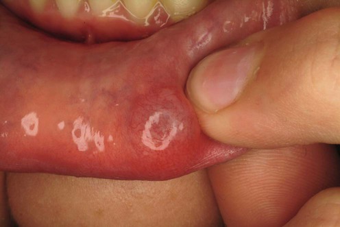
‘Superficial mucocele’, a variant of a mucocele, can manifest on the palate, retromolar pad, and posterior buccal mucosa. Superficial mucocles manifest as single or multiple vesicles, which can break down into an ulcer. Despite healing after a few days, superficial mucoceles recur often in the same location. A mucocele is termed a ranula when on the floor of the mouth, and epulis when on the gums.
Biopsy
Doppler ultrasonography
Color Doppler imaging
Toida M, Ishimaru JI, Hobo N. Int J Oral Maxillofac Surg 1993; 22: 353–5.
Twelve female and six male patients, with mucous cysts on the lower lip and the tip of the tongue, were treated by direct application of liquid nitrogen with a cotton swab. Each lesion was exposed to four or five cycles composed of freezing for 10–30 seconds and thawing for double the freezing time. No anesthesia was required. All lesions had disappeared completely 2 to 4 weeks after one or two treatment courses of cryosurgery. In all cases, neither scarring nor recurrence was noted during the 6 months to 5 years of follow-up.
Merida FMT. Med Cutan Ibero Lat Am 1976; 4; 15–18.
Eight cases of lip mucocele were treated with intralesional infiltration of triamcinolone acetonide (Kenacort). In three cases there was complete regression of the lesion after a variable period of time between the first and the fourth infiltration. In five cases the results were negative. The patients who reacted positively to treatment were followed-up for a year afterwards.
Bentley JM, Barankin B, Guenther LC. Clin Pediatr (Phila) 2003; 42: 475–82.
After a diagnosis is made, treatment is often not needed as smaller and more superficial mucoceles are likely to rupture and spontaneously heal. The patient’s future as it relates to mucoceles is good whether or not surgery is involved.
Mínguez-Martinez I, Bonet-Coloma C, Ata-Ali-Mahmud J, Carrillo-García C, Peñarrocha-Diago M, Peñarrocha-Diago M.J Oral Maxillofac Surg 2010; 68: 2468–71.
In a series of 89 cases, mucoceles were more commonly located on the lower lip; 43.8% resolved spontaneously and 8% of the surgically removed mucoceles reappeared.
Gill D. Australas J Dermatol 1996; 37: 220.
Punch is a useful technique for treating mucocele and has the added benefit of providing a histologically certain diagnosis.
Huang IY, Chen CM, Kao YH, Worthington P. J Oral Maxillofac Surg 2007; 65: 855–8.
Eighty-two patients with biopsy-confirmed mucoceles of the lower lip were treated with CO2 laser vaporization with no bleeding and minimal scar formation. Two recurrences occurred. Researchers noted mild discomfort and rare complications. At the operative site, one patient felt temporary numbness. Due to the fact that the operative time is shorter than with the excisional method, CO2 laser appears useful for children and for less cooperative mucocele patients
Yamasoba T, Tayama N, Syoji M, Fukuta M. Head Neck 1990; 12: 316–20.
Researchers reported 70 patients with lower lip mucoceles for patient characteristics, clinical features, and histopathologic findings. Patients were divided almost equally between males and females, with ages ranging from 2 to 63 years, with the highest incidence of lesions occurring in the second decade. Mucocele life span ranged from a few days to 3 years. Size did not affect outcome. The upper lateral incisor was the most commonly involved area. Of 70 biopsies, 68 were mucous extravasation cysts and two were mucous retention cysts. Surgical excision was the treatment of choice, with recurrence of the lesion in only two cases.
Bahadure RN, Fulzele P, Thosar N, Badole G, Baliga S. Eur J Paediatr Dent 2012; 13: 143–6.
Conventional surgical procedure with excision of minor salivary glands was effective in 23 patients and was considered with a 3-year follow-up to have a low recurrence rate.
Kang SK, Kim KS. Taehan Chikkwa Uisa Hykhoe Chi 1989; 27: 1059–71.
In a series of 112 patients, surgeons treated 107 mucoceles (95%) by excision and only five by marsupialization. Eighteen of 112 cases had recurrence with the recurrence rate being 16%. Only three of the 112 cases revealed an epithelial lining. This incidence indicates that the mucus extravasation by the damage of excretory duct rather than the ductal dilatation by mucus retention may play a critical role in the production of these lesions. In 81 cases (72.3%) minor salivary glands were included in the excision biopsy specimen.
Delbem AC, Cunha RF, Vieira AE, Ribeiro LL. Pediatric Dent 2000; 22: 155–8.
Micro-marsupialization requires neither injections nor surgery and was studied in 14 patients. Micro-marsupialization involves placing a topical anesthetic gel on the mucocele for 3 minutes, passing a 4-0 silk suture through the body of the mucocele, and tying a surgeon’s knot. The suture material is removed 7 days later, at which time the mucocele is resolved. The advantages of this technique include simplicity and relative lack of pain. Micro-marsupialization is not indicated for fibrotic lesions, lesions of the palate, or for lesions on the inside of the cheek (cheek mucosa). Of the original 14 patients treated by the micro-marsupialization, 12 presented full regression 1 week after treatment. Recurrence occurred in two cases.
Treatment of Skin Disease Comprehensive Therapeutic Strategies 4e
WhatsApp us


 Cryotherapy
 Intralesional corticosteroids
 No treatment (observation)
 Punch biopsy
 CO2 laser
 Surgical excision
 Marsupialization
 Micro-marsupialization