Chapter 86 Metabolic response to injury and infection
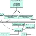
Injury and infection evoke in the host a hypermetabolic inflammatory response and a compensatory hypometabolic hypoimmune response. The magnitude of the response is proportional to the extent of injury. Additional components of illness, such as ischaemia and reperfusion or resuscitation, nutritional status, surgical procedures, transfusions, drugs and anaesthetic techniques, genetic polymorphisms and concurrent diseases, impact on the response. Some components of the metabolic response, or the failure to regulate the response, are destructive, and its modulation may improve patient survival.1
MEDIATORS OF THE METABOLIC RESPONSE
CYTOKINES
NEUROENDOCRINE MEDIATORS
Cytokine release from the site of injury or infection triggers vagal afferent impulses to the dorsal vagal complex (DVC) in the medulla oblongata. Synaptic connections with the rostroventral medulla and locus ceruleus, and the hypothalamic nuclei, activate the sympathetic nervous system and the HPA axis respectively.2 High circulating cytokine levels may also cross the blood–brain barrier, or affect neurons at circumventricular organs lacking a blood–brain barrier, such as the area postrema. In general, a biphasic response is observed following injury and infection: an initial neuroendocrine ‘storm’ followed by a decrease. The following are some neuroendocrine mediators involved in the response to stress:
THE METABOLIC RESPONSE
The metabolic response to injury and infection begins with the activation of receptors throughout the body by the above mediators. These receptors include toll-like receptors (TLR)-2 and TLR-4, and the receptor for advanced glycation end products (RAGE). Subsequent intracellular activation of the NF-κB pathway leads to gene induction and production of mRNA for the synthesis of proinflammatory cytokines. Catecholamines can initiate rapid functional changes via protein phosphorylation, which does not require gene induction. Behavioural effects such as anorexia, possibly due to elevated leptin levels, also affect the metabolic response. The metabolic effects may be described at three levels.
CELLULAR METABOLIC EVENTS
Intracellularly, heat shock protein (HSP) synthesis is induced. Many HSPs are also expressed constitutively. HSPs act as ‘chaperones’, assisting in the assembly, disassembly, stabilisation and internal transport of other intracellular proteins. HSPs facilitate translocation of the glucocorticoid-receptor complex from the cytosol to the nucleus, and inhibit NF-κB activity. HSPs have cellular protective roles in sepsis and ischaemia-reperfusion.5
Mitochondrial dysfunction limits cell metabolism and may be responsible for the ensuing multiorgan dysfunction of severe injury and infection. Electron transport chain complexes are inhibited by excessive nitric oxide and peroxynitrite generated during sepsis.6 Additionally, poly (ADP-ribose) polymerase-1 (PARP-1) is activated, reducing cell nicotinamide adenine dinucleotide (NADH) content, a substrate for ATP generation. The reduced ATP production may have functional consequences, such as diaphragmatic dysfunction.7 It has been hypothesised that this ‘metabolic shutdown’ is an adaptive response similar to hibernation.8
Apoptosis, or programmed cell death, may finally be induced when death receptors are engaged by their ligands, or by mitochondrial-mediated pathways.9 Death receptors include Fas and TNF receptor type I (TNFRI), and this pathway activates caspase. The mitochondrial pathway, mediated in part by glycogen synthase kinase-3β activation, causes activation of caspase. Caspases 8 and 9 activate caspase 3, which commits the cell to death. TNF-α, IL-10, cortisol and nitric oxide all induce apoptosis. Apoptosis further augments immunosuppression. It is probable that bioenergetic failure and the apoptotic death of immune cells are the major causes of death in late sepsis.
FACTORS AFFECTING THE METABOLIC RESPONSE
ENERGY BALANCE AND OXYGEN DELIVERY
Hypermetabolism increases oxygen demand and consumption. Capillary permeability increases, causing reduced intravascular filling, and inadequate oxygen delivery can lead to anaerobic metabolism and inadequate production of high-energy phosphates. In adequately resuscitated patients, aerobic glycolysis with lactate production rather than anaerobic glycolysis is characteristic of the metabolic response to stress.10 Subsequent cell swelling from injury and resuscitation activates phospholipase A2, augmenting the inflammatory cascade.11
SURGICAL PROCEDURES AND ANAESTHETIC TECHNIQUES
Total afferent neuronal blockade (somatic and autonomic block) (e.g. by epidural anaesthesia), and minimally invasive surgery, both of which greatly attenuate the metabolic changes of surgical injury, have been associated with reduced morbidity.12,13 Improved analgesia, avoidance of endotracheal intubation and maintenance of consciousness may also contribute to the effect.
STARVATION AND NUTRITION
Starvation alone produces adaptive hypometabolism with the use of fat as the primary fuel, sparing protein. In contrast, injury and infection result in hypermetabolism with prominent protein catabolism. Starvation in combination with the metabolic changes of stress produces a hypoalbuminaemic malnourished state. Malnutrition clearly contributes to morbidity and mortality. However, current forms of nutrition do not adequately reduce protein catabolism or promote protein synthesis, so that a positive energy balance often results in fat and fluid gain instead.14
DRUGS AND DISEASE
Adverse effects of steroids include increased infection rates and myopathy (particularly in conjunction with the use of neuromuscular blocking drugs). Commonly used intensive care unit (ICU) drugs such as the catecholamines, dopamine, corticosteroids, theophylline, calcium-channel blockers, antibiotics and blood transfusions all have immunomodulatory effects. Diseases such as diabetes will clearly impact on the metabolic response to stress. Apart from concurrent disease, the inciting organism may itself modulate its host response by affecting cytokine mediators, their production, secretion and elimination, or by interacting with their receptors.15
VALUE OF THE METABOLIC RESPONSE
The cytokines and neuroendocrine mediators reprioritise metabolic processes, increasing the supply of substrates to active tissues involved in defence against injury. Inflammation localises the area of injury. Cardiovascular changes divert blood flow to inflamed areas and vital organs, while salt and water retention maintains overall perfusion. Sympathectomised and adrenalectomised animals fare poorly when stressed. Etomidate, which blocks cortisol synthesis, was associated with increased mortality when used for ICU sedation.17
MODULATING THE METABOLIC RESPONSE
Antioxidants show promise in improving survival. The substitution of omega-3 fatty acids for omega-6 fatty acids in inflammatory mediators modulates the metabolic response. Clinically, enteral nutrition with eicosapentaenoic acid (EPA or fish oil), γ-linolenic acid (GLA or borage oil) and antioxidant vitamins18 has been shown to reduce mortality. Selenium prevents excessive oxidative damage, but was not shown to reduce mortality when supplemented in pharmacological doses, and a larger trial is needed.19 Ethyl pyruvate is another promising antioxidant able to directly neutralise peroxides and peroxynitrite non-enzymatically. Statins decrease the activation of the NF-κB pathway, and inhibit superoxide production. Experimentally, statins have been shown to prolong survival time in sepsis,20 but randomised controlled trials in humans are required.21
Protein catabolism leading to loss of muscle has functional consequences such as respiratory muscle weakness. A 25% body weight loss in the presence of injury may be fatal. Wound healing and multiple facets of host defence are impaired, increasing the risk of nosocomial infection. Parenteral glutamine supplementation has been reported to reduce mortality.22 Of the anabolic agents (GH, IGF-1 and testosterone derivatives), GH has received the most attention in the setting of critical illness. GH increased mortality in a heterogeneous group of critically ill patients.23 It is possible that patient selection, unmasking of glutamine deficiency and high GH doses (mean 0.1 mg/kg per day) resulted in the adverse outcome. Combined GH, IGF-1 and glutamine-supplemented parenteral nutrition has been shown to produce net protein gain.24
‘Intensive insulin therapy’ to attain a glucose of 4.4–6.1 mmol resulted in significant prevention of acute renal failure, and shortened time to weaning from mechanical ventilatory support.25 The mortality benefit shown in a previous trial could not be corroborated. Frequent hypoglycaemia may accompany the technique and further studies are underway.
A meta-analysis of steroid usage in septic shock found that high-dose steroids given over a short time increased mortality, while lower doses of hydrocortisone for 5–7 days and subsequently tapered resulted in improvement in survival and shock reversal.26 In the largest trial, the benefit was found only in patients who had a less than 9 μg/dl increase in cortisol after 250 μg tetracosactrin intravenously.27 Given the difficulties in free cortisol kinetics and definition of ‘relative adrenal insufficiency’ in the critically ill, this finding requires external validation.
The extracellular fluid is the aqueous medium within which the mediators of the metabolic response function, so that blood purification may modulate the intensity of the metabolic response and hence influence outcome. A randomised trial of 425 ICU patients with acute renal failure to ultrafiltration doses of 20 ml/hour per kg, 35 ml/hour per kg and 45 ml/hour per kg by continuous veno-venous haemofiltration resulted in survival at 15 days after treatment discontinuation of 41%, 57% and 58% respectively.28 Extension of the above approach leads to the inference that high volume haemofiltration (i.e. intensive plasma water exchange) may further improve survival.29
In an initial trial, activated protein C (APC) was found to reduce 28-day mortality in patients with severe sepsis. Longer term follow-up did not reveal an overall improvement in survival, although the study was not powered to detect this end-point.30 In less severely ill patients, APC did not influence outcome, and increased the rates of serious bleeding.31 APC is not indicated in paediatric severe sepsis.
The most logical approach is to monitor the host response so as to provide individualised therapy. Although IFN-γ did not reduce infection or mortality in burns patients,32 it is hypothesised that monitoring HLA-DR expression may be used to identify immunosuppressed patients who may benefit most from IFN-γ.33
1 Russell JA. Management of sepsis. N Engl J Med. 2006;355:1699-1713.
2 Webster JI, Tonelli L, Sternberg EM. Neuroendocrine regulation of immunity. Annu Rev Immunol. 2002;20:125-163.
3 Van den Berghe G, de Zegher F, Bouillon R. Acute and prolonged critical illness as different neuroendocrine paradigms. J Clin Endocrinol Metab. 1998;83:1827-1834.
4 Van den Berghe G, Baxter RC, Weekers F, et al. A paradoxical gender dissociation within the growth hormone/insulin-like growth factor I axis during protracted critical illness. J Clin Endocrinol Metab. 2000;85:183-192.
5 Bruemmer-Smith S, Stubber F, Schroeder S. Protective functions of intracellular heat-shock protein (HSP) 70-expression in patients with severe sepsis. Intensive Care Med. 2001;27:1835-1841.
6 Boulos M, Astiz ME, Barua RS, et al. Impaired mitochondrial function induced by serum from septic shock patients is attenuated by inhibition of nitric oxide synthase and poly(ADP-ribose) synthase. Crit Care Med. 2003;31:353-358.
7 Callahan LA, Supinski GS. Sepsis induces diaphragm electron transport chain dysfunction and protein depletion. Am J Respir Crit Care Med. 2005;172:861-868.
8 Singer M, De Santis V, Vitale D, et al. Multiorgan failure is an adaptive, endocrine mediated, metabolic response to overwhelming systemic inflammation. Lancet. 2004;364:545-548.
9 Hotchkiss RS, Osmon SB, Chang KC, et al. Accelerated lymphocyte death in sepsis occurs by both the death receptor and mitochondrial pathways. J Immunol. 2005;174:5110-5118.
10 Levy B, Gibot S, Franck P, et al. Relation between muscle Na+/K+ ATPase activity and raised lactate concentrations in septic shock: a prospective study. Lancet. 2005;365:871-875.
11 Cotton B, Guy J, Morris J, et al. The cellular, metabolic, and systemic consequences of aggressive fluid resuscitation strategies. Shock. 2006;26:115-121.
12 Rodgers A, Walker N, Schug S, et al. Reduction of postoperative mortality and morbidity with epidural or spinal anaesthesia: results from overview of randomised trials. BMJ. 2000;321:1493-1497.
13 Lacy AM, Garcia-Valdecasas JC, Delgado S, et al. Laparoscopy-assisted colectomy versus open colectomy for treatment of non-metastatic colon cancer: a randomised trial. Lancet. 2002;359:2224-2229.
14 Hart DW, Wolf SE, Herndon DN, et al. Energy expenditure and caloric balance after burn. Increased feeding leads to fat rather than lean mass accretion. Ann Surg. 2002;235:152-161.
15 Wilson M, Seymour R, Henderson B. Bacterial perturbation of cytokine networks. Infect Immunol. 1998;66:2401-2409.
16 Sørensen T, Nielsen G, Andersen P, et al. Genetic and environmental influences on premature death in adult adoptees. N Engl J Med. 1988;318:727-732.
17 Watt I, Ledingham IM. Mortality amongst multiple trauma patients admitted to an intensive therapy unit. Anaesthesia. 1984;39:973-981.
18 Pontes-Arruda A, Aragao A, Albuquerque J. Effects of enteral feeding with eicosapentaenoic acid, γ-linolenic acid, and antioxidants in mechanically ventilated patients with severe sepsis and septic shock. Crit Care Med. 2006;34:2325-2333.
19 Angstwurm M, Engelmann L, Zimmermann T, et al. Selenium in Intensive Care (SIC) study. Results of a prospective randomized, placebo-controlled, multiple-center study in patients with severe systemic inflammatory response syndrome, sepsis, and septic shock. Crit Care Med. 2007;35:118-126.
20 Merx M, Liehn E, Graf J, et al. Statin treatment after onset of sepsis in a murine model improves survival. Circulation. 2005;112:117-124.
21 Majumdar S, McAlister F, Eurich D, et al. Statins and outcomes in patients admitted to hospital with community acquired pneumonia: population based prospective cohort study. BMJ. 2006;333:999.
22 Novak F, Heyland DK, Avenell A, et al. Glutamine supplementation in serious illness: a systematic review of the evidence. Crit Care Med. 2002;30:2022-2029.
23 Takala J, Ruokonen E, Webster N, et al. Increased mortality associated with growth hormone treatment in critically ill adults. N Engl J Med. 1999;341:785-792.
24 Carroll P, Jackson N, Russell-Jones D, et al. Combined growth hormone/insulin-like growth factor I in addition to glutamine-supplemented TPN results in net protein anabolism in critical illness. Am J Physiol Endocrinol Metab. 2004;286:151-157.
25 Van den Berghe G, Wilmer A, Hermans G, et al. Intensive insulin therapy in the medical ICU. N Engl J Med. 2006;354:449-461.
26 Minneci P, Deans K, Banks S, et al. Meta-analysis: the effect of steroids on survival and shock during sepsis depends on the dose. Ann Intern Med. 2004;141:47-56.
27 Annane D, Sebille V, Charpentier C, et al. Effect of treatment with low doses of hydrocortisone and fludrocortisone on mortality in patients with septic shock. JAMA. 2002;288:862-871.
28 Ronco C, Bellomo R, Homel P, et al. Effects of different doses in continuous veno-venous haemofiltration on outcomes of acute renal failure: a prospective randomised trial. Lancet. 2000;355:26-30.
29 Ratanarat R, Brendolan A, Piccinni P, et al. Pulse high-volume haemofiltration for treatment of severe sepsis: effects on hemodynamics and survival. Critical Care. 2005;9:R294-302.
30 Angus D, Laterre P, Helterbrand J, et al. The effect of drotrecogin alfa (activated) on long term survival after severe sepsis. Crit Care Med. 2006;32:2199-2206.
31 Abraham E, Laterre P, Garg R, et al. Drotrecogin alfa (activated) for adults with severe sepsis and a low risk of death. N Engl J Med. 2005;353:1332-1341.
32 Wasserman D, Ioannovich JD, Hinzmann RD, et al. Interferon-gamma in the prevention of severe burns-related infections: a European phase III clinical trial. Crit Care Med. 1998;26:434-439.
33 Docke WD, Hoflich C, Davis K, et al. Monitoring temporary immunodepression by flow cytometric measurement of monocytic HLA-DR expression: a multicenter standardized study. Clin Chem. 2005;51:2341-2347.


