Maternal physiologic changes in pregnancy

Published on 07/02/2015 by admin

Filed under Anesthesiology

Last modified 22/04/2025

Print this page

rate 1 star rate 2 star rate 3 star rate 4 star rate 5 star
Your rating: none, Average: 0 (0 votes)

This article have been viewed 1199 times

Maternal physiologic changes in pregnancy

Gurinder M.S. Vasdev, MD and Barry A. Harrison, MD

Maternal physiologic changes in pregnancy begin approximately 5 weeks after implantation and may not return to normal until 8 weeks after delivery (Table 180-1).

Table 180-1

Maternal Physiologic Changes during Pregnancy

Parameter Change during Pregnancy Normal Pregnancy Value*
Cardiac    
Rate 75-95 beats/min
SV  
CO 3-8 L/min
MAP 80 mm Hg
SVR 1200-1500 dyn • s−1 • cm−5
Respiratory
Rate None  
VT 40%-45%
image 10.5 L/min
ERV 550 mL
FRC 1350 mL

Blood gas concentrations

None


7.4-7.45
25-33 mm Hg
92-107 mm Hg
16-22 mEq/L
Hematologic
Blood volume 4500 mL or 100 mL/kg
Plasma volume +45%
Erythrocyte volume +10%-15%
Hemoglobin 11.5-15 g/dl
Hematocrit 32%-36%
WBC count 6000-20,000/μL
Procoagulant factors  
Anticoagulant activity  
PAI 1 & 2  
Iron 30-193 μg/mL
TIBC 80.1 μmol/L
Electrolytes/Renal
Renal blood flow 700 mL/min
GFR 140 mL/min
Serum Cr 0.53-0.9 mg/dL
Serum BUN 8-10 mg/dL
image None 15-20 mEq/L
Na2+ 130-148 mEq/L
K+ None or ↓§ 3.3-5.0 mEq/L
Cl 97-109 mEq/L
Metabolic
Basal body temperature  
O2 consumption  
Insulin resistance  
Gastrointestinal
Lower esophageal sphincter tone  
Gastric emptying time None except during labor  
Gastric acid secretion  
Hepatobiliary system
Gallbladder emptying time  
Liver size None  
ALP Up to 2-4 × normal value
Bilirubin/AST/ALT None  
LDH 650-700 U/L
Prothrombin time None  
Albumin 2.3-4.2 g/dL
Lipids
Cholesterol 141-210/219-349 mg/dL
Triglycerides  

image

ALP, Alkaline phosphatase; ALT, alanine transaminase; AST, aspartate aminotransferase; BUN, blood urea nitrogen; CO, cardiac output; Cr, creatinine; ERV, expiratory reserve volume; FRC, functional residual capacity; GFR, glomerular filtration rate; LDH, lactate dehydrogenase; MAP, mean arterial pressure; PAI, plasminogen activator inhibitor; RBC, red blood cell; SV, stroke volume; SVR, systemic vascular resistance; TIBC, total iron-binding capacity; image
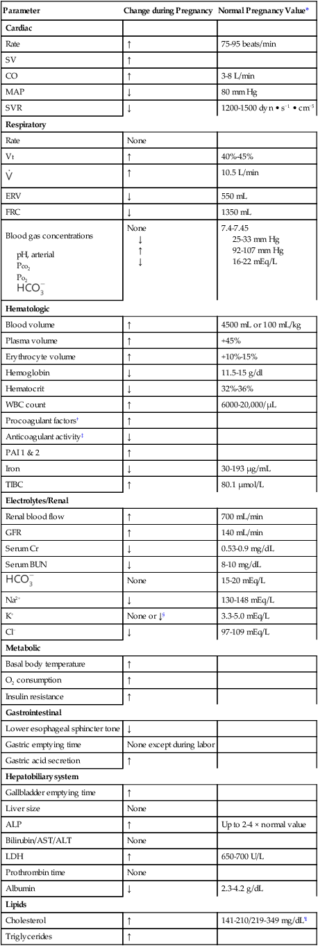
, minute ventilation; VT, tidal volume; WBC, white blood cell.

*Values are approximate and vary throughout pregnancy.

Factors XII:c, VII:c, VII, and V; von Willebrand factors; and fibrinogen.

Activated protein C and protein S.

§Although there are total body accumulations of Na+ and K+, because of the retention of fluid and increase in plasma volumes, concentrations decrease.
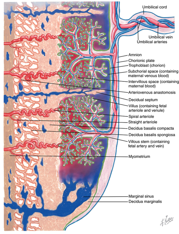
First trimester/third trimester.

Respiratory system

A variety of pregnancy-related changes occur in the respiratory system and may lead to maternal airway complications, which contribute to anesthesia-related maternal mortality risk in the United States. Increased vascularity of the upper airway and nares may result in edema of the upper airway, thus necessitating the use of a smaller tracheal tube. Decreased chest wall compliance may lead to early O2 desaturation, and decreased functional residual capacity (20%) and increased O2 demand (60%) may result in rapid O2 desaturation during apneic episodes.

Physiologic compensatory mechanisms in the mother yield improved fetal oxygenation. The mechanisms include a rightward shift in the maternal oxyhemoglobin dissociation curve (P50 = 30 mm Hg) and a 50% increase in minute ventilation. Progesterone is responsible for increasing the sensitivity of the central respiratory center to CO2, which would cause a respiratory alkalosis; however, because renal excretion of bicarbonate is increased in pregnant women, a neutral pH is maintained. Pregnant women also have a higher PO2 concentration, compared with the nonpregnant state.

Cardiovascular system

A myriad of cardiovascular changes occur during pregnancy. For example, maternal cardiac output increases 40% to meet the metabolic demands of both the mother and the fetus. The rise in cardiac output is initially due to an increase in stroke volume and, as pregnancy progresses, is maintained by an increase in heart rate as the stroke volume decreases. Progesterone decreases pulmonary and systemic vascular resistance, yet central venous and pulmonary artery occlusion pressures remain unchanged. Plasma volume increases up to 40% to 50% due to increased aldosterone production. Because red blood cell mass does not increase as much as does the blood volume, a dilutional anemia results.

When the pregnant woman is in the supine position, uterine compression of the inferior vena cava and aorta causes hypotension; however, this can be readily resolved by placing the parturient in a 15-degree left lateral tilt. Compression of the inferior vena cava and fluid retention also produce ankle edema and varicose veins and can enlarge portal-systemic shunts. Electrocardiographic changes (e.g., left-axis deviation, T-wave inversion in lead III) occur, with cardiac enlargement and rotation of the heart cephalad and leftward.

Gastrointestinal system

Pregnant women are more prone to developing symptomatic gastroesophageal reflux because their gastric pH is decreased, resulting from the production of gastrin by the placenta beginning at approximately 15 weeks of gestation and mechanical obstruction by the gravid uterus that delays gastric emptying and increases intragastric pressure. Lower esophageal sphincter competency is compromised when the gravid uterus causes the gastroesophageal junction to shift cephalad and posterior. Additionally, lower esophageal sphincter tone is altered by high progesterone and estrogen levels.

The risk of aspiration of gastric contents during induction of anesthesia is increased not only when the parturient undergoes emergency cesarean section (2% of deliveries in the United States) under general anesthesia, but, in reality, at any time after 18 to 20 weeks of pregnancy if the woman requires general anesthesia for a surgical procedure. Patients with larger intrauterine size (e.g., multiple gestations or polyhydramnios) are at risk even earlier during pregnancy.

Uterine physiology

Uterine blood flow and placental perfusion are affected by systemic vascular resistance, aortocaval compression, and uterine contraction. Placental blood supply is determined by spiral intervillous arteries, which are maximally dilated (Figure 180-1). They are supplied by arcuate and radial arteries, the ovarian arteries, and uterine arteries. Myometrial tension decreases the caliber of spiral arteries, reducing placental perfusion. Spiral arteries are maximally dilated and sensitive to the effects of α-adrenergic receptor agonists (e.g., phenylephrine). Vasoconstriction can cause dramatic changes in placental blood supply. The surface area and integrity of the placenta are affected by maternal and placental disorders.

Prolonged uterine contraction (hypertonia) can cause fetal asphyxia. Treatment options include fluids, bed rest, O2 and tocolytic agents (e.g., nitroglycerin and albuterol). After delivery, uterine contraction is potentiated with massage, oxytocin, methylergonovine maleate, and carboprost tromethamine. Approximately 500 mL of blood are added to the maternal circulation with uterine contraction.