Management of Respiratory Failure

Published on 12/06/2015 by admin

Filed under Pulmolory and Respiratory

Last modified 22/04/2025

Print this page

rate 1 star rate 2 star rate 3 star rate 4 star rate 5 star
Your rating: none, Average: 0 (0 votes)

This article have been viewed 3101 times

29

Management of Respiratory Failure

Supportive therapy aimed at maintaining adequate gas exchange is critical in the management of both acute respiratory failure and chronic respiratory insufficiency. In acute respiratory failure, survival depends on the ability to provide supportive therapy until the patient recovers from the acute illness that precipitated the need to support the respiratory system. In patients with chronic respiratory insufficiency, the goal is to maximize the patient’s function and minimize symptoms and cor pulmonale on a long-term basis. This chapter outlines the goals of supportive therapy and provides a discussion of the ways adequate gas exchange can be maintained, focusing on patients with acute respiratory failure. Because the principles for supportive management differ considerably in the two main categories of acute respiratory failure—acute respiratory distress syndrome (ARDS) and acute-on-chronic respiratory failure—these differences are emphasized in the course of the discussion. The chapter concludes with a consideration of two specific topics applicable to patients with chronic respiratory insufficiency: chronic ventilatory assistance and lung transplantation.

Goals of Supportive Therapy for Gas Exchange

Adequate uptake of O2 by the blood, delivery of O2 to the tissues, and elimination of CO2 all are parts of normal gas exchange. In terms of O2 uptake by the blood, almost all of the O2 carried by blood is bound to hemoglobin, and only a small portion is dissolved in plasma. It is apparent from the oxyhemoglobin dissociation curve that elevating PO2 beyond the point at which hemoglobin is almost completely saturated does not significantly increase the O2 content of blood. On average, assuming that the dissociation curve is not shifted, hemoglobin is approximately 90% saturated at a PO2 of 60 mm Hg. Increasing PO2 to this level is important, but a PO2 much beyond this level does not provide that much incremental benefit. In practice, patients with respiratory failure often are maintained at a PO2 slightly higher than 60 mm Hg to allow a “margin of safety” for fluctuations in PO2.

Oxygen delivery to the tissues, however, depends not only on arterial PO2 but also on hemoglobin level and cardiac output. In patients who are anemic, O2 content and thus O2 transport can be compromised as much by the low hemoglobin level as by hypoxemia (see Equation 1-3). In selected circumstances, blood transfusion may be useful in raising the hemoglobin and O2 content to more desirable levels.

Similarly, when cardiac output is impaired, tissue O2 delivery also decreases, and measures to augment cardiac output may improve overall O2 transport. Unfortunately, some of the measures used to improve arterial PO2 may have a detrimental effect on cardiac output. As a result, tissue O2 delivery may not improve (and even may worsen) despite an increase in PO2. Use of positive-pressure ventilation, particularly with positive end-expiratory pressure, is most important in this regard. This technique is discussed under Maintenance of Oxygenation.

Elimination of CO2 by the lungs is important for maintaining adequate acid-base homeostasis. However, achieving an acceptable pH value, not a “normal” PCO2, is the primary goal in managing respiratory failure and impaired elimination of CO2. In patients with chronic hypercapnia (and metabolic compensation), abruptly restoring PCO2 to normal (40 mm Hg) may cause significant alkalosis and thus risk precipitating either arrhythmias or seizures.

Maintenance of Carbon Dioxide Elimination

CO2 retention is an important aspect of respiratory failure in several types of patients. Most frequently, these patients have some degree of chronic CO2 retention, and their acute problem is appropriately termed acute-on-chronic respiratory failure. Patients with chronic obstructive lung disease, chest wall disease, and neuromuscular disease are all subject to the development of hypercapnia. Hypercapnia may be acute in certain groups of patients—individuals who have suppressed respiratory drive resulting from ingestion of certain types of drugs, for example, or occasional patients with severe asthma and status asthmaticus.

If the degree of CO2 retention is sufficiently great to cause a marked decrease in the patient’s pH (<7.25–7.30), ventilatory assistance with a mechanical ventilator is often necessary.* Similarly, if marked CO2 retention has impaired the patient’s mental status, ventilatory assistance is indicated. For the patient who has a good chance of rapid reversal of CO2 retention with therapy (assuming the level of CO2 retention is not life threatening), this therapy is often attempted first, with the hope of avoiding mechanical ventilation.

Measurements reflecting muscle strength and pulmonary function may be useful for the patient with acute or impending respiratory failure and can serve as an indirect guide to the patient’s ability to maintain adequate CO2 elimination. They also have been used as criteria for instituting ventilatory assistance or, conversely, for deciding when a patient aided by a mechanical ventilator might be weaned from ventilatory support. Although the decision to initiate mechanical ventilation is frequently based on clinical grounds, the objective measurements most commonly used as criteria for mechanical ventilation are (1) vital capacity (<10 mL/kg body weight) and (2) inspiratory force (<25 cm H2O negative pressure). The latter measurement, which is also called the maximal inspiratory pressure, is performed by having the patient inspire as deeply as possible through tubing connected to a pressure gauge. This technique quantifies the maximal negative pressure the patient can generate when the airway is occluded. These measurements are most useful in following patients with progressive neuromuscular weakness (e.g., myasthenia gravis) to determine when mechanical ventilation is necessary.

Although these and other specific measurements have been used to determine when a patient requires ventilatory assistance for eliminating CO2, none of the guidelines is absolute. Some of the many additional factors that enter into such decisions include the nature of the underlying disease, the tempo and direction of change of the patient’s illness, and the presence of other medical problems.

Maintenance of Oxygenation

Although hypoxemia is a feature of almost all patients with respiratory failure when breathing air (21% O2), the ease of supporting the patient and restoring adequate PO2 depends to a great degree on the type of respiratory failure. In most cases of acute-on-chronic respiratory failure, ventilation-perfusion mismatch and hypoventilation are responsible for hypoxemia. For these mechanisms of hypoxemia, administration of supplemental O2 is quite effective in improving PO2, and particularly high concentrations of inspired O2 are not necessary. Frequently O2 can be administered by face mask or nasal prongs to provide inhaled concentrations of O2 not exceeding 40%, and patients are able to achieve a PO2 greater than 60 mm Hg.

However, patients with chronic hypercapnia may be subject to further increases in PCO2 when they receive supplemental O2 (see Chapter 18). If PCO2 rises to an unacceptably high range, the patient may require intubation and assisted ventilation with a mechanical ventilator to maintain an acceptable PCO2. Fortunately, this complication is infrequent with judicious use of supplemental O2.

In the patient with hypoxemic respiratory failure such as ARDS, ventilation-perfusion mismatch and shunting are responsible for hypoxemia. When a large fraction of cardiac output is being shunted through areas of unventilated lung and therefore not oxygenated during passage through the lungs, supplemental O2 is relatively ineffective at raising PO2 to an acceptable level. In these cases, patients may require inspired O2 concentrations in the range of 60% to 100% and still may have difficulty maintaining PO2 greater than 60 mm Hg.

Such patients with ARDS also require ventilatory assistance, but generally for a different reason than patients with acute-on-chronic respiratory failure. In the latter, an unacceptable degree of CO2 retention is generally the indication for intubation and mechanical ventilation. In ARDS patients, oxygenation is extremely difficult to support, CO2 retention is much less frequent, and hypoxemia rather than hypercapnia is the primary indication for mechanical ventilation.

For patients with hypoxemic respiratory failure, inability to achieve a PO2 of 60 mm Hg or greater on supplemental O2 readily administered by face mask (generally in the range of 40%–60%) is often considered reason for intubation and mechanical ventilation. However, such decisions for ventilatory support are not based on just one number. Other factors taken into consideration include the nature of the underlying problem and the likelihood of a rapid response to therapy.

In the setting of ARDS, intubation and mechanical ventilation serve several useful purposes. First, high concentrations of O2 can be administered much more reliably through a tube inserted into the trachea than through a mask placed over the face. Second, administration of positive pressure by a ventilator relieves the patient of the high work of breathing (see Reducing Work of Breathing), allowing the patient to receive more reliable tidal volumes than he or she would spontaneously take, particularly because the poorly compliant lungs of ARDS promote shallow breathing and low tidal volumes. Finally, when a tube is in place in the trachea, positive pressure can be maintained in the airway throughout the respiratory cycle and not just during the inspiratory phase. In common usage, positive airway pressure maintained at the end of expiration in a mechanically ventilated patient is termed positive end-expiratory pressure (PEEP).

Why is positive pressure throughout the respiratory cycle beneficial for ARDS patients? Because they often have a great deal of microatelectasis resulting from fluid occupying alveolar spaces, low tidal volumes, and probably both decreased production and inactivation of surfactant. The resting end-expiratory volume of the lung (i.e., functional residual capacity [FRC]) is quite low in these patients but can be increased substantially by administration of PEEP. At the higher FRC, many small airways and alveoli that formerly were collapsed and received no ventilation are opened and capable of gas exchange. Therefore, blood supplying these regions no longer courses through unventilated alveoli and now can be oxygenated. Measurement of the “shunt fraction” shows that PEEP is quite effective at decreasing the amount of blood that otherwise would not be oxygenated during passage through the lungs.

When the shunt fraction is decreased by PEEP, supplemental O2 is much more effective at elevating the patient’s PO2 to an acceptable level. The concentration of inspired O2 then can be lowered, and the patient is less likely to experience O2 toxicity from extremely high concentrations of O2.

Reducing Work of Breathing

One pathophysiologic feature shared by most patients with respiratory failure is an imbalance in the work of breathing relative to the ability of the respiratory muscles to perform that work. In the case of acute-on-chronic respiratory failure in the patient with chronic obstructive lung disease, the flattened and mechanically disadvantaged diaphragm must cope with an increase in airway resistance. In neuromuscular disease in either the purely acute or the acute-on-chronic setting, respiratory muscle strength may be insufficient to handle even a relatively normal work of breathing. In the patient with ARDS, the noncompliant (i.e., stiff) lungs require an inordinately high work of breathing even though respiratory muscle strength may be intact.

Consequently, ventilatory assistance in the patient with respiratory failure is important not only for temporary support of gas exchange but also for mechanical support of inspiration, allowing the respiratory muscles to rest. Dyspnea is often alleviated when such support is provided and the patient no longer must expend so much energy on the act of breathing. Fatigued respiratory muscles are allowed to recover, and the relatively large amount of blood flow required by overworking respiratory muscles can be shifted to perfusion of other organ systems.

Mechanical Ventilation

Mechanical ventilators are critical to effective management of respiratory failure. By supporting gas exchange and assisting with the work of ventilation for as long a period as necessary, mechanical ventilators can keep a patient alive while the acute process precipitating respiratory failure is treated or allowed to resolve spontaneously. This section briefly describes the operation of mechanical ventilators, the available modes of ventilation, and the complications that can ensue from their use.

Ventilators currently used for management of acute respiratory failure are positive-pressure devices: they deliver gas under positive pressure during inspiration. Most commonly, the ventilator is used in a volume-cycled fashion, meaning each inspiration is terminated (and passive expiration allowed to occur) after a specified volume has been delivered by the machine. In contrast, in pressure-limited ventilation the assistance provided by the ventilator is targeted to a specified level of positive airway pressure. Volume cycling is much more reliable than pressure-limited ventilation in delivering constant specifiable tidal volumes. With the latter, changes in lung compliance, airway resistance, and the patient’s own inspiratory effort alter the volume of gas delivered as the specified target pressure is reached.

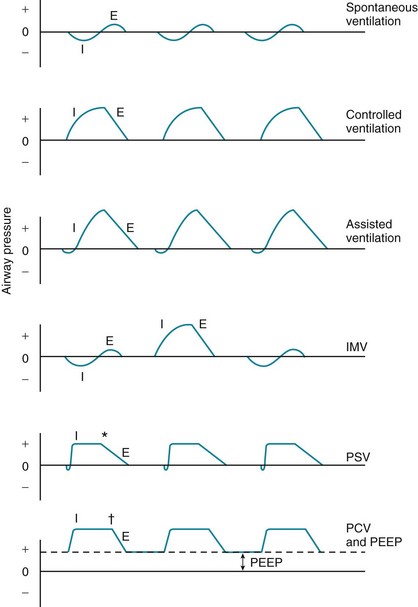
Several ventilatory patterns or modes are available with most mechanical ventilators when used in a volume-cycled fashion (Fig. 29-1). In controlled ventilation, ventilation is supplied entirely by the ventilator at a respiratory rate, tidal volume, and inspired O2 concentration chosen by the physician. If the patient attempts to take a spontaneous breath between the machine-delivered breaths, he or she does not receive any inspired gas. This type of ventilation is uncomfortable for the conscious patient capable of initiating inspiration and therefore can only be used for patients who are comatose, anesthetized, or unable to make any inspiratory effort.

In the assist-control mode of ventilation, the ventilator is able to “sense” when the patient initiates inspiration, at which point the machine assists by delivering a specified tidal volume. Although the tidal volume is set by the machine, the respiratory rate is determined by the number of spontaneous inspiratory efforts made by the patient. However, should the patient’s spontaneous respiratory rate fall below a specified level, the machine provides backup by delivering at least this minimal number of breaths. For example, if the backup rate set on the machine is 10 breaths per minute, the ventilator will automatically deliver a breath if and when 6 seconds have elapsed from the previous breath. Because the respiratory rate with this mode of ventilation is determined by the patient (after the rate exceeds the specified minimal level), fluctuations in minute ventilation can occur if the patient’s respiratory rate changes significantly.

A third ventilatory mode is intermittent mandatory ventilation (IMV). With IMV, the machine delivers a preset number of breaths per minute at a specified tidal volume and inspired O2 concentration. Between the machine-delivered breaths, the patient is able to breathe spontaneously from a gas source providing the same inspired O2 concentration given during the machine-delivered breaths. However, the machine does not assist the spontaneous breaths; therefore, the tidal volume for these breaths is determined by the patient. In a much more commonly used variant of IMV called synchronized IMV (SIMV), each of the machine-delivered breaths is timed to coincide with and assist a patient-initiated breath. If the patient being ventilated by IMV or SIMV modes changes his or her spontaneous respiratory rate significantly, the variation in minute ventilation theoretically is less than in the assist-control mode of ventilation, because each breath has not been supplemented by a comparatively large tidal volume delivered by the ventilator. In practice, both assist-control and SIMV are clinically useful and effective modes of ventilation. Little objective information supporting the use of one over the other is available.

Two types of pressure-limited ventilation are used commonly in certain clinical settings. The first is pressure support ventilation (PSV). With PSV, the ventilator senses when the patient initiates a breath, at which time the ventilator assists the patient’s efforts by providing a specified amount of positive pressure to the airway. This level of pressure support is reached rapidly and maintained throughout most of inspiration. The ventilator stops providing inspiratory assistance when the patient’s inspiratory flow rate falls below a specified target level, such as 25% of the peak inspiratory flow rate. The volume of each breath can be quite variable and is dependent on the preset level of inspiratory pressure support, the patient’s pattern of breathing, and the mechanical properties of the lungs. This type of ventilatory support is solely intended to assist a patient’s own breathing efforts; if the patient stops making inspiratory efforts, no backup level of support is provided by the ventilator. PSV may be the most comfortable form of mechanical ventilation in a conscious patient because he or she has the greatest freedom to determine the timing and depth of each breath.

In pressure-controlled ventilation (PCV), the targeted pressure level is set by the clinician and achieved rapidly, as is the case with PSV. However, in PSV the patient’s spontaneously initiated flow triggers the breath, and a decrease in flow terminates the breath. In contrast, with PCV the initiation of the breath, duration of inspiration, and duration of expiration are determined by the clinician and set on the ventilator; the patient’s breathing pattern does not influence the timing of the ventilatory assistance. Consequently, as for volume-cycled controlled ventilation, PCV is uncomfortable for the patient, who must be heavily sedated to tolerate the imposed ventilatory pattern. PCV is used primarily in patients with ARDS in whom problems with oxygenation and decreased lung compliance are particularly severe. In these cases, the clinician’s control of peak pressure and the relative timing of inspiration and expiration can facilitate improved oxygenation and minimize the risk of complications from high pressure delivered by the ventilator.

An important option available for the intubated patient with hypoxemic respiratory failure (especially when it is due to ARDS) is PEEP. When a patient is assisted by a mechanical ventilator without PEEP, airway (and alveolar) pressure falls during expiration from the positive level achieved at the height of inspiration down to zero. However, if the expiratory portion of the tubing is connected to a valve requiring a pressure of at least 10 cm H2O, for example, to open it, the valve closes and expiration ceases when the airway pressure falls to 10 cm H2O. Consequently, airway pressure at the end of expiration does not fall to zero but remains at the level determined by the specifications of the expiratory valve. On the basis of the pressure required to open the expiratory valve, the level of PEEP can be set as desired.

A variation of PEEP that works on the same principle is continuous positive airway pressure (CPAP). The term CPAP is used when the patient is breathing spontaneously (without a mechanical ventilator) and expiratory tubing is connected to a PEEP valve. To use CPAP, the patient can be either intubated or given a tightly fitting face mask. Although no positive pressure is provided by a mechanical ventilator during inspiration, inspired gas is delivered from a reservoir bag under tension or at a sufficiently high flow rate to keep airway pressure positive during inspiration as well as expiration.

With PEEP or CPAP, the benefit comes from the positive pressure within airways and alveoli at the end of expiration. FRC is increased by the positive pressure, and closure of airways and alveoli at the end of expiration is diminished.

In complicated cases of respiratory failure, such as patients with ARDS, a variety of ventilatory strategies are now used. Important goals of these particular strategies are to prevent closure of alveoli during expiration while simultaneously avoiding delivery of excessive volume and pressure to the airways and alveoli, with the potential for secondary complications (see later). A particularly common strategy is called a protective open lung strategy, in which sufficient PEEP is given to diminish airway closure during expiration, and relatively low tidal volumes (6 mL/kg) are used to protect the lung from higher volumes and pressures delivered during inspiration. In some cases, PCO2 may rise when these relatively low tidal volumes are used, but the elevation in PCO2 above normal levels is considered acceptable according to a strategy of permissive hypercapnia. By minimizing the need for high levels of ventilation, this strategy theoretically decreases the risks of developing high alveolar pressures and overdistention and injury of some alveolar units.

When the underlying problem that precipitated the need for mechanical ventilation has improved, ventilatory support is discontinued, typically after observing the patient during a short (30–120 minute) trial of spontaneous breathing with minimal or no positive pressure delivered by the mechanical ventilator. A useful guideline for assessing the patient’s initial response to the spontaneous breathing trial and predicting successful discontinuation of mechanical ventilation is provided by the rapid shallow breathing index. This index is the ratio of the patient’s respiratory rate divided by the tidal volume (expressed in liters) measured when the patient is not receiving assistance from the ventilator (i.e., during the spontaneous breathing trial). An index less than 105 is predictive of successful extubation, whereas an index greater than 105 is associated with a much higher likelihood of recrudescent respiratory failure after extubation.

Although the term weaning is still applied to discontinuation of mechanical ventilation, the older technique of slowly decreasing the amount of support provided by the ventilator is generally no longer used. As rational as it seems to wean the patient gradually from ventilatory support, an alternative and superior strategy is to perform a single daily trial of spontaneous breathing. If the patient tolerates the trial, then he or she is extubated; if the trial is unsuccessful, full ventilatory support is resumed for 24 hours until the time of the next daily trial.

Complications of Mechanical Ventilation

Intubation and mechanical ventilation of patients in respiratory failure are not without risks or complications (Table 29-1). The procedure of intubation can be complicated acutely by problems such as arrhythmias, laryngospasm, and malposition of the endotracheal tube (either in the esophagus or in a mainstem bronchus). When a tube remains in the trachea for days to weeks, complications affecting the larynx and trachea can occur. Vocal cord ulcerations and laryngeal stenosis and granulomas may develop. The trachea is subject to ulcerations, stenosis, and tracheomalacia (degeneration of supporting tissues in the tracheal wall) resulting from pressure applied by the inflated balloon at the end of the tube. As a precaution to decrease tracheal complications, tubes are made with cuffs that minimize the pressure exerted on the tracheal wall and the resulting pressure necrosis. For prolonged ventilatory support (weeks to months), a tracheostomy tube placed directly into the trachea through an incision in the neck has some advantages over prolonged orotracheal or nasotracheal intubation, including patient comfort and prevention of further vocal cord injury.

The presence of an endotracheal tube puts the patient at significant risk for nosocomial pneumonia, usually called ventilator-associated pneumonia. Several factors appear to contribute to the patient’s increased risk for developing pneumonia when intubated and receiving mechanical ventilation. They include bypassing of the normal anatomic barriers and upper airway clearance mechanisms that prevent organisms from reaching the lower respiratory tract, aspiration of oropharyngeal secretions around the endotracheal tube and into the lower respiratory tract, and bacterial contamination of the endotracheal tube or the ventilator circuitry connected to the endotracheal tube. Organisms causing ventilator-associated pneumonia are often relatively antibiotic-resistant bacteria, including gram-negative bacilli and Staphylococcus aureus, leading to significant increases in both duration of hospitalization and mortality.

Administration of positive pressure by a mechanical ventilator has its own attendant problems. Patients receiving positive-pressure ventilation are subject to barotrauma—traumatic changes such as pneumothorax or pneumomediastinum occurring as a result of high alveolar pressures. Because alveolar overdistention with rupture is currently thought to be the cause of these complications, the term volutrauma is now often used instead of barotrauma. Development of a pneumothorax in patients receiving mechanical ventilation can have catastrophic consequences if not detected and treated quickly. The ventilator continues to deliver gas under positive pressure, and the gas enters the pleural space through the rupture. The pressure in the pleural space and thorax increases, and a tension pneumothorax results (see Chapter 15), which severely diminishes venous return and cardiac output and causes rapid cardiovascular collapse. In such situations, a tube, catheter, or needle must be immediately inserted to decompress the pleural space, allow resumption of venous return, and enable reexpansion of the lung.

Besides barotrauma, another major adverse effect of positive-pressure ventilation is potential impairment of cardiovascular function. At least two mechanisms are thought to play a role. The first involves a decrease in venous return to the heart. Whereas the normally negative intrathoracic pressure during inspiration promotes venous return from the periphery, positive inspiratory pressure from a ventilator impedes venous return. The hemodynamic consequences of low cardiac output and blood pressure are even more likely when the patient is also receiving PEEP. In many cases, judicious administration of fluids can restore the effective intravascular volume and reverse the adverse hemodynamic consequences of positive-pressure ventilation.

The second mechanism involves an increase in pulmonary vascular resistance. When alveolar volume is increased with positive-pressure mechanical ventilation, alveolar vessels are compressed, compromising the overall cross-sectional area of the pulmonary vascular bed. As a result, pulmonary vascular resistance and the workload placed on the right ventricle increase. Right ventricular output is potentially compromised, and the right ventricle may dilate. This shifts the interventricular septum toward the left ventricular cavity, also impairing left ventricular filling and stroke volume.

Concern has been raised that the excessive volumes and high levels of pressure provided to the alveoli may be injurious to alveolar structures. Because lung injury in ARDS is often heterogeneously distributed (see Chapter 28), inspired gas is preferentially distributed to the more normal, more compliant alveoli than to the abnormal, less compliant alveoli. This puts the more normal alveoli at particular risk for overdistention. At the same time, more diseased alveoli are subject to collapse (atelectasis) during the expiratory phase of the respiratory cycle because of intraalveolar fluid and/or a disrupted or insufficient surfactant layer. Alveoli that are open during inspiration but collapse during expiration are subject to abnormal shear stresses during the repetitive process of opening and closing, a complication termed atelectrauma.

Both alveolar overdistention and atelectrauma may be accompanied by microscopic injury to cells of the alveolar wall and to intercellular attachments, leading to disruption of the normal permeability barrier provided by alveolar epithelial and capillary endothelial cells. In addition, proinflammatory cytokines may be released and perpetuate alveolar injury and inflammation. As a result, positive-pressure ventilation for respiratory failure, especially ARDS, can potentially compound or worsen the process for which it is being used. Therefore, it is currently believed the pattern of ventilation should avoid both alveolar closure (atelectasis) during expiration and overdistention during inspiration, the former by use of PEEP and the latter by limiting tidal volume to 6 mL/kg and alveolar distending pressure to no more than 30 cm H2O. Of note, the use of a low tidal volume ventilation strategy in patients with ARDS has been demonstrated to decrease mortality, whereas the use of higher levels of PEEP has not resulted in decreased mortality versus employing lower levels of PEEP.

Management of patients receiving positive-pressure ventilation, particularly those with hypoxemic respiratory failure who require PEEP, is complicated. Many factors interact in a complex way, specifically oxygenation, cardiac output, and fluid status. Optimal care requires both sophisticated patient monitoring and substantial expertise from the team responsible for patient care. Such care is necessary not only for proper support of vital functions but also for minimizing the complications of therapy.

Noninvasive Ventilatory Support for Acute Respiratory Failure

When patients with acute respiratory failure require mechanical ventilation, support traditionally has been provided by positive pressure administered through a tube placed into the trachea (i.e., endotracheal tube). However, use of the tube is associated with risks and complications, such as patient discomfort from the tube itself, injury to the airway mucosa, and development of lower respiratory tract infection (see Table 29-1). An alternative form of support for acute respiratory failure does not require use of an endotracheal tube; rather, positive pressure is provided through a tightly fitting mask placed over the face. This approach has been used for support of patients with a variety of types of acute respiratory failure, including patients with cardiogenic pulmonary edema, those with hypercapnic acute exacerbation of chronic obstructive pulmonary disease, and patients who are not considered suitable candidates for intubation. However, noninvasive ventilatory support is not appropriate if patients are unable to protect their airway; it is most useful when respiratory failure most likely is readily reversible and therefore of relatively short duration.

Selected Aspects of Therapy for Chronic Respiratory Failure

Chronic Ventilatory Support

Patients with chronic irreversible respiratory system disease who require continuous long-term ventilatory support pose a difficult problem. The first question is whether the patient wishes “to be on a machine” for the rest of his or her life. Some patients clearly wish to prolong life even if it means permanent ventilatory support; others make the decision that life is not worth living if they must be dependent on a ventilator for the remainder of their lives. When the patient chooses to be maintained on a ventilator, support usually is given by positive-pressure ventilation administered through a tracheostomy tube. The care of some patients can be handled at home with the proper support of family and visiting healthcare personnel. Management of other cases continues in chronic care hospitals or other facilities equipped to care for such patients.

A subgroup of patients with chronic respiratory insufficiency does not require continuous ventilatory support but benefits from nocturnal assistance with ventilation. These patients often have chronic neuromuscular or chest wall disease accompanied by chronic hypercapnia. Although the degree to which respiratory muscle fatigue contributes to the hypercapnia experienced by these patients is not clear, at least part of the rationale for using nocturnal ventilatory support is to afford these patients a number of hours each day when their inspiratory muscles are allowed to rest. After a period of nocturnal rest, the respiratory muscles presumably are better able to handle the work of breathing during the day, and daytime hypercapnia may be improved.

When ventilatory support is needed only during the night, it is generally preferable to avoid a chronic tracheostomy. Several options are available, and the most appropriate depends on the particular patient. Positive pressure can be administered at night through a mouthpiece or a mask (i.e., noninvasive positive-pressure ventilation [NIPPV]). Alternatively, lung inflation can be achieved by negative-pressure ventilation, intermittent negative pressure applied outside the chest wall, causing it to expand and the lungs to inflate. The original type of negative-pressure ventilator was the “iron lung,” used for ventilatory support from the 1930s through the polio epidemics of the 1950s. Currently, other negative-pressure devices are used more frequently. The two most common types are the raincoat (or “poncho”) ventilator and the cuirass (or “chest shell”) ventilator. Unlike the iron lung, which enclosed the entire body below the neck, raincoat and cuirass ventilators do not enclose or limit movement of the lower half of the body. These negative-pressure ventilatory support devices have been used most frequently for patients with chronic respiratory failure resulting from neuromuscular disease such as muscular dystrophy.

Lung Transplantation

First performed successfully in 1983, lung transplantation is a relatively recent form of therapy for management of severe and disabling chronic pulmonary disease. Availability of a lung transplant is limited, however, primarily because suitable donor organs are scarce, and difficulties with posttransplant infections and chronic rejection limit the long-term utility of the procedure.

Several types of transplantation can be performed, with selection generally based on the underlying clinical problem. Single-lung transplantation has been used effectively in patients with a number of end-stage diseases, including severe emphysema. For patients with cystic fibrosis, in whom chronic bilateral pulmonary infection complicates their lung disease, bilateral lung transplantation is generally performed to avoid infection of the new lung by spillover of infected secretions from a remaining diseased lung. When severe cardiac disease accompanies end-stage lung disease, combined heart-lung transplantation may be required. The most recent lung transplantation technique is lobar transplantation from living donors. In this technique, which is used primarily in patients with cystic fibrosis, the recipient is given bilateral implants of a lower lobe from each of two living donors.

In many ways, the lung transplant patient trades the primary lung disease for another disease: that of the transplant recipient. The major potential complications of lung transplantation fall under the general categories of rejection and infection. Because of the risk of rejection, patients are routinely given immunosuppressive drugs such as prednisone, mycophenolate mofetil (or azathioprine), and tacrolimus (or cyclosporine) as a regimen to prevent rejection. Nevertheless, either acute or chronic rejection can occur despite maintenance immunosuppression. Acute rejection is often characterized by fever, impairment of pulmonary function and gas exchange, and pulmonary infiltrates on chest radiograph. Episodes typically occur during the first several months after transplantation and are treated by acute intensification of the immunosuppressive regimen, especially with increased doses of corticosteroids. Chronic rejection is usually manifested as bronchiolitis obliterans, which is characterized by progressive inflammation, fibrosis, and obstruction of small airways. The physiologic consequence of this process is progressive airflow obstruction, which typically is unresponsive to augmentation of immunosuppressive therapy. As a result, bronchiolitis obliterans is the major cause of graft failure and death occurring later in the course after lung transplantation. Pharmacologic treatment of severe bronchiolitis obliterans has been disappointing, and the main treatment option for posttransplant patients with this syndrome is repeat transplantation.

The other major complication of lung transplantation is infection, the risk of which is greatly increased by the need for immunosuppressive therapy. In some cases, organisms (e.g., bacteria, cytomegalovirus) accompanied the donor organ, and development of a complicating infection was precipitated by immunosuppression and impairment of the recipient’s defense mechanisms. Patients are also subject to the variety of opportunistic infections common to patients with impaired cell-mediated immunity, including other viruses, fungi, and Pneumocystis.

Accompanying the growing experience with lung transplantation over the past decade has been a modest improvement in survival. Survival is approximately 75% to 80% at 1 year after transplantation and 60% at 3 years after transplantation. Lung transplantation is now an important but expensive therapeutic option for a highly selected group of patients. As more experience is gained with this form of treatment and as technology and immunosuppressive therapy evolve, we continue to refine our thinking about the role of transplantation in the overall management of patients with severe lung disease.

References

Mechanical Ventilation

Ambrosino, N, Vagheggini, G. Noninvasive positive pressure ventilation in the acute care setting: where are we? Eur Respir J. 2008;31:874–886.

Boles, J-M, Bion, J, Connors, A, et al. Weaning from mechanical ventilation. Eur Respir J. 2007;29:1033–1056.

Briel, M, Meade, M, Mercat, A, et al. Higher vs lower positive end-expiratory pressure in patients with acute lung injury and acute respiratory distress syndrome: systematic review and meta-analysis. JAMA. 2010;303:865–873.

Caples, SM, Gay, PC. Noninvasive positive pressure ventilation in the intensive care unit: a concise review. Crit Care Med. 2005;33:2651–2658.

Chandra, D, Stamm, JA, Taylor, B, et al. Outcomes of noninvasive ventilation for acute exacerbations of chronic obstructive pulmonary disease in the United States, 1998–2008. Am J Respir Crit Care Med. 2012;185:152–159.

Esan, A, Hess, DR, Raoof, S, et al. Severe hypoxemic respiratory failure: Part 1–Ventilatory strategies. Chest. 2010;137:1203–1216.

Fan, E, Needham, DM, Stewart, TE. Ventilatory management of acute lung injury and acute respiratory distress syndrome. JAMA. 2005;294:2889–2896.

Garpestad, E, Brennan, J, Hill, NS. Noninvasive ventilation for critical care. Chest. 2007;132:711–720.

Girard, TD, Bernard, GR. Mechanical ventilation in ARDS. A state-of-the-art review. Chest. 2007;131:921–929.

Khirani, S, Georgopoulos, D, Rossi, A, et al. Ventilator support in chronic obstructive pulmonary disease: invasive and noninvasive. Eur Respir Mon. 2006;11(monograph 38):401–429.

Liesching, T, Kwok, H, Hill, NS. Acute applications of noninvasive positive pressure ventilation. Chest. 2003;124:699–713.

Lorente, L, Blot, S, Rello, J. New issues and controversies in the prevention of ventilator-associated pneumonia. Am J Respir Crit Care Med. 2010;182:870–876.

MacIntyre, NR, Cook, DJ, Ely, EW, Jr., et al. Evidence-based guidelines for weaning and discontinuing ventilatory support: a collective task force facilitated by the American College of Chest Physicians; the American Association for Respiratory Care; and the American College of Critical Care Medicine. Chest. 2001;120(Suppl):375S–395S.

Malhotra, A. Low-tidal-volume ventilation in the acute respiratory distress syndrome. N Engl J Med. 2007;357:1113–1120.

McConville, JF, Kress, JP. Weaning patients from the ventilator. N Engl J Med. 2012;367:2233–2239.

Pinhu, L, Whitehead, T, Evans, T, et al. Ventilator-associated lung injury. Lancet. 2003;361:332–340.

Putensen, C, Theuerkauf, N, Zinserling, J, et al. Meta-analysis: ventilation strategies and outcomes of the acute respiratory distress syndrome and acute lung injury. Ann Intern Med. 2009;151:566–576.

Tobin, MJ. Advances in mechanical ventilation. N Engl J Med. 2001;344:1986–1996.

Tobin, MJ, Laghi, F, Jubran, A. Narrative review: ventilator-induced respiratory muscle weakness. Ann Intern Med. 2010;153:240–245.

Vlahakis, NE, Hubmayr, RD. Cellular stress failure in ventilator-injured lungs. Am J Respir Crit Care Med. 2005;171:1328–1342.

Chronic Ventilatory Support

Consensus Conference Report. clinical indications for noninvasive positive pressure ventilation in chronic respiratory failure due to restrictive lung disease, COPD, and nocturnal hypoventilation. Chest. 1999;116:521–534.

Hill, NS. Clinical applications of body ventilators. Chest. 1986;90:897.

Howard, RS, Davidson, C. Long-term ventilation in neurogenic respiratory failure. J Neurol Neurosurg Psychiatry. 2003;74(Suppl 3):iii24–30.

Kennedy, JD, Martin, AJ. Chronic respiratory failure and neuromuscular disease. Pediatr Clin North Am. 2009;56:261–273.

MacIntyre, NR, Epstein, SK, Carson, S, et al. Management of patients requiring prolonged mechanical ventilation: report of a NAMDRC consensus conference. Chest. 2005;128:3937–3954.

Make, BJ, Hill, NS, Goldberg, AI, et al. Mechanical ventilation beyond the intensive care unit. Report of a consensus conference of the American College of Chest Physicians. Chest. 1998;113(Suppl):289S–344S.

Nelson, JE, Cox, CE, Hope, AA, et al. Chronic critical illness. Am J Respir Crit Care Med. 2010;182:446–454.

Ozsancak, A, D’Ambrosio, C, Hill, NS. Nocturnal noninvasive ventilation. Chest. 2008;133:1275–1286.

Simonds, AK. Home ventilation. Eur Respir J Suppl. 2003;47:38s–46s.

Wallis, C, Paton, JY, Beaton, S, et al. Children on long-term ventilatory support: 10 years of progress. Arch Dis Child. 2011;96:998–1002.

Lung Transplantation

Ahmad, S, Shlobin, OA, Nathan, SD. Pulmonary complications of lung transplantation. Chest. 2011;139:402–411.

Kotloff, RM, Thabut, G. Lung transplantation. Am J Respir Crit Care Med. 2011;184:159–171.

Nicod, LP. Mechanisms of airway obliteration after lung transplantation. Proc Am Thorac Soc. 2006;3:444–449.

Orens, JB, Estenne, M, Arcasoy, S, et al. International guidelines for the selection of lung transplant candidates: 2006 update—a consensus report from the Pulmonary Scientific Council of the International Society for Heart and Lung Transplantation. Pulmonary Scientific Council of the International Society for Heart and Lung Transplantation. J Heart Lung Transplant. 2006;25:745–755.

Pierson, RN, III. Lung transplantation: current status and challenges. Transplantation. 2006;81:1609–1615.

Solomon, M, Grasemann, H, Keshavjee, S. Pediatric lung transplantation. Pediatr Clin North Am. 2010;57:375–391.

Todd, JL, Palmer, SM. Bronchiolitis obliterans syndrome: the final frontier for lung transplantation. Chest. 2011;140:502–508.