Management of end-stage heart failure: Heart transplantation and ventricular assist devices

Published on 07/02/2015 by admin

Filed under Anesthesiology

Last modified 22/04/2025

Print this page

rate 1 star rate 2 star rate 3 star rate 4 star rate 5 star
Your rating: none, Average: 0 (0 votes)

This article have been viewed 2564 times

Management of end-stage heart failure: Heart transplantation and ventricular assist devices

Doris B.M. Ockert, MD

End-stage heart failure

Heart failure (HF) is defined as insufficient cardiac output to meet the metabolic requirements of the tissues at normal cardiac-filling pressures. Cardiogenic shock is defined as sustained hypotension and tissue hypoperfusion. HF can be systolic (impaired contractility with impaired ejection fraction) or diastolic (decreased relaxation and compliance). Activation of the compensatory neurohormone system (renin-angiotensin-aldosterone system and release of natriuretic peptides, angiotensin II, norepinephrine, and endothelin) results in fluid retention, peripheral vasoconstriction, downregulation of β-adrenergic receptors, and ventricular remodeling. Eventually, left ventricular (LV) failure leads to pulmonary hypertension and right ventricular (RV) failure.

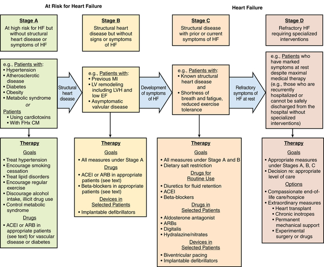
Echocardiography is used to assess ventricular function, to identify structural and functional cardiac abnormalities, and to guide therapy. The American Heart Association classification defines four stages of HF: A through D. Stage D is end-stage HF, (Figure 143-1). Coronary artery disease is the most common cause of both systolic and diastolic failure. Other causes include dilated nonischemic, restrictive, hypertrophic, and stress-induced cardiomyopathy. The most common cause of death is ventricular arrhythmia.

image
Figure 143-1 Stages in the development of heart failure and recommended therapy by stage. ACEI, Angiotensin-converting enzyme inhibitor; ARB, angiotensin II receptor blocker; EF, ejection fraction; FHx CM, family history of cardiomyopathy; HF, heart failure; LV, left ventricular; LVH, left ventricular hypertrophy; MI, myocardial infarction. (Modified from Jessup M, Abraham WT, Casey DE, et al. 2009 focused update: ACCF/AHA Guidelines for the Diagnosis and Management of Heart Failure in Adults: a report of the American College of Cardiology Foundation/American Heart Association Task Force on Practice Guidelines: developed in collaboration with the International Society for Heart and Lung Transplantation. Circulation 2009;119:1977-2016. © 2013 American Heart Association, Inc. All rights reserved.)

Patients with coronary artery disease or valvular heart disease should have medical therapy optimized and, depending on the anatomy, revascularization performed or valves repaired or replaced as appropriate. In patients with an ejection fraction of less than 30%, placement of an implantable cardioverter defibrillator (ICD), pacemaker resynchronization therapy, or both is recommended. Routine anticoagulation is not recommended. Surgical treatment options include placement of an intra-aortic balloon pump, ventricular assist device (VAD), or total artificial heart (TAH) or orthotopic heart transplantation.

Clinical indications for the use of a mechanical device before multisystem organ failure occurs include myocardial infarction, failed percutaneous coronary intervention, acute viral myocarditis, peripartum cardiomyopathy, cardiac contusion, postcardiotomy shock, chronic cardiomyopathy with acute decompensation, and intractable ventricular arrhythmias. Early intervention improves survival.

Ventricular assist devices

VADs to support the left, right, or both ventricles are either pulsatile or nonpulsatile pumps, located paracorporeally or intracorporeally, and are used as a bridge to recovery (short-term), a-bridge to transplantation, or destination therapy. The first-generation VADs use pulsatile pumps with valves that displace a given volume of blood with every beat. One pulsatile pump is still marketed in the United States, a paracorporeal VAD (pVAD; Thoratec, Pleasanton, CA) for short-term to intermediate-term use in patients who require bridge to transplantation or bridge to recovery (Figure 143-2). Approximately 10% of patients recover sufficient function to be weaned completely from mechanical support. Compared with previous devices, the pVAD allows for greater patient mobility (a portable device is available for patients who leave the hospital) and longer-term use (weeks to months and, in a few cases, years); its use is associated with lower rates of morbidity. Short-term anticoagulation is provided with heparin, whereas long-term anticoagulation requires warfarin and sometimes aspirin.

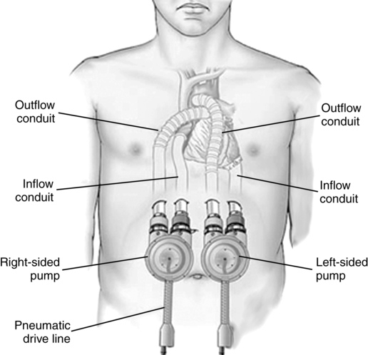
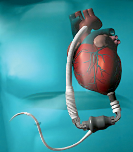
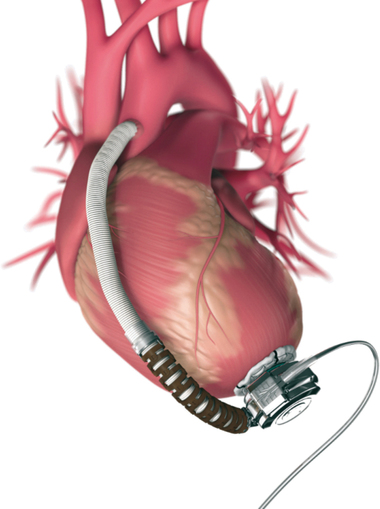
Second-generation VADs are smaller, intracorporeal, nonpulsatile, axial-flow pumps without valves. Third-generation VADs are bearingless and use a combination of magnetically and hydrodynamically suspended impellers. Currently available in the United States are the Heartmate II (Thoratec), a second-generation device approved by the U.S. Food and Drug Administration (FDA) for bridge to transplantation in 2008 and for destination therapy for patients who are not candidates for heart transplantation in 2010 (Figure 143-3), and the HeartWare ventricular assist system (Framingham, MA), a third-generation device approved in 2012 by the FDA as a bridge to transplantation (Figure 143-4).
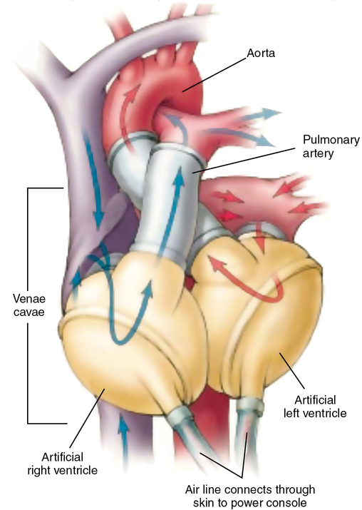
Left VADs (LVADs) drain blood from the LV through an inflow cannula to the pump and return blood via an outflow cannula into the proximal aorta. The Randomized Evaluation of Mechanical Assistance for the Treatment of Congestive Heart Failure (REMATCH) trial demonstrated that patients did better when they had a device implanted sooner rather than later (before they developed organ dysfunction, i.e., kidney failure). Long-term survival is approximately 80% at 1 year and more than 50% at 2 years after device implantation.

Anesthetic considerations for patients with end-stage heart failure requiring implantation of a VAD or TAH

Preoperative considerations

The preparation of these patients is similar to that required for other patients having cardiac surgical procedures involving cardiopulmonary bypass (CPB). However, patients who are receiving VADs, a TAH, or heart transplants are at greater risk for experiencing hemorrhage and are at risk for developing systolic dysfunction, ventricular arrhythmias, and sudden death prior to CPB.

A thorough preoperative evaluation should include an extensive review of the patient’s cardiac, pulmonary, renal, and metabolic history; a comprehensive examination of the patient; and a complete assessment of all laboratory results and imaging studies. All previously implanted devices such as pacemakers and ICDs should be interrogated. Packed red blood cells (cytomegalovirus free), fresh frozen plasma, and platelets must be available in the blood bank because these patients will have received anticoagulant therapy, have chronic anemia, and are at high risk of developing perioperative bleeding.

Intraoperative management

Patients receiving a VAD

In addition to standard American Society of Anesthesiologists monitoring devices, these patients should have a cannula placed in a radial or femoral artery prior to induction of anesthesia, if possible, for continuous measurement of arterial blood pressure. Central venous access should be achieved either with an 8.5F or 9F introducer through which a catheter may be inserted for measurement of central venous, pulmonary artery, and pulmonary artery occlusion pressures and cardiac output. Many clinicians prefer to perform these procedures prior to the induction of anesthesia because of the severity of the patient’s underlying heart disease. The pharmacologic agents used for induction and maintenance of anesthesia are similar to those used for any other patient with severe cardiomyopathy. After the trachea is intubated, a transesophageal echocardiographic (TEE) probe should be inserted. Patients with preexisting coagulopathy need baseline coagulation studies and possibly thromboelastography. Defibrillation pads must be placed prior to deactivation of defibrillation therapies.

Prior to CPB, a TEE examination should be performed to assess for the presence of a patent foramen ovale (PFO), aortic or mitral insufficiency, and intracardiac thrombi. Because the LVAD will create negative pressure at the tip of the cannula in the left ventricle, it is important to repair a PFO to prevent paradoxical embolism of air bubbles and thrombi and shunting of desaturated blood from right to left. The PFO may only be detected after CPB when the left heart is decompressed. Similarly, if the patient has aortic insufficiency, the device may create a flow loop, in which blood flowing from the device into the aortic root is drawn back through the incompetent aortic valve into the device and back into the root, etc., with insufficient flow to vital organs. In addition, if the LV continues to contract with the LVAD off-loading the LV and augmenting cardiac output, significant mitral regurgitation will decrease preload to the device and limit its effectiveness. Removing cardiac thrombi is critical to avoid their entry into the pump. The results of the intraoperative TEE examination should be shared with the surgeon so that he or she can assess the situation and decide whether any deficits should be corrected surgically. The midesophageal four-chamber or two-chamber TEE view may help guide the surgeon to the potential ventriculostomy site for the inflow cannula for the device. Because of the severity of the cardiomyopathy, any reduction in preload, heart rate, or contractility prior to CPB may produce sudden cardiovascular collapse. Vasoactive drugs such as phenylephrine, ephedrine, epinephrine, norepinephrine, or vasopressin may be required to maintain hemodynamic stability.

The primary cause for failure to wean the patient from CPB is inadequate LV preload. This may be caused by decreased intravascular volume, vasodilation, or RV failure, most likely secondary to pulmonary hypertension. Therapeutic options to improve LV preload include intravascular volume replacement, vasoconstrictor therapy (vasopressin, norepinephrine, phenylephrine), appropriate inotropic support in the case of RV failure (milrinone, epinephrine, dobutamine), and primary pulmonary vasodilator therapy (nitric oxide, prostaglandins).

After implantation of the device, a TEE exam is used to assess the inflow to and outflow from the LVAD. Once the patient is weaned from CPB, the most common causes of hypotension are decreased intravascular volume and decreased systemic vascular resistance.

Patients receiving a total artificial heart

A TAH may be an option for patients who have end-stage heart disease of sufficient severity that VADs fail to provide sufficient support and who have confounding factors that make heart transplantation unlikely or contraindicated. The anesthetic management for patients undergoing TAH implantation is similar to that previously described, except for the treatment of hypotension, and special care is needed to prevent air emboli. Because the native ventricles are excised, no benefit is gained with the use of inotropes for the treatment of hypotension. In addition to infusing fluids to increase intravascular volume and therefore venous return, vasopressors may be required to increase systemic vascular resistance and venous return. Nitric oxide and prostaglandins may be required to treat pulmonary hypertension. Following anastomosis of the artificial ventricles to the native atria, the mechanical ventricles must be primed. Therefore, the patient should be placed in Trendelenburg position and a TEE examination performed to detect air and monitor its evacuation as the mechanical ventricles begin to contract and eject. Once the mechanical ventricles are functioning and the patient’s hemodynamic values are satisfactory, the patient is weaned from CPB, bleeding is controlled, and the chest is closed and the patient may be transported to the intensive care unit.

Postoperative management

Complications of all devices include bleeding, thromboembolism, infection, hemolysis, device malfunction, and multiorgan failure. The management of the patient with an LVAD or both an LVAD and an RVAD is similar to the management of patients who have had a cardiac surgical procedure for which CPB is used: postoperative bleeding and hemodynamic values must be carefully monitored and stabilized, and the patient is weaned from mechanical ventilation. The possibility of RV dysfunction must be considered in a patient with only an LVAD in whom hypotension develops. Once the patient leaves the intensive care unit, the most likely cause of hypotension is decreased preload secondary to decreased intravascular volume.

Patients with VADs frequently require other surgical procedures. For unknown reasons, but probably related to the nonpulsatile blood flow with the second-generation and third-generation devices, approximately 40% to 50% of these patients will develop gastrointestinal bleeding because of arteriovenous malformations that form in the walls of the gastrointestinal tract. These patients will often require upper gastrointestinal endoscopy. Arteriovenous malformations are also occasionally seen in the wall of the urinary bladder. Arterial pulses are very weak, if present at all, in patients with axial flow pumps, making noninvasive blood pressure monitoring difficult. Measurement of blood pressure with either Doppler or an intra-arterial cannula is often necessary.

Considerations for patients for heart transplantation

Patients with end-stage heart disease are carefully screened for possible heart transplantation. They must be adherent with treatment, not abuse substances (including alcohol), have an adequate support system, be free of cancer, and have a body mass index of less than 38. Severe irreversible pulmonary hypertension is an absolute contraindication to heart transplantation (pulmonary vascular resistance > 6 Wood units or > 480 Dynes·sec−1·cm−5).

When the United Network for Organ Sharing is notified that a patient has been declared brain dead and the organs are available for transplantation, they identify potential recipients by matching the donor heart with potential recipients, based on the severity of the recipient’s disease, through HLA typing, ABO blood group compatibility, and body size. Once the best candidate is found, the transplant center is notified, and, if the transplant team and patient agree, the candidate is posted for transplantation.

These cases are always performed on an emergency basis. Donor-heart ischemic time should optimally be kept at less than 4 hours. Sterile technique is imperative because the patient will be immunosuppressed and at high risk of developing infection. Immunosuppression protocols vary. Commonly, 500 mg of methylprednisolone is administered after induction and again after release of the aortic cross-clamp. Other immunosuppressant drugs may be utilized depending on the preferences of the institution’s transplant service.

The surgical technique involves four major anastomoses: left and right atria and the end-to-end aortic and pulmonary anastomoses. The bicaval technique is used in some cases instead of the right atria anastomosis. Because the donor heart is denervated, only direct-acting β-adrenergic agents will increase heart rate.

RV failure is the most common cause for failure to wean from CPB after heart transplantation. Preventing hypoxia and hypercarbia are essential, and the use of pulmonary vasodilators (prostaglandin E1, nitric oxide, milrinone) and inotropes (epinephrine, dobutamine, milrinone) to support the RV may be necessary. Norepinephrine and vasopressin may be needed to support systemic vascular resistance. In the early postoperative period, patients are at risk for developing hyperacute and acute rejection, pulmonary and systemic hypertension, cardiac arrhythmias, respiratory failure, renal failure, and infection.

Allograft coronary artery disease is the major limiting factor to long-term survival following heart transplantation. This is diffuse disease that involves the vessels circumferentially. Cyclosporine and corticosteroids are the mainstays of long-term immunosuppression and may cause nephrotoxicity, hypertension, and malignant neoplastic disease. The survival rates for transplantation approach 90% for the first year and 75% at the seventh year.