Management of cerebral aneurysms

Published on 07/02/2015 by admin

Filed under Anesthesiology

Last modified 22/04/2025

Print this page

rate 1 star rate 2 star rate 3 star rate 4 star rate 5 star
Your rating: none, Average: 0 (0 votes)

This article have been viewed 1455 times

Management of cerebral aneurysms

Eric L. Bloomfield, MD, MS, MMI, FCCM

The prevalence of cerebral aneurysms in the general population in the United States is estimated to be 4% to 6%. The incidence of subarachnoid hemorrhage (SAH) resulting from rupture of a cerebral aneurysm is about 12 per 100,000 persons per year. From another perspective, aneurysms that have not ruptured carry a 1% to 2% per year risk of hemorrhage. Incidence increases with age, and the female-male ratio is 1.6:1. Cerebral aneurysms may also be seen in women during pregnancy, with an increased incidence during 30 to 40 weeks of gestation. However, delivery is rarely associated with aneurysmal rupture.

Predisposing factors for rupture include increased aneurysm size, weak aneurysm wall, history of previous rupture, and elevated transmural pressure gradient. The transmural pressure gradient is influenced by the difference between the pressure inside the aneurysm (mean arterial pressure) and the pressure outside the aneurysm (intracranial pressure [ICP]). Sudden changes in blood pressure or ICP may lead to rupture or rebleed of an aneurysm.

Aneurysm rupture

When an aneurysm ruptures, blood flows into the subarachnoid space. Patients may experience sudden onset of severe headache (often described as “the worst headache of my life”), altered level of consciousness, focal or global neurologic deficits, or coma, depending on the location and magnitude of the bleed. As the blood spreads in the subarachnoid space, signs of meningismus become evident. Obstructive hydrocephalus and increased ICP may occur. Categorizing the severity of rupture is achieved using the Hunt-Hess classification system, which is based on a 5-grade scoring scale. Grades 1 and 2 are associated with increasing headache, and grades 3 and 4, with increasing neurologic deficits; grade 5 signifies deep coma. Latter grades are associated with worse outcomes. Definitive diagnosis is made with imaging of the head (i.e., computed tomography or magnetic resonance imaging) or cerebral angiography.

Major causes of morbidity and death include rebleeding, cerebral vasospasm, and obstructive hydrocephalus. The worst among these causes continues to be vasospasm, the exact cause of which is unknown. If vasospasm is left untreated, permanent neurologic damage from ischemia is likely to occur. Vasospasm usually manifests at about 72 h after the rupture of the aneurysm. The initial clinical diagnosis of cerebral vasospasm is made with changes in neurologic status. Definitive diagnosis can be made with transcranial Doppler ultrasonography. Reference velocities are less than 120 cm/sec; velocities greater than this value are an indication that intracranial vessels are constricting.

Nimodipine is the standard drug used to manage vasospasm because it improves collateral blood flow (Table 134-1); however, it does not relieve the vasospasm of the main vessel. Optimal management entails the use of hypertension, hydration, and hemodilution (triple-H therapy) to overcome the vasospasm, which usually lasts for up to 14 days.

Table 134-1

Management of Aneurysms

  Aneurysm Category
Management Aspect Nonruptured Ruptured
Monitoring Standard Standard plus ICP
Brain protection No Probable
Vasospasm No Most likely
Triple-H therapy No Yes
Surgical treatment Elective Emergent
Surgical treatment versus endovascular coil placement Location-dependent Location-dependent
Nimodipine No Yes
Outcomes Good Depends on Hunt-Hess classification and severity of bleed

image

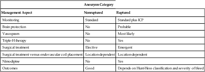
ICP, Intracranial pressure; triple-H therapy, use of hypertension, hydration, and hemodilution.

Other post-SAH problems include electrocardiographic changes, which may result from an intense sympathetic discharge. Some of these changes are benign; however, others may be indicative of myocardial damage. When these changes do occur, more intense invasive monitoring is necessary. If cardiac intervention is required, percutaneous endovascular therapy is probably the preferred treatment method.

Other problems associated with SAH include hyponatremia secondary to cerebral salt-wasting syndrome or syndrome of inappropriate antidiuretic hormone secretion (SIADH). The former results from secretion of atrial natriuretic hormone from the brain, causing a clinical triad of hyponatremia, volume contraction, and high urine sodium concentration. These patients may require fluid resuscitation. SIADH results from inappropriately excessive release of antidiuretic hormone and subsequent excessive free-water retention. In contrast with the treatment of cerebral salt-wasting syndrome, the treatment of SIADH is fluid restriction. However, because of vasospasm, restricting fluid may not be possible because fluid administration, rather than fluid restriction, is a primary component of triple-H therapy. In this situation, the administration of hypertonic saline should be considered.

Surgical treatment

Previously, when an aneurysm ruptured and an SAH ensued, the patient was observed for 10 to 14 days to allow time for resolution of both vasospasm and cerebral edema; however, the incidence of rebleeding was extremely high, with resultant morbidity and death. Today, treatment is early surgical intervention with either a surgical clip or an endovascular coil. Placement of either of these devices makes it easier to administer triple-H therapy should vasospasm occur. These interventions also decrease the incidence of rebleeding.

Aneurysms smaller than 7 mm have a low incidence of rupture. In aneurysms of this size, the principal treatment was previously to place a clip around the neck of the aneurysm. This treatment was in use until about 15 years ago, when Guglielmi detachable coils were introduced as a minimally invasive endovascular alternative. The International Subarachnoid Aneurysm Trial was conducted to evaluate surgery versus endovascular coiling. Some patients in the trial were observed for up to 7 years. Major results showed a decreased death rate in the endovascular coiling group (23.5% vs. 30.9%; P <0.0001) and less epilepsy but, compared with the surgical group, a higher incidence of rebleeding and less than optimal occlusion rate. However, as endovascular coiling techniques have improved, an increasing number of patients are treated with endovascular therapy.

Anesthetic management

Surgical and anesthetic management go hand in hand when caring for patients with an aneurysm. When compared with individuals with a higher grade of aneurysm, patients with a Hunt-Hess grade 1 or 2 aneurysm have a lower incidence of having increased ICP. Major blood loss is a potential adverse outcome during surgery, and brain relaxation probably will be necessary. With intracranial hemorrhage, cerebral autoregulation is lost. Consequently, the anesthesia care team must closely monitor, and maintain stable, the transmural pressure gradient.

In former times, intraoperative bleeding was restrained with controlled hypotension. Now, most surgeons prefer to have the blood pressure at baseline to maintain cerebral perfusion pressure. Temporary clipping of the main feeder vessel to the aneurysm may be needed. With this clipping, it may be necessary to temporarily increase the patient’s blood pressure to improve collateral circulation. Although evidence of barbiturate-mediated brain protection in humans is lacking, this technique may be requested by the surgical team. If nothing else, the barbiturates are effective in decreasing ICP and may help “relax” the brain during clip placement. If the temporary clip is removed, any dramatic increase in blood pressure that could lead to bleeding should be avoided.

During the operation, the use of standard American Society of Anesthesiologists monitors plus invasive arterial blood pressure monitoring are indicated. A central venous catheter may be helpful but is not necessary, depending on the patient’s comorbid conditions. Monitoring of the electroencephalogram and sensory-evoked potentials may be used, but no human trials have shown consistent benefit. If hydrocephalus is present, the neurosurgeon may opt to place a ventriculostomy for monitoring ICP and cerebrospinal fluid drainage. Brain bulk can also be decreased with intravenously administered mannitol.

Anesthesia induction can be accomplished with propofol and maintained with an opioid, such as fentanyl citrate, and an inhalation agent, such as isoflurane. It may become necessary to avoid an anesthesia level that exceeds 1 MAC (minimum alveolar concentration) of an inhalation agent to avoid cerebral vasodilation and subsequent increases in ICP. If the brain is extremely edematous, use of a total intravenous anesthetic technique may be considered.

Aside from avoiding hyperglycemia and fever during periods when the brain is at risk for developing ischemic injury, definitive evidence for brain-protective interventions are devoid in the human literature (see Chapter 131). Despite this, many physicians use barbiturates or propofol to achieve burst suppression (4-6 bursts/min) during critical periods of aneurysm operations. The Intraoperative Hypothermia for Aneurysm Surgery Trial did not show any benefit of a mild temperature decrease (to 33° C) during operations. No other research results have been published on this topic recently.