Malignant melanoma

Published on 19/03/2015 by admin

Filed under Dermatology

Last modified 22/04/2025

Print this page

rate 1 star rate 2 star rate 3 star rate 4 star rate 5 star
Your rating: none, Average: 0 (0 votes)

This article have been viewed 1366 times

Malignant melanoma

Andrea Hui, Philip Friedlander and Orit Markowitz

Evidence Levels:  A Double-blind study  B Clinical trial ≥ 20 subjects  C Clinical trial < 20 subjects  D Series ≥ 5 subjects  E Anecdotal case reports

image

Melanoma is the leading cause of death among all cutaneous diseases in the US. In 2010, it was estimated that 68,130 new cases of melanoma were diagnosed in the United States, and nearly 8,700 patients died from this cancer.

Management strategy

Early detection of melanoma is paramount in its management. Physicians and patients must become familiar with the ABCDE signs of melanoma: asymmetry, border irregularity, color variegation, diameter >6 mm, and evolution. A specificity of 88% and sensitivity of 73% has been reported if two out of three of the following characteristics are noted on physical exam: irregular outline, diameter >6 mm, and color variegation. Even expert clinicians misdiagnose melanoma in up to one-third of cases, and only biopsy and histologic examination can provide a definitive diagnosis.

Important in the management of melanoma is primary prevention (risk reduction) and secondary prevention (early detection). Educating the public about the risks of sun exposure is a crucial part of primary prevention. Secondary prevention involves teaching high-risk patients about the ABCDE signs and how to perform total body self-examinations. The NIH stresses the importance of screening programs and regular skin examinations by health professionals. The American Academy of Dermatology Task Force recommends follow-up one to four times a year for 2 years after a diagnosis of melanoma, depending on the thickness of the lesion and other risk factors, such as a family history of melanoma, and then once or twice a year thereafter. Other modalities such as dermoscopy and the recently FDA-approved MelaFind are non-invasive tools aiding in identification of high-risk pigmented lesions.

The first step in the diagnosis of suspected melanoma is to obtain a biopsy. The ideal biopsy of a suspected primary cutaneous melanoma is a full-thickness excision with at least a 2 mm margin of normal-appearing surrounding skin to ensure that the lesion is not transected. If total excision is not practical, a full-thickness incisional biopsy usually suffices.

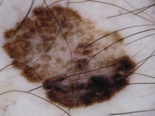
Staging of melanoma is crucial because it not only assigns patients into well-defined risk groups, it also aids in clinical decision-making as reviewed in the latest National Comprehensive Cancer Network (NCCN) clinical practice guidelines on melanoma. The goal of surgical management of primary cutaneous melanoma is to achieve negative histological margins to prevent recurrence and metastases. The current surgical margin guidelines from the NCCN are based on Breslow depth (Table 143.1). The standard treatment for stage IA melanoma is wide local excision. Total excision of primary melanoma with wide margins offers the best chance for cure. However, melanoma cells may extend non-contiguously for several millimeters beyond the visible lesion.
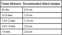
Table 143.1

Current melanoma excisional margin guidelines

Tumor thickness Recommended clinical margins
In situ 0.5 cm
≤1.0 mm 1.0 cm
1.01–2 mm 1–2 cm
2.01–4 mm 2.0 cm
>4 mm 2.0 cm

image

The NCCN suggests that sentinel lymph node (SLN) biopsy be discussed and offered to patients in those with melanomas at least 1 mm thick, or with ulceration or additional adverse features. The SLN is detected by preoperative lymphoscintigraphy followed by intraoperative injection of a dye and/or a radiocolloid around the primary lesion. The presence of melanoma in the SLN is the most significant factor in the prognosis of a patient with localized cutaneous melanoma undergoing SLN biopsy. If the SLN is histologically positive, a selective lymphadenectomy is recommended. The overall effect of selective lymphadenectomy following a positive SLN has yet to show survival benefit. An elective lymphadenectomy may also be performed, which involves removal of regional lymph nodes without preceding clinical or microscopic evidence of nodal metastases. This procedure, which results in significant morbidity, remains controversial.

For patients with stage III melanoma, the number of positive nodes and clinical node palpability are the most important prognostic factors. The standard treatment is wide local excision with nodal dissection. The only adjuvant therapy approved by the FDA at present is high-dose interferon (IFN)-α2b. For patients with clinically positive lymph nodes, a diagnostic nodal biopsy (e.g., fine-needle biopsy) is recommended and, if positive, a therapeutic lymphadenectomy is performed. Traditionally, patients with satellitosis or in-transit metastases on an extremity have been candidates for treatment with isolated limb hyperthermic perfusion/infusion with chemotherapeutic agents such as melphalan.

Stage IV melanoma has a very poor prognosis, with patients demonstrating a 5-year survival below 5%. The presence of BRAF mutations has transformed the systemic management of stage IV melanoma. Until 2011, only two treatments, dacarbazine and high-dose interleukin-2 (HD-IL-2), were FDA-approved for the treatment of stage IV melanoma. HD-IL-2 requires administration as an in-patient and can carry significant toxicity risks including capillary leak syndrome, renal failure, and neurologic toxicity. Given its toxicity profile, only a very limited number of patients are appropriate candidates for this treatment.

It has become increasingly evident that melanoma is a molecularly heterogeneous disease. Recently, sequencing of components of the MAPK pathway in a panel of cancer cell lines identified activating mutations in the downstream BRAF protein in 50–60% of melanomas. Over 90% of the mutations in BRAF occur at position 600, with the most common being a V600E mutation.

Vemurafenib is a potent inhibitor of V600E mutated BRAF. A randomized phase III study of first-line systemic therapy in patients with stage IV melanoma demonstrated a statistically significant overall survival benefit following vemurafenib treatment when compared to dacarbazine treatment. However, vemurafenib’s median progression-free survival is only 5.3 months. As such, elucidating resistance mechanisms is extremely important. Inhibition of the MAPK pathway downstream of BRAF by using the MEK inhibitor trametinib also confers a significant overall survival benefit relative to treatment with chemotherapy in patients with V600-mutated melanoma.

Approximately 15–30% of patients treated with BRAF inhibitors develop cutaneous squamous cell carcinomas. These carcinomas are believed to result from paradoxical activation of the MAPK pathway in cells containing both wild-type BRAF and active RAS.

A limitation of BRAF inhibition is its duration of benefit. Immunotherapies offer the potential for longer lasting benefit as demonstrated by the small subset of patients who develop durable responses to HD-IL-2. Cytotoxic T-lymphocyte-associated antigen 4 (CTLA-4) is an immunomodulatory protein which downregulates T-cell activity. Inhibition of CTLA-4 using the monoclonal antibody ipilimumab has antitumor activity in melanoma. Treatment of stage IV melanoma patients with ipilimumab has an overall survival benefit when compared to treatment with a peptide vaccine. An overall survival benefit is also seen following treatment with the combination of ipilimumab and dacarbazine when compared to treatment with single agent dacarbazine.

Both vemurafenib and ipilimumab were FDA-approved in 2011 for the treatment of stage IV melanoma. There is a pressing need to develop strategies to improve the durability of molecularly targeted therapy and the response rate of immunomodulatory therapy. Combination approaches including dual therapy with ipilimumab and vemurafenib are under active investigation. Therapies that combine BRAF and MEK inhibition are being investigated with the theory that dual pathway inhibition will delay the onset of resistance and decrease the incidence of cutaneous toxicity. Targeting the subset of melanomas that contain a mutation in KIT as opposed to BRAF with oral inhibitors of KIT generates responses in patients, but also with limited durability.

Antibody-mediated inhibition of immunomodulatory proteins other than CTLA-4 may prove efficacious. Targeting either the programmed death 1 protein (PD-1) or its ligand PD-L1 produced durable responses in a subset of patients with melanoma. PD-1 is an inhibitory receptor on T cells whose immunosuppressive ligand PD-L1 is expressed on many melanomas. The interaction of PD-1 to its ligand is particularly important in regulating immune activity in the tumor microenvironment. Strategies that combine CTLA-4 and PD-1 blockade are currently in clinical trial. Learning how to optimally modulate the immune system and to durably apply targeted therapies can potentially change the landscape of melanoma therapy with the goal that treatment becomes less palliative and more curative in its intent.

Specific investigations

National Comprehensive Cancer Network. Clinical practice guidelines in oncology. V 1.2013.

Coit D, et al. Available online: http://www.nccn.org/professionals/physician_gls/pdf/melanoma.pdf

Mitotic rate was added to the initial stratification of stage I and II melanoma. The extent of initial work-up is controversial. Most agree that chest radiography and blood work are not necessary for stage IA melanoma. For stage IB and stage IIA melanomas, a baseline chest radiograph is optional because it is not sensitive nor specific. Other imaging studies, such as CT, MRI, and/or PET scans, should be performed only to evaluate specific signs or symptoms, or for stages III and IV. Fine-needle aspiration is indicated for stage III with macrometastases or in-transit metastases, and stage IV. LDH is only indicated for stage IV. These guidelines also discuss frequency of follow-up visits and appropriate follow-up tests, both of which depend on the stage of the melanoma as well as other risk factors.

The role of sentinel lymph node biopsy for melanoma: evidence assessment.

Johnson TM, Sondak VK, Bichakjian CK, Sabel MS. J Am Acad Dermatol 2006; 54: 19–27.

This is a comprehensive review of 1198 articles identified by a search for melanoma sentinel node in the National Institutes of Health National Library of Medicine. The paper discusses the evidence base behind the sentinel node hypothesis. The authors state that the available evidence overwhelmingly supports SLN status as the most powerful independent factor predicting survival, with highest sensitivity and specificity of any nodal staging test. They also discuss that SLN biopsy results in improved regional disease control. The article claims that, based on available evidence, there is a potential subset survival benefit for subclinical detection of nodal disease followed by immediate CLND. The article reviews the MSLT-1 interim results and discusses the morbidities of SLN biopsy. It also reviews other aspects of SLN biopsy, including the evidence for its use in candidates for adjuvant therapy. The authors conclude that current evidence supports the use of SLN biopsy in the management of melanoma.

First-line therapies

image Surgical excision B
image Selective lymphadenectomy C
image Elective lymphadenectomy C

Second-line therapies

image Pegylated interferon alfa-2b A
image Paclitaxel and carboplatin D
image Imiquimod E
image Interleukin-2 B
image Isolated limb perfusion E
image Bcl-2 antisense and dacarbazine A
image Dacarbazine A
image Dacarbazine A
image Vemurafenib A
image Trametinib B
image Imatinib B

Adjuvant therapy with pegylated interferon alfa-2b versus observation alone in resected stage III melanoma: final results of EORTC 18991, a randomised phase III trial.

Eggermont AM, Suciu S, Santinami M, Testori A, Kruit WH, Marsden J, et al. Lancet 2008; 372: 117–26.

This study assessed whether long-term (5 years) adjuvant pegylated IFN-α2b therapy could be well tolerated and while maintaining recurrence-free survival. A total of 1256 patients with resected stage III melanoma were randomly assigned to an observation group (n=629) or pegylated IFN-α2b group (n=627) for 5 years. Relapse-free survival was significantly better in the IFN treatment group when compared to the observation group (45.6% vs 38.9%), but there was no significant benefit to overall survival. Subsequently, pegylated IFN-α2b was approved by the FDA in 2011 as adjuvant therapy for patients with stage III melanoma.

High-dose recombinant interleukin 2 therapy for patients with metastatic melanoma: analysis of 270 patients treated between 1985 and 1993.

Atkins MB, Lotze MT, Dutcher JP, Fisher RI, Weiss, et al. J Clin Oncol 1999; 17: 2105–16.

In this intention-to-treat analysis, 270 patients with metastatic melanoma were entered into eight clinical trials conducted between 1985 and 1993. These patients received at least one dose of high-dose recombinant IL-2 by IV infusion every 8 hours for up to 14 consecutive doses over 5 days as clinically tolerated, with a second cycle performed after 6 to 9 days of rest. Additional courses were repeated every 6 to 12 weeks as tolerated in stable or responding patients. The overall response rate was 16%, with 6% of patients achieving complete responses. The median response duration in partial responders was 5.9 months. Severe toxicities reversed rapidly after therapy was completed.

Improved survival with ipilimumab in patients with metastatic melanoma.

Hodi FS, O’Day SJ, McDermott DF, Weber RW, Sosman JA, Haanen JB, Gonzalez R, et al. N Engl J Med 2010; 363: 711–23.

The FDA approved ipimumab, a monoclonal antibody against the immune checkpoint receptor cytotoxic T lymphocyte antigen-4 (CTLA-4) based on this randomized phase III trial. A total of 676 patients with unresectable stage III or IV melanoma were assigned to three groups: ipilimumab plus glycoprotein 100 peptide vaccine (gp100) (n=403), ipilimumab alone (n=137), or gp100 alone (n=136). Ipilimumab, with or without gp100 vaccine, compared with gp100 alone, significant improved overall survival in patients with previously treated metastatic melanoma (10.1 months for ipilimumab alone, 10.0 months for combination therapy, versus 6.4 months for gp100 alone).

Inhibition of mutated, activated BRAF in metastatic melanoma.

Flaherty KT, Puzanov I, Kim KB, Ribas A, McArthur GA, Sosman JA, et al. N Engl J Med 2010; 363: 809–19.

This landmark multicenter, phase I, dose-escalation trial of PLX4032, an oral inhibitor of V600E-mutated BRAF, demonstrated complete or partial tumor regression in the majority of patients with metastatic melanoma. Fifty-five patients (49 had melanoma) were enrolled in the dose-escalation phase, with an additional 32 patients with V600E mutated BRAF melanomas were enrolled in the extension phase allowing the maximum dose that could be administered without adverse effects. In the dose-escalation cohort, among 16 patients with V600E mutated BRAF melanoma, 10 patients demonstrated partial response and one had a complete response. In the extension cohort, 24 patients had a partial response and two had a complete response. Median progression-free survival for all patients was over 7 months. Interestingly, responses to PLX4032 were seen as early as 14 days after initiating treatment, as seen on FDG-labeled PET imaging.

Improved survival with MEK inhibition in BRAF-mutated melanoma.

Flaherty KT, Robert C, Hersey P, Nathan P, Garbe C, Milhem, et al. N Engl J Med 2012; 367: 107–14.

In previous trials, MEK inhibition by trametinib showed promise. In this phase III open-label trial, trametinib, as compared with chemotherapy, significantly improved rates of progression-free and overall survival among patients with metastatic melanoma containing the V600E or V600K BRAF mutation. A total of 322 patients were randomly assigned to receive either oral trametinib daily or chemotherapy in a 2 : 1 ratio (intravenous dacarbazine or paclitaxel) every 3 weeks. Patients in the chemotherapy cohort who demonstrated disease progression were permitted to cross over to receive trametinib. Median progression-free survival was 4.8 months in the trametinib group and 1.5 months in the chemotherapy group. At 6 months, the overall rate of survival was 81% in the trametinib group and 67% in the chemotherapy group despite crossover.

KIT as a therapeutic target in metastatic melanoma.

Carvajal RD, Antonescu CR, Wolchok JD, Chapman PB, Roman RA, Teitcher J, Panageas K, et al. J Am Med Assoc 2011; 305: 2327–34.

In this single-group, open-label, phase II trial, treatment with the KIT inhibitor imatinib showed significant clinical responses in patients with advanced melanoma harboring KIT mutations. Patients (n=205) with metastatic melanoma were screened for the presence of KIT mutations. Fifty-one patients were identified with this mutation, and 28 patients from this group were treated. These 28 patients had advanced unresectable melanoma arising from acral, mucosal, and chronically sun-damaged areas. Imatinib was administered orally twice daily. The overall durable response rate was 16% with a median time to progression of 12 weeks and median overall survival of 46.3 weeks.

Third-line therapies

image Radiation therapy B
image Mohs micrographic surgery B

Share this: