Malignant atrophic papulosis

Published on 18/03/2015 by admin

Filed under Dermatology

Last modified 18/03/2015

Print this page

rate 1 star rate 2 star rate 3 star rate 4 star rate 5 star
Your rating: none, Average: 2 (1 votes)

This article have been viewed 1018 times

Malignant atrophic papulosis

Noah Scheinfeld

Evidence Levels:  A Double-blind study  B Clinical trial ≥ 20 subjects  C Clinical trial < 20 subjects  D Series ≥ 5 subjects  E Anecdotal case reports

image

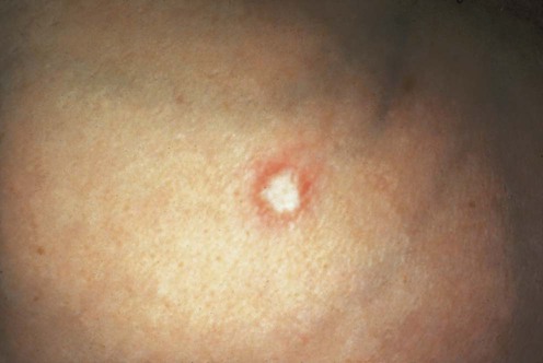
(From Lebwohl MG. The Skin and Systemic Disease: A Color Atlas, 2nd edn. Churchill Livingstone 2003, with permission of Elsevier.)

Malignant atrophic papulosis (MAP), eponymically known as Degos disease (DD), possesses purely cutaneous and systemic variants. These variants are starkly divided by their prognosis, with purely cutaneous DD being wholly benign in course and systemic DD usually being fatal within a few years of onset. Cutaneous DD can precede the development of systemic DD and no factor has yet been defined that will predict which persons with cutaneous DD will go on to develop systemic DD.

About 300 cases of DD have been reported. It is likely that the purely cutaneous type of DD is under-diagnosed as it is so banal and so benign. DD affects all ages and both sexes. A familial variant appears to exist. The disease is more commonly reported in men and young adults but this may simply be sampling error.

Cutaneous and systemic DD have similar cutaneous eruptions. In the skin, DD occurs as erythematous, pink or red papules (2–15 mm), which evolve into scars with central, porcelain white atrophic centers. The papules of MAP usually have a peripheral telangiectatic rim and can be domed or atrophic. The papules can have central crusts. Urticaria, ulceropustular and/or gumma-like nodules can manifest. All areas of the body, with the exception of the face, can be involved.

It is important to stress that the mere appearance in the skin of DD is not itself an event that will shorten life. Purely cutaneous DD has a benign course and can wax and wane with no effect on mortality over a lifetime. Systematic DD presents a dire prognosis. The cause of death in systemic DD is related to perforation of blood vessels and bleeding, usually intestinal perforation. Systemic MAP can involve the nervous, ophthamologic, gastrointestinal, cardiothoracic, and hepatorenal systems. Death usually occurs within 2 to 3 years from the onset of systemic involvement.

Virus-like inclusions, observable with an electron microscope are frequently present in the endothelial cells and fibroblasts of patients with DD. C3 deposits are sometimes noted. There can be intracytoplasmic cylinders in the histiocytes. In a series of three patients with MAP, electron microscopy demonstrated an increased number of Weibel–Palade bodies and a more intense staining of von Willebrand factor in endothelial cells.

Pathologically, the tissue affected by DD reveals an occlusive arteriopathy involving small-caliber vessels which results in tissue infarction. The bland appearance of MAP’s vasculopathy (or an endovasculitis) on histological examination does not equate with the serious nature of systemic DD.

No specific laboratory test can be used to diagnose DD. Cases of DD can manifest antiphospholipid antibodies and antinuclear antibodies but these seem to be correlative rather than causative antibodies. Some reports note increased plasma fibrinogen levels, increased platelet aggregations, and a decrease of local and systemic fibrinolytic activity. There are cases of systemic lupus erythematosus and other collagen vascular diseases that display DD-like atrophic porcelain white papules; however, these cases likely do not represent cases of DD.

Histopathologically, DD manifests with a wedge-shaped degeneration of collagen with a prominent interface reaction with squamatization of the dermoepidermal junction, melanin incontinence, and epidermal atrophy.

MAP is no longer an incurable disease. Reports have noted that initiation and continuing treatment with eculizumab can put patients with systemic MAP into remission. Eculizumab is a monoclonal antibody directed against the complement protein C5. Eculizumab is currently only approved for treatment of paroxysmal nocturnal hemoglobinuria. While the sample size of the use of eculizumab is small, and most of the cases are unpublished, it seems that eculizumab is the treatment of choice for systemic MAP. The dosing for this treatment is still not clear but is likely at the high end of published doses (900 mg intravenously or perhaps higher given continuously for treatment of MAP). The success of eculizumab suggests that MAP is a vascular rather than an immune disease; research into how MAP might be triggered continues and the etiology of the inception of MAP remains to be defined. As eculizumab is a very expensive complex treatment, its use is not recommended for purely cutaneous DD.

The role of testing is to establish if systemic DD is present so that treatment with eculizumab can be considered. A patient with purely cutaneous DD should be given regular stool guaiac tests to test for internal bleeding. Tissue specimens can help to buttress the diagnosis of DD but many clinical and histological features of DD can rarely be found in patients with lupus; therefore a patient’s clinical, histological and laboratory data must be integrated to definitively support the diagnosis of DD.