[level-membership-for-dermatology-category]
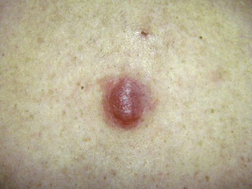
Lymphocytoma cutis
Specific investigations


First-line therapies
 Excision
 Topical corticosteroids
 Intralesional corticosteroids
 Oral antibiotics (if positive Borrelia serology)
 CO2 or Nd:YAG laser (if occurring within a tattoo)
 Antimalarials
 Sun avoidance/sun block (light exacerbated)
Second-line therapies
 Superficial radiotherapy
 Intralesional interferon-α
 Argon laser
 Cryotherapy
 Topical 0.1% tacrolimus ointment
 Intralesional rituximab
 Topical photodynamic therapy
 Subcutaneous interferon-α2b
 Thalidomide
[/level-membership-for-dermatology-category][not-level-membership-for-dermatology-category]
Lymphocytoma cutis
Specific investigations
Differential diagnosis of cutaneous infiltrates of B lymphocytes with follicular growth pattern.
Leinweber B, Colli C, Chott A, Kerl H, Cerroni L. Am J Dermatopathol 2004; 26: 4–13.
The histopathologic, immunophenotypic, and molecular features of Borrelia burgdorfori
Buy Membership for Dermatology Category to continue reading. Learn more here
[/not-level-membership-for-dermatology-category]
