Published on 18/03/2015 by admin
Filed under Dermatology
Last modified 22/04/2025
This article have been viewed 1840 times
Robert E. Lee, Giuseppe Micali and Robert A. Schwartz
Evidence Levels: A Double-blind study B Clinical trial ≥ 20 subjects C Clinical trial < 20 subjects D Series ≥ 5 subjects E Anecdotal case reports
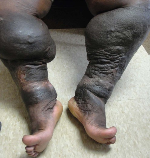
Lymphedema is a chronic, progressive, and sometimes debilitating condition. It is due to the abnormal accumulation of protein-rich lymphatic fluid in interstitial spaces as a result of ineffective drainage. Lymphedema is classified into primary and secondary forms. Primary lymphedema is caused by a developmental malformation of the lymphatic system. Primary congenital lymphedema (Milroy disease) is an uncommon autosomal dominant disorder due, in some families, to missense mutations that interfere with vascular endothelial growth factor receptor-3 signaling, resulting in abnormal lymphatic vascular function. Primary lymphedema may be further classified by age of onset into congenital lymphedema (before age 2 years), lymphedema praecox (between age 2 and 35 years), and lymphedema tarda (after age 35 years). Secondary lymphedema is due to obstruction or damage of an otherwise normal lymphatic system. In the United States the most common causes are malignancy, surgical manipulation, or radiation damage. Globally, the most common cause is filariasis. Acquired lymphedema may predispose to recurrent cellulitis and an aggressive type of angiosarcoma, also known as Stewart–Treves syndrome. Chronic lymphedema may result in verrucous and proliferative changes resembling elephant skin (elephantiasis).
Lymphedema must be distinguished from cutaneous edema of cardiac, hepatic, or renal etiology. It is characterized clinically by brawny, non-pitting edema. Lymphoscintigraphy (isotope lymphography) is a first-line imaging modality to evaluate and diagnose disorders of the lymphatic vasculature. A conservative approach with medical and physical therapy is the primary treatment for lymphedema. The main management strategy is to reduce stasis of protein-rich lymph in the extravascular tissue and to improve the outflow of lymphatic circulation.
Complex decongestive therapy (CDT) represents an effective treatment plan. It is a four-component therapeutic modality composed of multilayer compression bandaging, manual lymphatic drainage, skin care, and exercise. The therapeutic efficacy of CDT is highly dependent upon patient compliance. Therefore, strict adherence to the treatment regimen should be encouraged. Pneumatic compression therapy was widely used to control lymphedema. However, owing to its poor outcome as a monotherapy, its current use has been limited. Unfortunately, recent advances in these devices have showed beneficial results. Medications, such as diuretics, have shown limited or no effect on lymphedema. Meticulous skin care and hygiene may prevent secondary bacterial and fungal infections. Topical or systemic antibiotics should be initiated at the first sign of infection. This is especially important as recurrent infections may lead to further lymphatic injury.
Surgical approaches are reserved for cases refractory to conservative medical management. Microsurgical lymphatic–venous anastomoses have yielded promising results. Excisional surgical therapy has been performed to reduce limb size and to improve mobility in chronic advanced cases of lymphedema.
Lymphoscintigraphy
MRI
CT
Indocyanine green lymphography
Witte CL, Witte MH, Unger EC, Williams WH, Bernas MJ, McNeill GC, et al. Radiographics 2000; 6: 1697–16719.
An excellent review article illustrating multiple clinical cases in which lymphoscintigraphy, MRI, and CT were useful in the evaluation and diagnosis of patients with primary or secondary lymphedema.
The advantages and limitations of each imaging modality are reviewed.
Mihara M, Hara H, Araki J, Kikuchi K, Narushima M, Yamamoto T, et al. PLoS One 2012; 7: e38182.
This study suggests that ICG lymphography combined with MRI may have a higher sensitivity in diagnosing early upper limb lymphedema in comparison to other diagnostic imaging modalities.
Karadibak D, Yavuzsen T, Saydam S. J Surg Oncol 2008; 97: 572–7.
A prospective study of 62 women with breast cancer-related lymphedema demonstrating the effectiveness of complete decongestive therapy by decreasing limb volume and fear of activity.
Godoy Mde F, Pereira MR, Oliani AH, de Godoy JM. Int J Med Sci 2012; 9: 280–4.
A randomized controlled trial of 20 women resulting in the reduction of lymphedematous arms from breast cancer treatment by combining compression therapy with controlled exercises using a facilitating device.
Schook CC, Mulliken JB, Fishman SJ, Grant FD, Zurakowski D, Greene AK. Plast Reconstr Surg 2011; 127: 2419–31.
Pediatric patients with primary lymphedema can be successfully managed with compression garments without the need for surgical intervention.
Godette K, Mondry TE, Johnstone PA. J Soc Integr Oncol 2006; 4: 8–12.
There are several contraindications to performing complete decongestive therapy (CDT). These include hypertension, paralysis, diabetes mellitus, bronchial asthma, acute infections, and congestive heart failure. Malignant disease is also widely considered a contraindication to CDT. However, this opinion is not unequivocally supported by current cancer research.
Adams KE, Rasmussen JC, Darne C, Tan IC, Aldrich MB, et al. Biomed Opt Express 2010; 1: 114–25.
This study supports the effectiveness of pneumatic compression device treatment by showing lymphatic function improvement in both control subjects and breast cancer-related lymphedema patients.
Campisi C, Bellini C, Campisi C, Accogli S, Bonioli E, Boccardo F. Microsurgery 2010; 30: 256–60.
A retrospective study of 1800 subjects undergoing microsurgery for the treatment of peripheral lymphedema resulting in marked improvement in 83% of patients.
Suami H, Chang DW. Plast Reconstr Surg 2010; 126: 1853–63.
A review of surgical procedures for the treatment of breast cancer-related lymphedema and current issues in the management of lymphedema with surgical treatment.
Campisi C, Da Rin E, Bellini C, Bonioli E, Boccardo F. Microsurgery 2008; 28: 138–42.
Microsurgical methods may provide successful and long-lasting results, with both derivative lymphaticovenous anastomoses and reconstructive lymphaticovenous–lymphatic anastomoses. Better long-term results are obtained in earlier stages, before tissue fibrosis and sclerosis ensue.
Ahmed Omar MT, Abd-El-Gayed Ebid A, El Morsy AM. J Surg Res 2011; 165: 82–90.
Fifty patients were studied, 25 receiving laser versus 25 with placebo. Low level laser therapy reduced limb volume and increased shoulder mobility and hand grip strength in 93% of fifty women with breast cancer-related lymphedema.
Treatment of Skin Disease Comprehensive Therapeutic Strategies 4e
WhatsApp us


 Complex (or complete) decongestive therapy
 Pneumatic compression therapy
 Surgery
 Low level laser therapy