Lung Defense Mechanisms

Published on 12/06/2015 by admin

Filed under Pulmolory and Respiratory

Last modified 22/04/2025

Print this page

rate 1 star rate 2 star rate 3 star rate 4 star rate 5 star
Your rating: none, Average: 4 (1 votes)

This article have been viewed 2470 times

22

Lung Defense Mechanisms

In the process of exchanging thousands of liters of air each day for O2 uptake and CO2 elimination, the lung is exposed to a multitude of foreign substances transported with the inhaled air. Some of these are potentially injurious; others are relatively harmless. Inhaled air is not the only source of foreign material. Secretions from the mouth and pharynx frequently are aspirated into the tracheobronchial tree, especially during sleep, even in healthy individuals. This myriad of foreign substances is perhaps best classified into three major categories: small particulate material, noxious gases, and microorganisms. Because the oropharynx is rich with bacteria, aspirated secretions are particularly important as a source of unwanted bacteria entering the airways.

To protect itself against potentially toxic inhaled material, the respiratory system has evolved complex protective mechanisms that can be dissected into different components. Each component appears to have a distinct role, but a tremendous degree of redundancy and interaction exists among different components. That the distal lung parenchyma is normally sterile (and is not in a state of constant inflammation) serves as testimony to the effectiveness of the defense system. However, the protective mechanisms can break down, resulting in respiratory infection. Such a breakdown in defense can occur as a result of certain diseases, a large inoculum of microorganisms that overwhelms a normal host, an especially virulent organism, or frequently as a consequence of medical treatment that impairs the immune system.

Before the discussion of infectious disorders of the respiratory system in Chapters 23 through 26, it is appropriate to first consider how the lung protects itself against the infectious agents to which it is exposed. Although this chapter focuses on protective mechanisms against infection, defenses against noninfectious substances, especially inhaled particulate material, also are addressed. The major categories of defense mechanisms to be discussed include (1) physical or anatomic factors relating to deposition and clearance of inhaled material, (2) antimicrobial peptides, (3) phagocytic and inflammatory cells that interact with the inhaled material, and (4) adaptive immune responses, which depend on prior exposure to and recognition of the foreign material. The chapter concentrates on the aspects of the host defense system specific to the lung and then proceeds with a discussion of several ways the system breaks down, resulting in an inability to handle microorganisms and an increased risk for certain types of respiratory tract infection. The chapter concludes by briefly considering how we can activate or augment specific immune responses through immunization, thus enhancing defenses against selected respiratory pathogens.

Physical or Anatomic Factors

The pathway from the mouth or nose down to the lung parenchyma requires that inhaled air traverse a series of progressively branching airways. The laminar flow of air through the airways is disrupted at the branch points (subcarinae), thus enhancing deposition of particulate material at these locations. Hence, inhaled particulates frequently are deposited at various points in the airway, never reaching the most distal region of lung, the alveolar spaces. Particle size is an important determinant of deposition along the airway and thus affects the likelihood of a particle’s reaching the distal parenchyma. When an inhaled particle is greater than 10 µm in diameter, it is likely to settle high in the upper airway (e.g., in the nose). For particles 5 to 10 µm in diameter, settling tends to occur somewhat lower, in the trachea or the conducting airways, but not down to the level of the small airways and alveoli. The particles most likely to reach the distal lung parenchyma range in size from 0.5 to 5 µm. Many bacteria fall within this size range, so deposition along the airways is not very effective for excluding bacteria from the lower respiratory tract. However, large particles of dust and other inhaled material are effectively excluded from the distal lung parenchyma by virtue of their size. Of note, the target size for particles of inhaled medications such as bronchodilators is less than 5 µm so that the medication can bypass the conducting airways and reach the more distal lung.

When particles are deposited in the trachea or bronchi, two major processes, cough and mucociliary transport, are responsible for physical removal of these particles from the airways. Cough is an important protective mechanism, frequently triggered by stimulation of airway irritant receptors, which are most prominent in the proximal airways and are activated by inhaled or aspirated foreign material. Rapid acceleration and high flow rates of air achieved by a cough often are effective in clearing irritating foreign material from the airways.

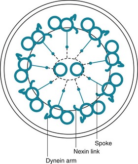
The term mucociliary transport or mucociliary clearance refers to a process of waves of beating cilia moving a blanket of mucus (and any material trapped within the mucus) progressively upward along the tracheobronchial tree. From the trachea down to the respiratory bronchioles, the most superficial layer of epithelial cells lining the airway has cilia projecting into the airway lumen. These cilia have a structure identical to that of cilia found elsewhere in the body, consisting of longitudinal microtubules with a characteristic architecture. Specifically, a cross-sectional view of cilia shows two central microtubules surrounded by nine pairs of microtubules arranged around the periphery (Fig. 22-1). Small projecting side arms from each doublet, called dynein arms, are crucial to the contractile function of the microtubules and hence to the beating of the cilia.

Strikingly, the movement of cilia on a particular cell and the movement between cells are quite coordinated, producing actual “waves” of ciliary motion. Exactly how such a pattern of ciliary motion is coordinated from cell to cell or even within the same cell is not known. What this wavelike motion accomplishes is movement of the overlying mucous layer in a cephalad direction (i.e., from distal to more proximal parts of the tracheobronchial tree) at an estimated speed of 6 to 20 mm/min in the trachea. If inhaled particles are trapped in the mucous layer, they too are transported upward and eventually are either expectorated or swallowed.

Two layers comprise the mucous blanket bathing the epithelial cells. Directly adjacent to the cells is the sol layer, within which the cilia are located. The aqueous sol layer contains a number of molecules in solution that are part of the innate immune system and are discussed in the Antimicrobial Peptides section. Superficial to the sol layer is the more viscous gel layer, which is produced by both submucosal mucous glands and goblet cells. Picture the viscous gel layer floating on top of the sol layer and being propelled upward as the cilia are able to beat more freely within the less viscous sol layer.

Antimicrobial Peptides

The sol layer contains a number of substances that are important in innate immunity. The innate immune system can be thought of as a fast-acting system that is ready to quickly protect the lungs without prior sensitization and ideally avoid activation of the adaptive immune system (discussed in the Adaptive Immune Responses section). In addition to mucociliary clearance, the innate immune system is composed of small molecules, proteins, and cells that are able to respond to inhaled particles in a way that does not require any previous exposure to the particle. These molecules are generally highly conserved in evolution and are present in many invertebrate species as well as in humans. They are able to immediately interact with microorganisms through recognition of conserved structures on the microbes, and they can act directly to kill the invader and stimulate a further host immune response. They provide a fast, energy-efficient, effective frontline defense, with broad overlap in actions. There are many components of innate immunity in the lung, and a full description is beyond the scope of this chapter. The interested reader is referred to the in-depth reviews listed in the references. For the reader to get a sense of the system, this chapter focuses on a few of the best described of these molecules: lysozyme, lactoferrin, defensins, collectins (surfactant protein A [SP-A] and surfactant protein D [SP-D]), and immunoglobulin (Ig)A.

Lysozyme is present throughout the respiratory tract but is most prominent in the proximal airways. It is synthesized by respiratory epithelial cells, serous glandular cells, and macrophages. As the name implies, lysozyme causes bacterial cell death by inducing lysis. It is most active against gram-positive organisms. Decreased levels of lysozyme have been correlated with increased susceptibility to acute bronchitis.

Lactoferrin is present in airway fluid. It is produced by serous cells and neutrophils. Lactoferrin acts to agglutinate and kill bacteria, enhance neutrophil adherence, and prime neutrophil superoxide production. Its name derives from the fact that lactoferrin also functions to block iron from supporting bacterial metabolism. Lactoferrin binds to bacteria through recognition of highly conserved carbohydrate moieties on the microbial cell surface.

Defensins are a family of small proteins with intrinsic antimicrobial activity that are found in the lung and on other mucosal surfaces, including the gastrointestinal and reproductive tracts. The two most important types of defensins in the lung are α-defensins and β-defensins. α-Defensins are synthesized by resident neutrophils; β-defensins are made by respiratory epithelial cells. Defensins have broad antimicrobial activity against both gram-positive and gram-negative organisms. They act by making the microbial cell wall permeable, thus causing release of microbial cell contents and destruction of the membrane potential. The activity of defensins is highly sensitive to salt concentrations, and they are inactivated in the abnormal milieu in the lungs of patients with cystic fibrosis.

SP-A and SP-D are members of the collectin family of proteins. Their antimicrobial function is a result of binding and aggregating microbes and facilitating interaction with phagocytic cells. They also appear to be important in regulation of pulmonary macrophage activity and cytokine production. Animal models indicate that defects in either of these proteins increase the susceptibility to respiratory infection; however, human disease related to a genetic mutation or deletion has not been identified.

Respiratory IgA can be considered part of the innate immune system because it is also constitutively produced by the respiratory epithelium and does not require prior exposure. IgA is further discussed in the section on humoral immune mechanisms.

Phagocytic and Inflammatory Cells

Pulmonary Alveolar Macrophages

In the airways and at the level of the alveoli, particles and bacteria can be scavenged by mononuclear phagocytic cells called pulmonary alveolar macrophages. These cells constitute a major form of defense against material that has escaped deposition in the upper airway and has reached the intrathoracic airways or the alveolar structures.

Pulmonary alveolar macrophages are large mobile cells approximately 15 to 50 µm in diameter. They are descendants of circulating monocytes derived from bone marrow. These cells adhere to the alveolar epithelium. Their cytoplasm contains a variety of granules of various shapes and sizes, many of which are packages of digestive enzymes that can dispose of ingested foreign material. Alveolar macrophages have a major role in killing microorganisms that have reached the lower respiratory tract. They also release chemoattractant cytokines (chemokines) that recruit other inflammatory cells.

When an alveolar macrophage is exposed to inhaled particles or bacteria, attachment of the foreign material to the surface of the macrophage is the first step in the processing sequence. The particles or bacteria are engulfed within the plasma membrane, which invaginates and pinches off within the cell to form a cytoplasmic phagosome containing the now isolated foreign material. In some circumstances, this sequence of attachment and phagocytosis is facilitated by opsonins, which coat the foreign material. Opsonins are proteins that bind to extracellular materials and make them more adherent to phagocytic cells and more amenable to engulfment or ingestion. Opsonins can be specific for the particular foreign substance, such as antibodies directed against antigenic material, or they may demonstrate nonspecific binding to a variety of substances. Particularly important specific opsonins are antibodies of the IgG class directed against antigenic foreign material, either bacteria or other antigenic particles. Nonspecific opsonins in the lung include secretory IgA, complement, and fibronectin. All these opsonins greatly promote attachment to and ingestion by macrophages.

After bacteria or other foreign material is isolated within phagosomes, a process of intracellular digestion occurs within the macrophage. Often the phagosomes combine with lysosomes to form phagolysosomes, in which proteolytic enzymes supplied by the lysosome digest, detoxify, or destroy the phagosomal contents. In addition to lysosomal enzymes, a variety of oxidation products, such as hydrogen peroxide and other intermediate products of oxidative metabolism, are toxic to bacteria and may play a role in the ability of the macrophage to kill ingested microorganisms.

After they are activated, the resident pulmonary macrophages participate in orchestrating further immune responses. Macrophages release inflammatory mediators such as tumor necrosis factor (TNF)-α and interleukin (IL)-1β, as well as other cytokines and chemokines that are active in recruiting additional inflammatory cells.

The macrophage does not always kill or totally eliminate inhaled foreign material to which it is exposed. In some cases, such as with inhaled silica particles, the ingested material is toxic to the macrophage and eventually may kill the cell. In other cases, ingested material is inert but essentially indigestible and may persist indefinitely in the form of an indigestible residue. Organisms that are especially capable of persistent infection of macrophages without being killed or deactivated include Mycobacterium tuberculosis and the human immunodeficiency virus (HIV).

An increasingly appreciated role of the alveolar macrophage is to suppress inflammation in the lung. The lung is unique in that it is constantly exposed to inhaled foreign substances but at the same time must maintain an exquisitely delicate gas-exchange apparatus. Even a small amount of inflammation within the alveolar wall would have a negative effect on gas exchange, and a fine balance keeps the distal airways sterile, but not in a state of constant harmful inflammation. Alveolar macrophages are able to process a large amount of inhaled substances without inciting an immune response external to the macrophage itself. It is estimated that the normal pool of alveolar macrophages can handle up to 109 inhaled bacteria before the bacteria overwhelm the macrophages and cause infection in the alveoli. In addition, alveolar macrophages, through complex signaling mechanisms, function to keep dendritic cell and T-cell activation in check. The detailed working of this fine equilibrium between inflammation and quiescence in the lung is an area of active research.

Dendritic Cells

Dendritic cells are present throughout the body in various forms. They are bone marrow–derived cells that, in the lung, are located in the airway epithelium as well as in alveolar walls and peribronchial connective tissue. These cells have long and irregular cytoplasmic extensions that form a contiguous network. The primary function of dendritic cells is to sample the airway microenvironment, ingest and process antigens, and then migrate to regional lymph nodes. In the lymph nodes, dendritic cells present antigen to T cells, a critical step for the later immunologic defense provided by lymphocytes. Langerhans cells, a type of dendritic cell with a particular ultrastructural appearance, are the cells with abnormal proliferation that appear to be responsible for Langerhans cell histiocytosis of the lung (also called eosinophilic granuloma; see Chapter 11).

Polymorphonuclear Leukocytes

Another important cell involved in pulmonary defense is the polymorphonuclear leukocyte (PMN). The PMN is a particularly important component of the defense mechanism for an established bacterial infection of the lower respiratory tract. Normally, few PMNs reside in the small airways and alveoli. When bacteria overwhelm the initial defense mechanisms already discussed, they may replicate within alveolar spaces, causing a bacterial pneumonia. Examination of the histologic features of a bacterial pneumonia reveals that a prominent component of the inflammatory response is an outpouring of PMNs into the alveolar spaces. These cells probably are attracted to the lung by a variety of stimuli, particularly products of complement activation and chemotactic factors released by alveolar macrophages.

The eventual movement of PMNs out of the vasculature and into the lung parenchyma depends on the initial adherence of PMNs to the vascular endothelium. A variety of factors mediate this process of adhesion, including integrins (on the surface of the PMNs) and adhesion molecules (on the surface of the vascular endothelial cells).

When PMNs are involved, they play a crucial role in phagocytosis and killing of the population of invading and proliferating bacteria. Neutrophil granules contain several antimicrobial substances, including defensins, lysozyme, and lactoferrin. In addition, neutrophils are capable of generating products of oxidative metabolism that are toxic to microbes.

Natural Killer Cells

Natural killer (NK) cells are part of the rapid initial response and are capable of killing microorganisms without prior sensitization. These cells lack surface markers characteristic of either T or B lymphocytes (discussed in the section on cellular immune mechanisms). NK cells are important in rapid protection against viral infections. They act by recognizing and killing virus-infected cells that have been transformed and no longer express certain markers of cellular health on the cell surface. NK cells also are important in surveillance for neoplasms, and they use the same methods to detect and kill malignantly transformed cells.

Adaptive Immune Responses

The final category of defense mechanisms for the respiratory system is the adaptive immune response, which involves recognizing and responding to specific antigenic material after prior sensitization. Bacteria, viruses, and other microorganisms are perhaps the most important antigens to which the respiratory tract is repetitively exposed. Presumably, immune defense mechanisms are particularly important in protecting the individual against these agents. The processes of the adaptive immune response are not unique to the lung, and only a superficial discussion of general principles is provided here as a basis for understanding the adaptive immune responses in the lung. For more detailed information, the reader is referred to specialized texts and review articles on immunology.

The two major components of the adaptive immune system are humoral (or B-lymphocyte related) and cellular (or T-lymphocyte related). Humoral immunity involves activation of B lymphocytes (which do not require the thymus for differentiation) and production of antibodies by plasma cells (which are derived from B lymphocytes). Cellular immunity refers to activation of T lymphocytes (which depend on the thymus for differentiation) and execution of certain specific T-lymphocyte functions, including the production of soluble mediators or cytokines. The two lymphocyte systems are not independent of each other. In particular, T lymphocytes appear to have an important role in regulating immunoglobulin or antibody synthesis by the humoral immune system.

Both humoral and cellular immunity are important in protection of the respiratory system against microorganisms. For certain infectious agents, humoral immunity is the primary mode of protection. For other agents, cellular immunity appears to be paramount. In the lung and in blood, T lymphocytes are more numerous than B lymphocytes, but both systems are essential for effective defense against the spectrum of potentially harmful microorganisms.

Lymphocytes can be found in many locations within the respiratory tract, extending from the nasopharynx down to distal regions of the lung parenchyma. True lymph nodes are present around the trachea, carina, and at the hilum of each lung in the region of the mainstem bronchi. These lymph nodes receive lymphatic drainage from most of the airways and lung parenchyma. Lymphoid tissue is present in the nasopharynx, and collections of lymphocytes arranged in nodules are found along medium to large bronchi. These latter collections are called bronchus-associated lymphoid tissue and may be responsible for intercepting and handling antigens deposited along the conducting airways. Smaller aggregates of lymphocytes can be found in more distal airways and even scattered throughout the pulmonary parenchyma.

Humoral Immune Mechanisms

Humoral immunity in the respiratory tract appears in the form of two major classes of immunoglobulins: IgA and IgG. Antibodies of the IgA class are particularly important in the nasopharynx and upper airways, where they constitute the primary antibody type. The form of IgA present in these areas is secretory IgA, which includes a dimer of IgA molecules (joined by a polypeptide) plus an extra glycoprotein component termed the secretory component. Secretory IgA appears to be synthesized locally, and the quantities of IgA are much greater in the respiratory tract than in the serum.

Evidence suggests that secretory IgA plays an important role in the respiratory defense system. By virtue of its ability to bind to antigens, IgA may bind to viruses and bacteria, preventing their attachment to epithelial cells. In addition, IgA is efficient in agglutinating microorganisms; the agglutinated microbes are more easily cleared by the mucociliary transport system. Finally, IgA appears to have the ability to neutralize a variety of respiratory viruses as well as some bacteria.

In contrast to IgA, IgG is particularly abundant in the lower respiratory tract. It is synthesized locally to a large extent, although a fraction also originates from serum IgG. It has a number of biological properties, such as agglutinating particles, neutralizing viruses and bacterial toxins, serving as an opsonin for macrophage phagocytosis of bacteria, activating complement, and causing lysis of gram-negative bacteria in the presence of complement.

The overall role of the humoral immune system in respiratory defenses includes protecting the lung against a variety of bacterial and, to some extent, viral infections. The clinical implications of this role and the consequences of impairment in the humoral immune system are discussed in the section on defects in the adaptive immune system.

Cellular Immune Mechanisms

Cellular immune mechanisms, those mediated by thymus-dependent (T) lymphocytes, also operate as part of the overall defense system of the lungs. Sensitized T lymphocytes produce a variety of soluble biologically active mediators called cytokines, some of which (e.g., interferon [IFN]-γ) have the ability to attract or activate other protective cell types, particularly macrophages. T lymphocytes also are capable of interacting with the humoral immune system and modifying antibody production.

Two important types of T lymphocytes have been well characterized on the basis of specific cell surface markers and functional characteristics. One type consists of cells that are positive for the CD4 surface marker, commonly called CD4+ or helper T cells. CD4+ cells, in turn, are divided into TH1 and TH2 subsets, which mediate cellular immune defense and allergic inflammation, respectively. The other major type of T lymphocyte consists of cells that are positive for the CD8 surface marker. These CD8+ cells include suppressor and cytotoxic T cells. On exposure to specific antigens, both CD4+ and CD8+ cells produce a variety of cytokines that interact with other components of the immune system, particularly B lymphocytes and macrophages.

One important role for the cellular immune system is to protect against bacteria that have a pattern of intracellular growth, especially M. tuberculosis (see discussion of tuberculosis in Chapter 24). In addition, the cellular immune system has a critical role in the handling of many viruses, fungi, and protozoa.

Although separating the immune protection of the lung into different categories is important for discussion purposes, all of these limbs are deeply intertwined, and dysfunction in one aspect likely will cause problems in other parts of the system. Development of a respiratory infection generally indicates that a number of defense mechanisms have been overcome by the infecting organism.

Failure of Respiratory Defense Mechanisms

Clinically important deficiencies have been recognized for each of the major categories of respiratory defense mechanisms. As a result, respiratory infections may ensue, and analysis of the specific types of infections associated with each type of defect is clinically useful.

Impairment of Physical Clearance

The simplest impairment of physical clearance to understand is the inability to cough effectively. Three factors are required to generate the high velocities of an effective cough: (1) a large inspiration, (2) an increase in intrathoracic pressure against a closed glottis, and (3) a coordinated expiratory blast during which the glottis opens. Considering each of these steps, it becomes easier to appreciate why certain patients have difficulty with clearing inhaled particles and respiratory secretions. The patient with a weakened or paralyzed diaphragm will not be able to take a deep breath. The patient with weak expiratory muscles, such as the person with quadriplegia, will not be able to generate the large increase in intrathoracic pressure. The patient with a chronic tracheostomy or paralyzed vocal cord will not be able to effectively close the glottis to increase intrathoracic pressure. All these patients are prone to respiratory tract infections, even if the underlying immune systems are normal.

Other physical or anatomic factors that influence deposition and clearance of particles include genetic abnormalities and environmental factors affecting the mucociliary transport system. Especially interesting information has been provided by a genetic abnormality termed the dyskinetic cilia syndrome, also sometimes called the immotile cilia syndrome. In this disorder, a defect in ciliary structure and function leads to absent or impaired ciliary motility and hence to ineffective mucociliary clearance. More than 20 types of defects are recognized, but the most common is absence of dynein arms on the microtubules. Clinically, the impairment in mucociliary clearance is associated with chronic sinusitis, chronic bronchitis, and bronchiectasis. In males, the sperm tail, which has a structure similar to that of cilia, is abnormal, resulting in poor sperm motility and infertility. The disorder called Kartagener syndrome, which consists of the triad of chronic sinusitis, bronchiectasis, and situs inversus, is a variant of the dyskinetic cilia syndrome (see Chapter 7). Normal ciliary motion in a specific direction is believed to be responsible for the proper rotation of the heart and positioning of intraabdominal organs during embryogenesis. When ciliary function is significantly disturbed, positioning of the heart and intraabdominal organs becomes random, thus accounting for the situs inversus found in approximately 50% of patients with dyskinetic cilia syndrome.

Viral respiratory tract infections frequently cause temporary structural damage to the tracheobronchial mucosa. Functionally, alteration of the mucosa is associated with impaired mucociliary clearance, which may retard the transport of invading bacteria out of the tracheobronchial tree. This is only one of the mechanisms by which viral respiratory tract infections predispose the individual to complicating bacterial superinfections.

Environmental factors also may cause impairment of mucociliary clearance. Exposure to cigarette smoke is the most important clinically and probably contributes to the predisposition of heavy smokers to recurrent respiratory tract infections. Some atmospheric pollutants, such as sulfur dioxide (SO2), nitrogen dioxide (NO2), and ozone (O3), appear to depress mucociliary clearance, but the clinical consequences are not entirely clear. High concentrations of O2, such as 90% to 100% inhaled for more than several hours, appear to be associated with impaired mucociliary function. Here the consequences may be relevant to patients with respiratory failure who require these extremely high concentrations. In addition, general anesthesia with inhalational drugs administered during surgery is associated with short-term ciliary dysfunction and contributes to the increased risk of pneumonia in patients during the postoperative period.

Management of patients with respiratory failure often involves insertion of a tube into the trachea (an endotracheal tube) and support of gas exchange with a mechanical ventilator (see Chapter 29). Endotracheal tubes pose a significant risk for bacterial infection of the lower respiratory tract, often called ventilator-associated pneumonia, in part by preventing glottic closure, a critical component of the sequence of events leading to an effective cough. In addition, the endotracheal tube provides a direct conduit into the trachea for any bacteria that have colonized or contaminated the ventilator tubing or the endotracheal tube itself.

Impairment of Phagocytic and Inflammatory Cells

Clinical problems result from deficiencies in the number or function of the two major phagocytic and inflammatory cell types: alveolar macrophages and PMNs. One of the more important ways in which macrophage function can be impaired is by viral respiratory tract infections. These infections may paralyze the ability of the macrophage to kill bacteria, another reason why patients with viral infections are more susceptible to superimposed bacterial bronchitis or pneumonia.

Cigarette smoking depresses the ability of alveolar macrophages to take up and kill bacteria. Hypoxia, starvation, alcoholism, and cold exposure similarly appear to be conditions in which impaired bacterial killing is at least partly due to depressed macrophage function. Treatment with corticosteroids, given for myriad diseases, seems to depress migration and function of macrophages, and this may compound additional adverse effects of steroids on lymphocytes and the immune system. Some data suggest that macrophage migration is impaired in AIDS, possibly complicating the other host defense defects recognized in the disease (see Chapter 26).

PMNs are reduced in number in several clinical circumstances, generally as a result of an underlying bone marrow disease (e.g., leukemia) or as a result of treatment administered. Chemotherapeutic agents used to treat malignancy commonly destroy rapidly proliferating cells of the bone marrow, resulting in temporary loss of PMN precursors and marked depression in the number of circulating PMNs. When PMNs are present at a concentration less than approximately 1000/mm3 of blood, the risk of bacterial infection begins to rise, becoming particularly marked when the count drops below 500/mm3. Although opportunistic fungal infections are generally associated with impairment of cellular immunity rather than with neutropenia, the fungus Aspergillus is an important respiratory pathogen in the neutropenic patient.

Defects in the Adaptive Immune System

The adaptive immune system is subject to defects in function that affect its humoral and cellular components. In comparison with innate immunity, there is much less redundancy in the adaptive immune system, and as a general principle, defects in adaptive immunity result in a much greater risk of infection. Deficiencies in the humoral immune system, such as decreased or absent immunoglobulin production (i.e., hypogammaglobulinemia or agammaglobulinemia), are associated with recurrent bacterial respiratory infections, often leading to bronchiectasis. The risk of infection is best defined for individuals with IgG or global immunoglobulin deficiency. Although some individuals with selective IgA deficiency seem to have an increased risk of respiratory infections, either viral or bacterial, this risk may be at least partly related to a coexisting deficiency of one of the four recognized IgG subclasses.

Cellular immunity is disturbed most frequently by treatment with corticosteroids, cytotoxic agents, or other immunosuppressive drugs and in some well-defined disease states, such as Hodgkin lymphoma and AIDS. A number of congenital immunodeficiency syndromes are characterized by profound impairments in cellular immunity as well. Unlike most other deficits in respiratory defenses, problems with cell-mediated immunity may lead to infection with a special group of microorganisms, including intracellular bacteria (especially mycobacteria), fungi, Pneumocystis, and certain viruses, particularly cytomegalovirus. Some of these organisms, such as Pneumocystis and several of the fungi, rarely affect individuals with normal cellular immunity, whereas other organisms, such as M. tuberculosis, can affect individuals without any defined defects in cellular immunity.

In summary, the defense mechanisms available to protect the respiratory tract from invading microorganisms are varied and complex. People are capable of thwarting these defenses by exposing themselves to damaging influences such as cigarette smoke and ethanol. Just as important, physicians often manage patients with pharmacologic agents or other modalities that disrupt host defense mechanisms, making it essential that physicians be aware of the potential infectious complications of therapy.

In the clinical setting, deficiencies in immunoglobulins and PMNs are strongly associated with an increased risk of bacterial infections. Although problems with mucociliary clearance and macrophage function are somewhat less well defined in terms of the specific infectious risk, bacterial infections also appear to be prominent in these settings. In contrast, disturbances in cellular immunity are characterized by an increased risk of a different subset of infections, especially infections caused by mycobacteria, Pneumocystis, fungi, and certain viruses.

Augmentation of Respiratory Defense Mechanisms

In contrast to disease and the actions of physicians in treating patients that can lead to impairment of normal lung defense mechanisms, there are important opportunities to augment defense mechanisms and protect against some forms of respiratory tract infection. Immunization against certain respiratory pathogens has induced production of antibodies against the organisms and has conferred either relative or complete protection against infection by these microbes.

Perhaps the most notable examples are immunization against toxins of the bacteria that cause pertussis (whooping cough) and immunization against influenza viruses and many subtypes of the common bacterium Streptococcus pneumoniae (pneumococcus). Universal immunization against pertussis is recommended during childhood, and as of 2006, the U.S. Centers for Disease Control and Prevention (CDC) recommend a booster vaccine for all adults. Immunization with influenza and pneumococcal vaccines had been generally targeted to individuals believed to be at relatively high risk for contracting or developing complications from these infections. However, in 2010, the U.S. Advisory Committee on Immunization Practices changed their recommendation for influenza vaccination to include all individuals 6 months of age and older. Likewise, pneumococcal vaccination is now recommended universally both for young children and for adults over 65 years of age. Pneumococcal vaccination is also recommended for individuals outside of those age groups who are at increased risk of invasive pneumococcal disease.

References

Pulmonary Host Defenses

Bals, R. Epithelial antimicrobial peptides in host defects against infection. Respir Res. 2000;1:141–150.

Barbato, A, Frischer, T, Kuehni, CE, et al. Primary ciliary dyskinesia: a consensus statement on diagnostic and treatment approaches in children. Eur Respir J. 2009;34:1264–1276.

Boyton, RJ, Openshaw, PJ. Pulmonary defences to acute respiratory infection. Br Med Bull. 2002;61:1–12.

Curtis, JL. Cell-mediated adaptive immune defense of the lungs. Proc Am Thorac Soc. 2005;2:412–416.

Fahy, JV, Dickey, BF. Airway mucus function and dysfunction. N Engl J Med. 2010;363:2233–2247.

Hiemstra, PS. The role of epithelial beta-defensins and cathelicidins in host defense of the lung. Exp Lung Res. 2007;33:537–542.

Holt, PG. Pulmonary dendritic cells in local immunity to inert and pathogenic antigens in the respiratory tract. Proc Am Thorac Soc. 2005;2:116–120.

Kim, S, Shao, MXG, Nadel, JA. Mucous production, secretion and clearance. In: Mason RJ, Broaddus VC, Murray JF, et al, eds. Textbook of pulmonary medicine. ed 4. Philadelphia: Elsevier Saunders; 2005:330–354.

Kingma, PS, Whitsett, JA. In defense of the lung: surfactant protein A and surfactant protein D. Curr Opin Pharmacol. 2006;6:277–283.

Lambrecht, BN. Alveolar macrophage in the driver’s seat. Immunity. 2006;24:366–368.

Martin, TR, Frevert, CW. Innate immunity in the lungs. Proc Am Thorac Soc. 2005;2:403–411.

Mason, CM, Nelson, S. Pulmonary host defenses. Implications for therapy. Clin Chest Med. 1999;20:475–488.

Mizgerd, JP. Acute lower respiratory tract infection. N Engl J Med. 2008;358:716–727.

Mizgerd, JP. Respiratory infection and the impact of pulmonary immunity on lung health and disease. Am J Respir Crit Care Med. 2012;186:824–829.

Moore, BB, Moore, TA, Toews, GB. Role of T- and B-lymphocytes in pulmonary host defences. Eur Respir J. 2001;18:846–856.

Opitz, B, van Laak, V, Eitel, J, et al. Innate immune recognition in infectious and noninfectious diseases of the lung. Am J Respir Crit Care Med. 2010;181:1294–1309.

Pilette, C, Ouadrhiri, Y, Godding, V, et al. Lung mucosal immunity: immunoglobulin-A revisited. Eur Respir J. 2001;18:571–588.

Riches, DW, Fenton, MJ. Monocytes, macrophages and dendritic cells of the lung. In: Mason RJ, Broaddus VC, et al, eds. Textbook of pulmonary medicine. ed 4. Philadelphia: Elsevier Saunders; 2005:355–376.

Tsai, KS, Grayson, MH. Pulmonary defense mechanisms against pneumonia and sepsis. Curr Opin Pulm Med. 2008;14:260–265.

Underwood, MA, Bevins, CL. Defensin-barbed innate immunity: clinical associations in the pediatric population. Pediatrics. 2010;125:1237–1247.

Vareille, M, Kieninger, E, Edwards, MR, et al. The airway epithelium: soldier in the fight against respiratory viruses. Clin Microbiol Rev. 2011;24:210–229.

Vermaelen, K, Pauwels, R. Pulmonary dendritic cells. Am J Respir Crit Care Med. 2005;172:530–551.

Augmentation of Respiratory Defense Mechanisms

Centers for Disease Control and Prevention (CDC). Prevention and control of influenza with vaccines: recommendations of the Advisory Committee on Immunization Practices (ACIP), 2011. MMWR Morb Mortal Wkly Rep. 2011;60:1128–1132.

Centers for Disease Control and Prevention (CDC); Advisory Committee on Immunization Practices. Updated recommendations for prevention of invasive pneumococcal disease among adults using the 23-valent pneumococcal polysaccharide vaccine (PPSV23). MMWR Morb Mortal Wkly Rep. 2010;59:1102–1106.

Huss, A, Scott, P, Stuck, AE, et al. Efficacy of pneumococcal vaccination in adults: a meta-analysis. CMAJ. 2009;180:48–58.

Lambert, LC, Fauci, AS. Influenza vaccines for the future. N Engl J Med. 2010;363:2036–2044.

Tosh, PK, Jacobson, RM, Poland, GA. Influenza vaccines: from surveillance through production to protection. Mayo Clin Proc. 2010;3:257–273.