Lumbar Radiculopathy

Published on 03/03/2015 by admin

Filed under Neurology

Last modified 22/04/2025

Print this page

rate 1 star rate 2 star rate 3 star rate 4 star rate 5 star
Your rating: none, Average: 5 (1 votes)

This article have been viewed 14829 times

62 Lumbar Radiculopathy

Clinical Vignette

A 53-year-old man had a history of occasional, severe episodes of low back pain radiating down his buttock and posterior left thigh that began with an athletic injury at age 17. Typically, he experienced exacerbations every few years that lasted a few days. Precipitating factors included sitting for prolonged periods, activities such as jogging, or playing hockey. In general he “toughed out” these exacerbations by forcing himself out of bed in the morning despite the pain, and continuing with his usual activities and being careful not to suddenly bend over. If his symptoms persisted, it was necessary to use simple analgesics, low-dose muscle relaxants, and to “take it easy.” After a weekend of skiing, he developed severe left sciatica that progressively worsened over a 3-day period. The pain was excruciating, kept him up at night and did not respond to the usual medications. Getting out of bed in the morning was very painful and he had to force himself up despite the “paralyzing” pain. He noticed left foot drop with paresthesiae over his great toe. Straining or coughing further exacerbated the discomfort. He went to see a neurosurgeon; en route, routine jolting of the car significantly exacerbated the pain. Neurologic exam demonstrated a left foot drop, marked lumbosacral paravertebral muscle spasm, diminished lumbar lordosis, and an inability to tolerate straight leg raising on the left. Magnetic resonance imaging (MRI) demonstrated an extruded disc fragment at L4–5 interspace with compression of the left L5 root. A micro-hemi-laminectomy was performed, the disc fragment removed, and the nerve root decompressed. The sciatic pain was relieved the next morning.

Comment: This patient’s course was typical for an intermittent, recurrent, subacute lumbosacral radiculopathy; his intermittent symptoms had always improved with conservative therapy. The sudden onset of an acute severe radiculopathy secondary to disc extrusion with excruciating pain and the rapid development of a foot drop over a few days led to successful surgical intervention.

Lumbosacral radiculopathy, frequently called “sciatica,” is one of the most common neurologic afflictions, typically affecting 1% of the population/year. Most individuals with sciatica experience some degree of chronic low back pain. These symptoms are a major cause of disability and are the primary cause of workers’ compensation disability in the United States.

Clinical Presentation

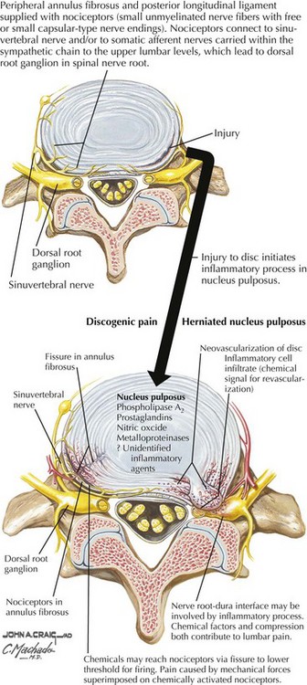
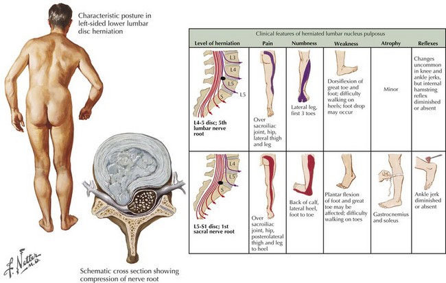
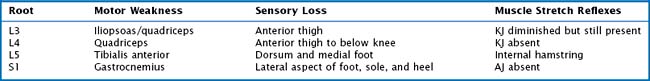
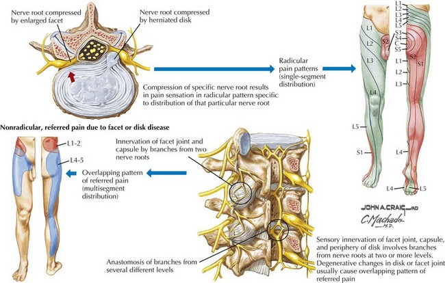
Sciatic pain may occur acutely or evolve more gradually; when the onset is sudden, it may be spontaneous or related to a specific incident, sometimes a seemingly trivial event, such as bending over to make a bed. The symptoms may be minor and clinically inconsequential or significant requiring urgent evaluation and treatment (Fig. 62-1). Depending on the specific nerve root involved, the pain may be classic sciatica with radiation down the posterior aspect of the leg into the foot, as is seen with compression of the L5 or S1 roots (Figs. 62-2 and 62-3). At higher levels, with L3 or L4 root compression, the pain may radiate to the anterior thigh. The clinical signs of lumbar radiculopathy are due to the specific level of involvement (Table 62-1), and the most common levels of nerve root irritation are L5 and S1 roots, followed less commonly by L4 and L3 roots. It is rare to have involvement of the higher roots (L1 and L2).

In the adult, the spinal cord ends between L1 and L2; therefore the nerve root compressed by disc herniation depends upon whether the lesion is medial in the spinal canal or lateral in the neural foramen. The exiting root passes around the pedicle cephalad to the disc space. Therefore a lesion occurring at the disc space in the spinal canal compresses the passing root, the root with the next lower number. For example, a medial disc rupture in the spinal canal at L4–5 will compress the L5 root, whereas less commonly the disc rupturing laterally in the neural foramen will compress the L4 root.

Etiology

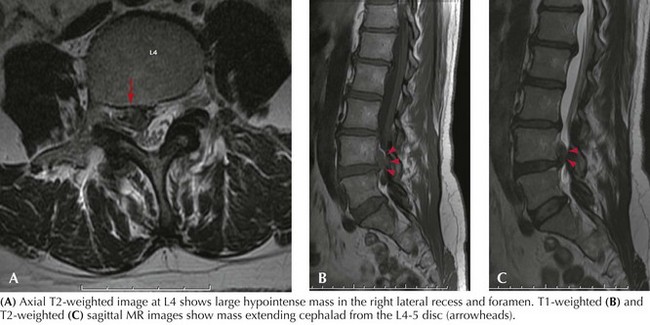
The most frequent cause of lumbar radiculopathy is a herniated lumbar disc, due to herniation of the nucleus pulposus, usually occurring with an equal frequency at the lowest two levels, L4–5 and L5–S1 (Figs. 62-4 and 62-5; see Fig. 62-1). Only ~5% of lumbar disc herniations occur at higher levels. Herniation is the last manifestation of disc degeneration that is an ongoing process in all humans. Hence, disc herniation is uncommon in youth, although occasionally teenagers and rarely toddlers have symptomatic herniations. Disc herniation occasionally occurs with spinal stenosis and may be the cause of rapid deterioration. Most lumbar radiculopathies are unilateral; bilateral sciatica has an ominous significance, suggesting compression of the cauda equina; these patients are at risk for loss of sphincter functions as well as sexual function in males. Early recognition is essential, as even after expeditious decompression, sphincter control and potency may not always return. Rarely spondylosis with foraminal encroachment resulting from disc degeneration may cause radiculopathy.

Differential Diagnosis

Spinal stenosis is becoming more prevalent with the increasingly aging population. It rarely occurs before age 60 years, although individuals with achondroplasia or other congenital processes, with narrow spinal canals caused by shortened pedicles, are predisposed to premature spinal stenosis. Spondylosis is the primary pathologic process, characterized by hypertrophy of the ligaments and facet joints (Fig. 62-6). Patients may develop single or multilevel spinal canal compression of the lumbosacral nerve roots; L3–4 and L4–5 interspaces are the most commonly affected levels; it is rare at L5–S1, unless there is subluxation of the vertebral bodies. Characteristically, the patient has a neurogenic claudication pain pattern mimicking arteriosclerotic occlusive (ASO) disease of the legs. Most individuals become symptomatic with standing or ambulating (Fig. 62-6). They are able to walk a set distance and then feel the need to sit; relief is usually rapid with sitting. Characteristically patients are more comfortable flexed at the waist; thus walking uphill may be easier than walking downhill, as spinal hyperextension associated with walking downhill may precipitate symptomatology. Patients may also be more comfortable leaning forward on a walker or grocery cart. Often the patient has a normal neurologic exam; occasionally, with long-standing symptomatic spinal stenosis, there may be neurologic deficits.

The primary issue is often the differentiation of spinal stenosis from vascular claudication. The demographic population for both conditions is similar. Individuals with spinal stenosis tend to have pain of a more dysesthetic burning character in contrast to the squeezing tight discomfort typical for ASO. Another useful differentiating point on history is that those with spinal stenosis can ride a bicycle long distances, whereas arteriosclerotic patients are limited as if they are walking. Unlike patients with disc herniations, patients with spinal stenosis are typically comfortable at rest, showing no signs of paraspinal spasm, difficulty with straight leg raising testing, or problems bending forward. Plain radiographs provide a good inexpensive means to recognize severe spondylotic changes. MRI is the diagnostic modality of choice. Computed tomographic (CT) myelography is helpful if the patient cannot tolerate MRI. For individuals who have relatively modest symptomatology, there is no urgency to proceed with surgery. However, once the patient is limited in walking, or is uncomfortable even when seated, a wide decompressive laminectomy and foraminotomy at appropriate spinal levels brings significant relief for a high percentage of patients. For patients whose stenosis may be associated with spondylolisthesis, spinal fusion is indicated.

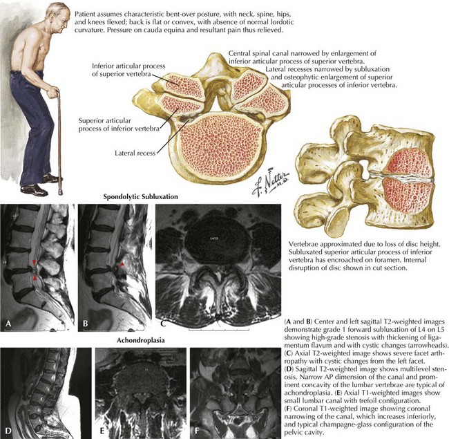
Spondylolisthesis, the anterior slippage of the superior vertebral body with respect to the inferior is another common cause of lumbar root compression, resulting in low back pain (Figs. 62-6A and B), radiculopathy symptoms, and sometimes cauda equina syndrome. The two common causes of spondylolisthesis involve spinal degenerative (spondylotic) changes and congenital defects of the vertebral pars interarticularis. Patients with degenerative spondylolisthesis tend to be older, whereas those with a pars defect usually present in their third or fourth decade with significant lumbar and root pain, usually related to postural change.

Although relatively uncommon, synovial cysts may produce symptoms identical to disc herniation. The cysts develop from hypertrophy of synovial tissue in the facet joint. Neurosurgeons may encounter these cysts pushing into the paraspinal muscles when reflecting them for exposure of the spine. In this location, they indicate degenerative joint disease, but by themselves are not symptomatic. When cysts become intraspinal they may compress the nerve root; synovial cysts create a surrounding inflammatory reaction and therefore must be carefully dissected from the dura of the nerve roots. Resection of lumbar epidural synovial cysts usually relieves patients’ pain.

Epidural infections occur secondary to disc surgery or via hematogenous spread. Unlike metastatic tumors that primarily involve the vertebral bodies, abscess involves the disc space, with secondary spread to the adjacent vertebral bodies. Back pain from disc space and vertebral body involvement and secondary nerve root pain are usually very severe. Common causative organisms in the United States are coagulase-positive and -negative Staphylococcus from surgical or hematogenous spread. Gram-negative organisms from urinary sepsis may also be causative agents. Intravenous drug users and immunocompromised patients have higher incidences of epidural abscess. Worldwide, the most common cause of spinal infection is tuberculosis. The incidence of spinal TB appears to be increasing with the increasing incidence of HIV positivity in susceptible populations.

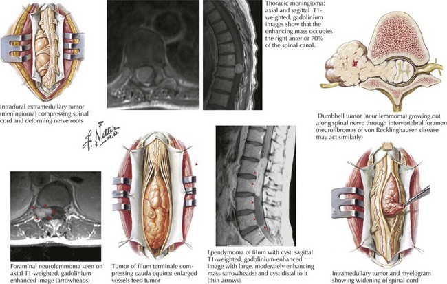
Neoplasms may be a cause of lumbosacral pain. Metastatic extradural cancers to the spine are the most common tumors. Primary bone tumors and intradural primary and metastatic tumors may also mimic discogenic disorders. The common cancers that metastasize include prostate, breast, lung, melanoma, and myeloma. Usually symptoms begin with spine pain that worsens gradually; root pain starts once neural elements become involved and may worsen rapidly. Evaluation and treatment in this situation is urgent, as recovery after treatment may not be complete. Schwannoma, meningioma, myxopapillary ependymoma, and lipoma are the common lumbar spinal intradural tumors (Fig. 62-7). The symptoms of schwannoma, meningioma, and ependymoma are gradually progressive. Patients with a lipoma, a congenital tumor, may have a history of baseline neurologic deficits with a slow, later progression.

Diagnostic Approach

The evaluation of patients having their initial bout of acute sciatica or low back pain does not routinely require any diagnostic testing. Most individuals recover spontaneously. However, for those who do not fit the classic pattern of nerve root compression or acute low back strain, and for patients who do not improve, neurodiagnostic testing is necessary.

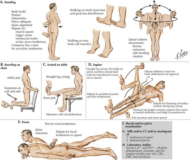
Lumbar spine plain radiographs, including lateral flexion and extension views, serve two purposes: the anatomy of the spine with its degenerative changes is demonstrated, as are subluxations and instability and destructive lesions in the vertebral bodies, and disc space can be seen. MRI is the primary spinal imaging modality (Figs. 62-5 and 62-6). Good-quality MRI demonstrates disc herniation or spinal stenosis and identifies the rare tumor or infection. However, on occasion, for technical reasons, MR imaging may not be successful; for example, the patient may have moved during imaging; obesity or claustrophobia may be other impediments. Myelogram with CT continues to be a valuable adjunct to the diagnostic repertoire, especially when MRI is contraindicated (e.g., pacemaker) or not tolerated. Myelography with water-soluble contrast, followed by axial CT scanning, can demonstrate nerve root filling or lack thereof with more clarity than MRI. Sagittal and coronal reconstructions of CT data give excellent additional information. Electrodiagnostic studies are invaluable in those situations where data from imaging are difficult to interpret or where other superimposed conditions, for example polyneuropathy, coexist.

Treatment

Treatment of lumbar radiculopathy is usually successful if the history, physical exam, and imaging correlate. Most acute episodes of back pain or nerve root pain, without significant neurologic deficit, only require judicious rest and simple analgesics. Strict bed rest is not necessary because it may lead to rapid deconditioning; it may also predispose to more serious complications, such as deep venous thrombosis, pulmonary embolism, and rarely fatal paradoxical cerebral emboli. Patients need to be encouraged to get up as much as possible, but to avoid activities that exacerbate their symptoms. When patients have recovered from their acute symptoms, they can begin a judicious exercise program, graduating to a full fitness exercise program. Analgesics and anti-inflammatory medication including occasional use of steroids may help patients. With this approach, 80% of patients improve within 3 months. This is the natural history of discogenic nerve root compression, and care is advised when evaluating therapeutic claims for other treatment modalities such as chiropractic manipulations or acupuncture. For patients with more chronic, nondisabling pain, lifestyle changes with weight loss and health club membership are the best approach, although unfortunately, few patients successfully change their behavior patterns.

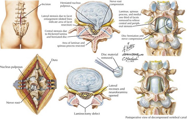
When acute symptoms do not improve, or the chronic degenerative disc-related pain persists, surgery is an option (Fig. 62-8). An important indication for surgery is the presence of a significant persistent neurologic deficit such as foot drop. However, severe or chronic unrelenting nerve root pain that disrupts a patient’s life is a common reason to proceed with nerve root decompression. When advising patients who are making decisions about surgery, they should be clear that postponing surgery would not place them in neurologic jeopardy, but that the discomfort will likely persist. Surgical goals for patients with degenerative disease or disc rupture relate to the pain’s origin. If the patient has root pain with corresponding root compression on imaging, the root or roots should be decompressed, and the herniated disc or synovial cyst removed. Surgery for an extruded disc requires removal of the extruded fragment with freeing of the compressed nerve root. With this technique, >90% of patients obtain symptomatic relief.

If posture-related lumbar pain is the primary symptom, root decompression alone will not resolve the symptoms. Spinal segmental instability, with abnormal spinal motion, can also cause significant pain secondary to intermittent compression of nerve roots. It can also increase degeneration around the facet joints and disc annulus, causing primary back pain. In these uncommon instances, spinal fusion is a reasonable consideration.

The treatment of a patient with a tumor depends on the tumor histology and the extent of neurologic involvement. If the initial presentation of a metastatic tumor is in the spine, needle biopsy of the spinal tumor or biopsy of an obvious tumor demonstrated in the lung, breast, prostate, or skin can provide the diagnosis. Radiotherapy is appropriate when the tumor is radiosensitive, the spine is stable, and there is relatively minimal neural compression. However, if these considerations are not met, surgery is indicated. A major destructive lesion involving most of the vertebral body and both pedicles usually requires a 360-degree decompression and fusion. Neurologic deterioration can be rapid, and once paresis has occurred, the patient may not recover, even after emergency surgery.

Appropriate therapy of an epidural infection is controversial. Some reports demonstrate good results with antibiotic treatment. However, because of the potential for rapid loss of neurologic function, surgical drainage of frank pus is advisable to reduce pain and to prevent paraplegia. Rapid deterioration can occur in patients treated nonoperatively with antibiotics.