27 Lower Extremity
Saphenous Nerve Block
Anatomy of the Saphenous Nerve
The saphenous nerve is the largest cutaneous branch of the femoral nerve. Sensory fibers from L3 and L4 levels contribute to this pure sensory nerve.1,2 The saphenous nerve branches off the posterior division of the femoral nerve and descends anteroinferiorly through the femoral triangle, lateral to the femoral sheath, anterior to the femoral artery and posterior to the aponeurotic covering of the adductor canal (also known as Hunter canal, subsartorial canal).1,2 It then exits from the adductor canal, descends under the sartorius muscle, and then courses around the posterior edge of the sartorius muscle at its tendon portion. In the medial aspect of the knee, the saphenous nerve descends vertically behind the sartorius muscle, pierces the fascia lata and becomes subcutaneous.1,2 At this level, the infrapatellar branch pierces the sartorius muscle and courses inferiorly to the infrapatellar region and innervates the skin in front of the patella. The infrapatellar branch communicates with the anterior cutaneous branches of the femoral nerve above the knee, and with other branches of saphenous nerve below the knee, as well as branches of the lateral femoral cutaneous nerve on the lateral side of the knee, forming the plexus patellae.1
The remaining course of the saphenous nerve then passes down along the medial aspect of the tibia, accompanied by the great saphenous vein and, divides into two branches at the lower third of the leg.1 One of the branches of the descending portion of the saphenous nerve courses along the medial border of the tibia and ends at the ankle, whereas the other branch passes anteriorly to the ankle and is distributed to the medial aspect of the foot, reaching as far as the metatarsophalangeal joint of the great toe and communicating with the medial branch of the superficial peroneal nerve (Fig. 27-1).1 The saphenous nerve supplies sensory innervations to the anterior medial aspect of the leg, including the medial malleolus (Fig. 27-2).1,2
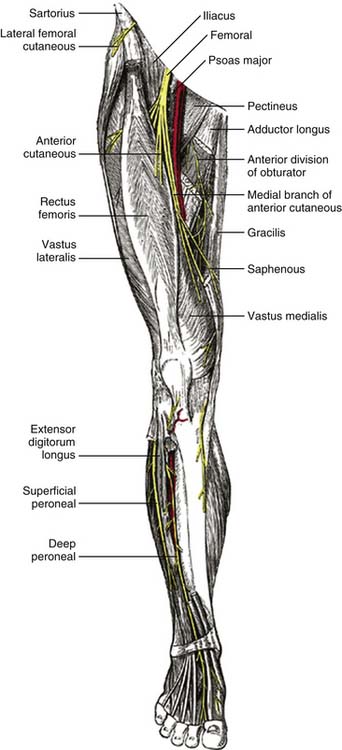
Figure 27-1 Anatomy of saphenous nerve.
(From Williams PL, Warwick R: Gray’s Anatomy, 36th ed. Philadelphia, WB Saunders; 1980.)
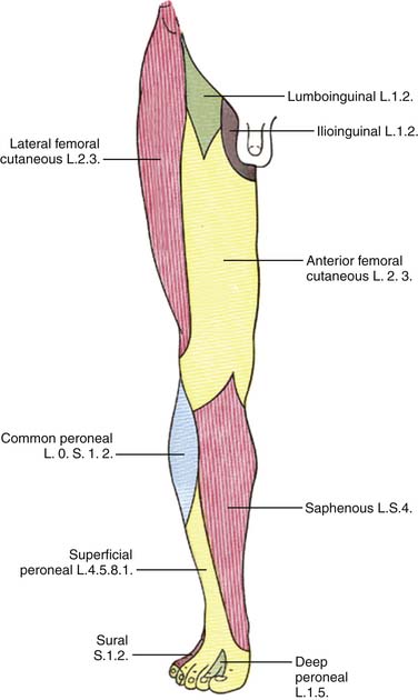
Figure 27-2 Saphenous nerve innervation area.
(From Williams PL, Warwick R: Gray’s Anatomy, 36th ed. Philadelphia, WB Saunders; 1980.)
The adductor canal, the entrapment site for the saphenous nerve, is located approximately 10 centimeters proximal to the medial femoral condyle. It is an aponeurotic tunnel in the middle third of the thigh, extending from the apex of the femoral triangle to the opening in the adductor magnus. It courses between the anterior compartment of the thigh and the medial compartment of the thigh. The vastus medialis muscle lies anteriorly and laterally, whereas the adductor longus lies posteriorly. The content in the canal includes the femoral artery, femoral vein, and branches of the femoral nerve, the saphenous nerve, and the nerve to the vastus medialis muscle). It is covered by a strong aponeurosis that extends from the vastus medialis, across the femoral vessels to the adductor longus and magnus. The sartorius muscle lies on the aponeurosis.2
The adductor canal can be located by palpating along the anteromedial aspect of the vastus medialis muscle and then sliding posteriorly until the edge of the sartorius muscle is felt. The adductor canal is located directly beneath this point.2,3
Pathophysiology of Saphenous Nerve Entrapment and Symptoms
The pathophysiology of saphenous nerve neuropathy involves two different mechanisms: acute compression with high pressure and chronic intermittent compression by contraction and relaxation of the fibrous tissue that impinges the nerve. Demyelination of the involved nerve seems to be a common pathologic finding.3,4
Saphenous nerve entrapment typically manifests as burning pain after prolonged walking or standing in the medial knee and leg. Pain may also be present at rest. Stair climbing may aggravate pain. Sensory symptoms typically involve paresthesia, hypoesthesia, with variations due to the length of nerve compression at the entrapment site. Hyperalgesia, allodynia, and numbness may also be present in the distribution of the saphenous nerve innervation area. The pain associated with this neuropathy may be referred to the lower medial aspect of the knee, or it may radiate down the medial aspect of the lower extremity to the ankle and foot.3,4 There is no motor compromise because the saphenous nerve is a purely sensory nerve. This is important in differentiating this syndrome from radiculopathy in the L4 dermatome distribution.
Clinical criteria for the diagnosis of saphenous nerve entrapment neuropathy include pain in the distribution of the saphenous nerve, normal motor function, and tenderness to palpation over the entrapment site. Entrapment site tenderness is a key feature of saphenous nerve neuropathy. Vigorous palpation at the exit point for the saphenous nerve may result in local pain and referred pain in the nerve’s distribution.3,4
Electrodiagnostic studies are a valuable tool in diagnosing saphenous nerve entrapment. Changes in latency and amplitude of the sensory nerve action potential (SNAP) can be seen. However, in some cases SNAP of the saphenous nerve may be difficult to record, even in the unaffected leg. In this event, cortical sensory evoked potential recording may be used to help diagnose this condition.5
Indications for Saphenous Nerve Block
Contraindications for saphenous nerve block of this procedure include overlying infection, severe bleeding disorder or coagulopathy and preexisting neurologic damage in the affected area.
Technique
Block of saphenous nerve for entrapment neuropathy is often performed at the adductor canal because this is often the site of entrapment. A transsartorial approach is most frequently used.6 The patient lies in the supine position, the sartorius muscle is palpated just above the knee with the leg extended and actively elevated. A skin wheal is raised over the sartorius muscle belly. The needle is inserted at 1-finger width above the patella slightly posterior to the coronal plane and slightly caudal, through the muscle belly of the sartorius until a loss of resistance identifies the subsartorial adipose tissue. The depth of insertion is typically between 1.5 and 3 cm.7 After negative aspiration for blood, 5 mL of local anesthetics (lidocaine or bupivacaine) and 20 to 40 mg of triamcinolone is injected.
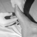
The saphenous nerve can also be located by eliciting paresthesia in the distribution of the nerve.8 In this approach, the patient is placed in the lateral position with the leg slightly flexed. The medial condyle of the femur is palpated. A point just in front of the posterior edge of the medial condyle is then identified and prepped. A ½ inch, 25-gauge needle in then advanced through this point toward the medial condyle of the femur until a paresthesia in the distribution of the saphenous nerve is elicited (Figs. 27-3 and 27-4). When a paresthesia in the distribution of the saphenous nerve is elicited, the needle is withdrawn 1 mm and the patient is observed to be sure that there is no persistent paresthesia. The mix of local anesthetics and corticosteroid can then be injected.8
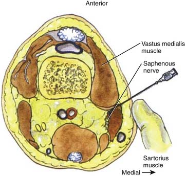
Figure 27-3 Saphenous nerve block at knee at the subsartorial space.
(From Brown DL. Atlas of Regional Anesthesia, 2nd ed. Philadelphia: WB Saunders; 1999.)
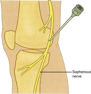
Figure 27-4 Saphenous nerve block at the femoral medial condyle.
(From Waldman S. Atlas of Interventional Pain Management. 2nd ed. Philadelphia: WB Saunders; 2003.)
Ultrasound-guided saphenous nerve block technique has been adopted in recent years.9–11 In this approach, the patient is placed supine with the leg slightly externally rotated. Using a linear transducer with the appropriate frequency range (10 to 12 MHz), the distal one third of the thigh is scanned distally toward the knee. In the distal thigh, the saphenous nerve is predominantly hyperechoic and located posterior to the sartorius muscle in the subsartorial compartment (Fig. 27-5). The vastus medialis muscle lies medially to the sartorius muscle. The transducer is placed over the medial aspect of the right distal thigh transverse to the course of the saphenous nerve. A 5 to 8 cm 22-gauge needle is inserted and advanced in parallel to the transducer and the ultrasound beam in the lateral to medial direction until it reaches the fascial plane between the sartorius and vastus medialis muscles. After negative aspiration, the mix of local anesthetics and corticosteroid can then be injected.9–11

Complications
Saphenous nerve block is a relatively simple procedure with few complications. Incidence of hematoma from vascular injury can be minimized by avoiding multiple needle insertions and avoiding insertion of the needle through superficial veins. Nerve injury, can be minimized by avoiding injection if high pressures are felt on injection or if patient reports paresthesia or pain in the distribution of the nerve on injection, suggesting intraneural placement of the needle tip.
1. Williams P.L., Warwick R. Gray’s Anatomy, 36th ed. Philadelphia: WB Saunders; 1980.
2. Sauerland E.K., Patrick W., Tank P.W. Grant’s Dissector. Hagerstown, Md: Lippincott Williams & Wilkins; 2005.
3. Romanoff M.E., Cory P.C., Kalenak A., et al. Saphenous nerve entrapment at the adductor canal. Am J Sports Med. 1989;17:478-481.
4. Worth R.M., Kettelkamp D.B., Defalque R.J., Duane K.U. Saphenous nerve entrapment—A cause of medial knee pain. Am J Sports Med. 1984;12:80-81.
5. Tranier S., Durey A., Chevallier B., Liot F. Value of somatosensory evoked potentials in saphenous entrapment neuropathy. J Neurol Neurosurg Psychiatry. 1992;55:461-465.
6. van der Wal M., Lang S.A., Yip R.W. Transsartorial approach for saphenous nerve block. Can J Anaesth. 1993;40(6):542-546.
7. Brown D.L. Atlas of regional anesthesia, 2nd ed. Philadelphia: WB Saunders; 1999.
8. Waldman S. Atlas of Interventional Pain Management, 2nd ed. Philadelphia: WB Saunders; 2003.
9. Lundblad M., Kapral S., Marhofer P., Lönnqvist P.A. Ultrasound-guided infrapatellar nerve block in human volunteers: Description of a novel technique. Br J Anaesth. 2006;97:710-714.
10. Krombach J., Gray A.T. Sonography for saphenous nerve block near the adductor canal. Reg Anesth Pain Med. 2007;32:369-370.
11. Marhofer P. Ultrasound Guidance for Nerve Blocks: Principle and Practical Implementation. Oxford University Press; 2008.