Published on 19/03/2015 by admin
Filed under Dermatology
Last modified 22/04/2025
This article have been viewed 2826 times
Neil J. Korman
Evidence Levels: A Double-blind study B Clinical trial ≥ 20 subjects C Clinical trial < 20 subjects D Series ≥ 5 subjects E Anecdotal case reports
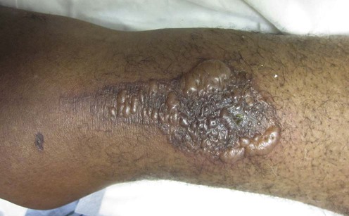
Linear IgA bullous dermatosis is an acquired autoimmune blistering disease of the skin and mucous membranes. The skin lesions consist of papulovesicles or blisters that may have an arcuate pattern, with a ‘cluster of jewels’ grouping of blisters along with urticarial plaques. Involvement of the oral mucous membranes is common and ocular involvement, with subsequent scarring of the conjunctiva, may uncommonly occur. Although originally believed to be a distinct entity, it is now clear that chronic bullous disease of childhood is the childhood counterpart of adult linear IgA bullous dermatosis. Direct immunofluorescence studies demonstrate that all patients have linear IgA deposits at the epidermal basement membrane zone, and the diagnosis of linear IgA bullous dermatosis is dependent upon this finding. The target antigens involved are 97 kDa and, less commonly, 290 kDa. The 97 kDa antigen is an anchoring filament protein that is part of the 180 kDa bullous pemphigoid antigen-2, and antibodies directed against the 290 kDa protein represent an IgA response directed against type VII collagen. Several reports stress the association with ulcerative colitis. Drug-induced disease is a well-recognized entity, and vancomycin is the most commonly implicated agent.
If drug-induced disease is considered, the suspect trigger drug must be withdrawn. Treatment of linear IgA bullous dermatosis is dictated by the severity of disease and the areas of involvement. All patients should be evaluated by an ophthalmologist to ensure the absence of ocular disease. Because linear IgA bullous dermatosis tends to be chronic it is important to be aware of the potential not only for short-term, but also for long-term, toxicities in any treatment used. In addition, treatment of children with chronic bullous disease of childhood (the childhood counterpart of linear IgA bullous dermatosis of adults) requires special consideration to ensure that any medications used have no specific contraindications in children.
The majority of patients with disease limited to the skin will respond well to treatment with dapsone, and this is the first-line therapy for patients with linear IgA bullous dermatosis. Dapsone generally works quite rapidly, with responses often occurring in the first few days of starting the drug. It is most effective for the skin lesions of linear IgA bullous dermatosis, with the mucous membrane lesions being more resistant.
Because of a dose-related oxidant stress on normal aging red blood cells, all patients treated with dapsone will experience some degree of hemolysis that is usually dose-dependent. A reduction of approximately 2–3 g of hemoglobin is often observed. As long as this decrease is relatively gradual and patients have no history of cardiovascular disease or anemia, this is usually well tolerated. It is important to measure levels of glucose-6-phosphate dehydrogenase (G6PD) in patients to be treated with dapsone because those with a deficiency in this enzyme can develop severe hemolysis. Methemoglobinemia, which is also dosage dependent, occurs in most patients but is usually asymptomatic. More worrisome toxicities include bone marrow suppression and even agranulocytosis, which usually occurs early in the course of therapy, and a dapsone-induced neuropathy, which occurs more commonly in patients treated for several years with more than 200 mg of dapsone daily. Less commonly, hepatitis, nephritis, pneumonitis, erythema multiforme, and the dapsone hypersensitivity syndrome have all been reported.
For those patients who fail to achieve satisfactory control of their disease with dapsone as first-line therapy, it is often of value to add systemic corticosteroids. This combination is considered second-line therapy. The dosage of prednisone required is often in the 20–40 mg daily range. Often the addition of prednisone will not only cause significant clinical improvement, but it may also allow the dosage of dapsone to be reduced, thereby minimizing its potential toxicity.
Other viable second-line therapies include colchicine, sulfapyridine, and the combination of tetracycline and niacinamide: sulfapyridine at doses of approximately 1–3 g daily, and colchicine has been reported to be beneficial at doses of 1.0–1.5 mg daily. The combination of tetracycline and niacinamide, usually at doses of 1.5 g of niacinamide and 2 g of tetracycline, has been used with success. Tetracycline should not be used in children under 9 years of age because it can permanently stain teeth.
Third-line therapies include sulfamethoxypyridazine, dicloxacillin, erythromycin, mycophenolate mofetil, azathioprine, cyclosporine, methotrexate, interferon-α, and intravenous immunoglobulin (IVIG). Toxicity profiles and financial considerations favor using erythromycin, dicloxacillin, or sulfamethoxypyridazine prior to treatment with the immunosuppressive agents or IVIG.
Skin biopsy of blister for histology
Perilesional skin biopsy for direct immunofluorescence
Indirect immunofluorescence
Consider G6PD level before dapsone use
Consider thiopurine methyl transferase estimation before azathioprine use
Ophthalmology consult
Leonard JN, Haffenden GP, Ring NP, McMinn RM, Sidgwick A, Mowbray JF, et al. Br J Dermatol 1982; 107: 301–16.
Kelly SE, Frith PA, Millard PR, Wojnarowska F, Black MM. Br J Dermatol 1988; 119: 161–70.
Webster GF, Raber I, Penne R, Jacoby RA, Beutner EH. J Am Acad Dermatol 1994; 30: 355–7.
Wojnarowska F, Marsden RA, Bhogal B, Black MM. J Am Acad Dermatol 1988; 19: 792–805.
Fortuna G, Marinkovich MP. Clin Dermatol 2012; 30: 38–50.
The above are excellent reviews of the clinical and immunological features of linear IgA bullous dermatosis.
Baden LA, Apovian C, Imber MJ, Dover JS. Arch Dermatol 1988; 124: 1186–8.
Litt’s Drug Eruption Reference Manual, 10th edn (Taylor and Francis, New York, 2004) implicates acetaminophen, aldesleukin, amiodarone, ampicillin, atorvastatin, candesartan, captopril, carbamazepine, cefamandole, ceftriaxone, co-trimoxazole, cyclosporine, diclofenac, furosemide, glyburide, granulocyte–macrophage colony-stimulating factor (GM–CSF), ibuprofen, interferon-α, lithium, metronidazole, naproxen, penicillins, phenytoin, piroxicam, rifampin, sulfamethoxazole, and vancomycin as drug triggers for linear IgA disease. The extent of disease may sometimes simulate toxic epidermal necrolysis.
The diagnosis of linear IgA bullous dermatosis requires routine histologic studies as well as direct and indirect immunofluorescence studies. Once the diagnosis is confirmed, laboratory studies to be obtained will depend upon the specific treatment anticipated.
Wojnarowska F. J Roy Soc Med 1980; 73: 371–3.
One of the first reports of this condition and response to dapsone. Since then, most series have demonstrated dapsone to be the most effective first-line monotherapy.
Banodkar DD, Al-Suwaid AR. Int J Dermatol 1997; 36: 213–16.
Eight patients were given colchicine, five of whom showed complete remission within 4 to 6 weeks. The remaining three also responded but required concurrent steroids to maintain remission.
Chaffins ML, Collison D, Fivenson DP. J Am Acad Dermatol 1993; 28: 998–1001.
Yomoda M, Komani A, Hashimoto T. Br J Dermatol 1999; 141: 608–9.
There are four reports of successful use of 2 g of tetracycline and 1.5 g of nicotinamide daily in adults only.
McFadden JP, Leonard JN, Powles AV, Rutman AJ, Fry L. Br J Dermatol 1989; 121: 759–62.
Reports of sulfamethoxypyridazine (0.25–1.5 g daily) use as monotherapy in four patients with linear IgA disease who were intolerant of dapsone.
Skinner RB, Totondo CK, Schneider MA, Raby L, Rosenberg EW. Pediatr Dermatol 1995; 12: 65–6.
Siegfried EC, Sirawan S. J Am Acad Dermatol 1998; 39: 797–800.
Powell J, Kirtschig G, Allen J, Dean D, Wojnarowska F. Br J Dermatol 2001; 144: 769–74.
Peterson JD, Chan LS. Clin Exp Dermatol 2007; 32: 756–8.
Cooper SM, Powell J, Wojnarowska F. Clin Exp Dermatol 2002; 27: 677–9.
Alajlan A, Al-Khawajah M, Al-Sheikh O, Al-Saif F, Al-Rasheed S, Al-Hoqail I, et al. J Am Acad Dermatol 2006; 54: 652–6.
Antibiotics have been used largely in childhood disease and are a reasonable option because of their low toxicity.
Farley-Li J, Mancini AJ. Arch Dermatol 2003; 139: 1121–4.
Lewis MA, Yaqoob NA, Emanuel C, Potts AJ. Oral Surg Oral Med Oral Pathol Oral Radiol Endod 2007; 103: 483–6.
The adult dose is usually 35–45 mg/kg, which is about 1.5 g twice daily for a 75 kg adult.
Burrows NP, Jones RR. J Dermatol Treat 1992; 3: 31–3.
Young HS, Coulson IH. Br J Dermatol 2000; 143: 204–5.
Therapy-resistant blistering responding to cyclosporine 4 mg/kg daily.
Chan LS, Cooper KD. Lancet 1992; 340: 425.
Kroiss MM, Vogtt T, Landthaler M, Stolz W. Br J Dermatol 2000; 142: 582–4.
Goebeler M, Seitz C, Rose C, Sitaru C, Jeschke R, Marx A, et al. Br J Dermatol 2003; 149: 912–4.
Gluth MB, Witman PM, Thompson DM. Int J Pediatr Otorhinolaryngol 2004; 68: 965–70.
A newborn with skin involvement had life-threatening respiratory compromise from disease affecting the larynx, subglottis, trachea, and esophagus. Management with both tracheostomy and gastrostomy tube placement was necessary. Treatment included systemic corticosteroids, dapsone, and IVIG.
Segura S, Iranzo P, Martínez-de Pablo I, Mascaró JM Jr, Alsina M, Herrero J, et al. J Am Acad Dermatol 2007; 56: 960–7.
As the use of intravenous immunoglobulin (IVIG) therapy for autoimmune blistering diseases has evolved, it has become clear that to obtain optimal results patients should receive treatment with high-dose IVIG, defined as 2 g/kg per cycle, typically given at 4-week intervals.
Madnani NA, Khan KJ. Indian J Dermatol Venereol Leprol 2010; 76: 427–9.
An 8-year-old boy demonstrated a dramatic response with thalidomide with total clearance in 1 month, and while on treatment he was disease-free for 1 year. The authors postulate that thalidomide works by inhibition of interleukin-12 which is a potent pro-inflammatory cytokine. Development of neuropathy is a concern with long-term use.
Kasperkiewicz M, Meier M, Zillikens D, Schmidt E. Dermatology 2010; 220: 259–63.
Immunoadsorption has been previously successfully applied in severe and/or otherwise treatment-resistant IgG-mediated immunobullous disorders.
Treatment of Skin Disease Comprehensive Therapeutic Strategies 4e
WhatsApp us


 Dapsone
 Dapsone and prednisone
 Sulfapyridine
 Colchicine
 Tetracycline and niacinamide
 Sulfamethoxypyridazine
 Dicloxacillin
 Flucloxacillin
 Erythromycin
 Trimethoprim–sulfamethoxazole
 Methotrexate
 Interferon-α
 Mycophenolate mofetil
 Azathioprine
 Cyclosporine
 IVIG
IVIG Thalidomide
 Immunoabsorption
