Published on 19/03/2015 by admin
Filed under Dermatology
Last modified 22/04/2025
This article have been viewed 4889 times
Anwar Al Hammadi and Eric Berkowitz
Evidence Levels: A Double-blind study B Clinical trial ≥ 20 subjects C Clinical trial < 20 subjects D Series ≥ 5 subjects E Anecdotal case reports
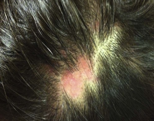
Lichen planopilaris (LPP), also known as follicular lichen planus, is a clinical syndrome consisting of lichen planus (LP) associated with cicatricial scalp alopecia. The condition is more common in women, and presents with perifollicular erythema and keratotic plugs at the margins of the expanding alopecia. The follicular involvement is limited to the infundibulum and the isthmus, both demonstrating lichenoid inflammation. The main complications of follicular lichen planus are atrophy and scarring, with permanent hair loss. Three forms of LPP are recognized, including classic LPP; Graham–Little syndrome, characterized by the triad of multifocal scalp cicatricial alopecia, non-scarring alopecia of the axilla and/or groin, and keratotic follicular papules; and frontal fibrosing alopecia that affects mainly postmenopausal women and appears as cicatricial alopecia of the frontoparietal hairline and is associated with non-scarring alopecia of the eyebrows.
Therapeutic management for LPP is challenging. However, if the associated inflammation can be controlled in its early stages, follicular units may be preserved and hair regrowth may be possible. A good therapeutic response would include a reduction in associated symptoms along with stabilization of the disease and some regrowth of hair in the active perimeter of the alopecic patch. For the most part, therapeutic reports are anecdotal. Oral antihistamines may be used to control pruritus, and high-potency topical corticosteroids are used to control the inflammation in early lesions. Intralesional injections of 3–5 mg/mL of triamcinolone acetonide are effective in well-developed lesions. Hydroxychloroquine may be efficacious. Retinoids have demonstrated some effect in the treatment of LP and therefore provide a possible alternative to corticosteroid treatment. Other agents that have been reported to be of use are cyclosporine and mycophenolate mofetil. There is some rationale for trying biologic agents such as tumor necrosis factor (TNF)-blocking agents for this condition.
Skin biopsy
Immunofluorescence studies
Dermoscopy
Tandon YK, Somani N, Bergfeld WF. J Am Acad Dermatol 2008; 59: 91–8.
Characteristic features of LPP include: the absence of arrector pili muscles and sebaceous glands; a perivascular and perifollicular lymphocytic infiltrate; mucinous perifollicular fibroplasia with absence of interfollicular mucin; and superficial perifollicular wedge-shaped scarring.
Ioannides D, Bystryn JC. Arch Dermatol 1992; 128: 214–16.
Direct immunofluorescence studies were performed on biopsied lesions of patients with LPP and LP. All of the LPP studies demonstrated abnormal linear deposits of immunoglobulin, consisting of IgG or IgA restricted to the basement membrane. The biopsies from those with LP demonstrated fibrillar deposits.
These different appearances suggest different disease processes for LPP and LP.
Micali G, Lacarrubba F, Massimino D, Schwartz R. J Am Acad Dermatol 2011; 64: 1135–46.
Video dermatoscopy is performed with a video-camera equipped with lenses providing magnification ranging from ×10 to ×1000, and shows the reduction to total absence of orifices, hyperkeratotic perifollicular scales, and erythema. In addition, perifollicular arborizing vessels, pigmented networks, and white pale or blue-gray dots in dark-skinned individuals, corresponding to focal decrease in melanin content, can also be observed
Chieregato C, Zini A, Barba A, Magannini M, Rosina P. Int J Dermatol 2003; 42: 342–5.
This clinical trial reports good therapeutic benefit from early treatment with high-potency topical steroids. Only four patients did not respond to the therapy and needed other treatments.
Newton RC, Hebert AA, Freese TW, Solomon AR. Dermatol Clin 1987; 5: 603–18.
High-potency topical corticosteroids may be used to control inflammation in early scalp lesions. Intralesional injections with triamcinolone acetonide at concentrations of 3–5 mg/mL are more effective in well-developed lesions. Additionally, oral corticosteroids have been used in short tapering dosages to control severe disease.
Kossard S, Lee MS, Wilkinson B. J Am Acad Dermatol 1997; 36: 59–66.
Oral corticosteroids and antimalarials were found to slow the course of LPP.
Mehregan DA, Van Hale HM, Muller SA. J Am Acad Dermatol 1992; 27: 935–42.
This large study of 45 patients involved multiple different therapeutic modalities. High-potency topical steroids and oral steroids (30–40 mg daily) for 3 months demonstrated the highest success rates.
Mahrle G, Meyer-Hamme S, Ippen H. Arch Dermatol 1982; 118: 97–100.
This paper reports the comparative results of the effects of the aromatic retinoid etretinate on various skin disorders.
Retinoids have been found to have a significant impact in cases of LP and have therefore been tried with some success in cases of LPP. Isotretinoin may be preferred over acitretin, as the latter agent has been associated with hair loss, but long-term therapy with low-dose acitretin has been studied in psoriasis and is well tolerated.
Cevasco NC, Bergfeld WF, Remzi BK, de Knott HR. J Am Acad Dermatol 2007; 57: 47–53.
In this retrospective study of 29 patients, researchers found that the most commonly prescribed treatments for the reduction of primary symptoms associated with LPP were ketoconazole shampoo (86%), topical steroids (83%), multivitamins with minerals (76%), intralesional steroids (69%), topical minoxidil (41%), and tetracycline (38%). The most common treatment combination was topical steroid, ketoconazole shampoo, tetracycline, and multivitamins with minerals (14%).
Although patient response to any of these therapies was minimal, tetracycline (1 g/day) was the only treatment associated with a significant number of positive responses (six of 11 patients).
Chiang C, Sah D, Cho BK, Ochoa BE, Price VH. J Am Acad Dermatol 2010; 62: 387–92.
This retrospective study of 40 adult patients showed significant efficacy of hydroxychloroquine in the treatment of LPP. After 6 months, 69% of patients (26 of 38) were full or partial responders. After 12 months, 83% of patients (33 of 40) were full or partial responders. Of the nine responders at 12 months, six were able to discontinue hydroxychloroquine and had a sustained remission with no systemic medication for at least 1 year.
Cho BK, Sah D, Chwalek J, Roseborough I, Ochoa B, Chiang C, et al. J Am Acad Dermatol 2010; 62: 393–7.
In this retrospective study of 16 adult patients mycophenolate mofetil was effective at reducing the signs and symptoms of active LPP in 83% of patients (10 of 12) who had failed multiple prior treatments after at least 6 months of treatment. Five of 12 patients were complete responders, five of 12 patients were partial responders, and two of 12 patients were treatment failures. Four patients withdrew from the trial because of adverse events.
Mirmirani P, Willey A, Price V. J Am Acad Dermatol 2003; 49: 667–71.
The authors present three patients with recalcitrant LPP, unresponsive to hydroxychloroquine, high-potency topical steroids, and intralesional steroids, who were treated with cyclosporine. The duration of treatment ranged from 3 to 5 months, with resolution of symptoms, no progression of hair loss, and no evidence of disease activity. One of the patients had a mild recurrence, which resolved with topical 0.1% tacrolimus in Cetaphil lotion.
Boyd AS, King LE Jr. J Am Acad Dermatol 2002; 47: 967–8.
The authors describe the case of a patient who was diagnosed with LPP and treated with hydroxychloroquine, azathioprine, isotretinoin, cyclophosphamide, dapsone, cyclosporine, levamisole, chloroquine, acitretin, methotrexate, clofazamine, and enoxaparin, all without any benefit. The patient was then started on thalidomide 50 mg orally twice a day, with resolution of the symptoms. Side effects limited the ability to continue the medication. Initially, the patient only complained of fatigue and constipation; however, at 4 months, mild numbness and tingling of the fingers and toes developed and the patient experienced significant depression, necessitating discontinuation of the medication.
Thalidomide’s mechanism of action in this disorder may involve TNF inhibition, suggesting that medications such as adalimumab, etanercept, or infliximab may be beneficial. There has, however, been a case report of LPP developing in a patient treated with etanercept.
Navarini AA, Kolios AG, Prinz-Vavricka BM, Haug S, Trüeb RM. Arch Dermatol 2011; 147: 1325–6.
In this small study of 13 patients with biopsy-proven active classic LPP or variants, pruritus and pain improved in all patients who had these complaints prior to the treatment. Erythema reduced in five patients and decreased inflammatory lesions in seven patients. An increased growth of hair after 4 to 8 weeks was observed in three patients, with stable remission in two patients.
Mirmirani P, Karnik P. Arch Dermatol 2009; 145: 1363–6.
A 47-year-old man with LPP recalcitrant to therapy responded to pioglitazone at a dose of 15 mg/day.
Because of the theoretical basis for the use of PPAR agonists in LPP, further studies are warranted to determine if this treatment is genuinely efficacious for the disorder.
Treatment of Skin Disease Comprehensive Therapeutic Strategies 4e
WhatsApp us


 High-potency corticosteroids
 Intralesional corticosteroids
 Oral corticosteroids
 Retinoids
 Antimalarials
 Tetracycline
 Mycophenolate mofetil
 Cyclosporine
 Tacrolimus
 Thalidomide
 TNF-blocking drugs
 Low-dose excimer 308-nm laser
 Pioglitazone