Published on 16/03/2015 by admin
Filed under Dermatology
Last modified 22/04/2025
This article have been viewed 1853 times
Suhail M. Hadi and Ahmed S. Hadi
Evidence Levels: A Double-blind study B Clinical trial ≥ 20 subjects C Clinical trial < 20 subjects D Series ≥ 5 subjects E Anecdotal case reports
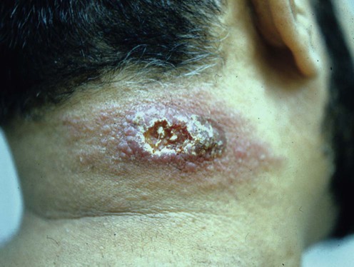
Leishmaniasis is a flagellate protozoan disease caused by many species of the genus Leishmania. It can be classified into three clinical forms: visceral (kala azar), which is the most severe, mucocutanous, which can lead to extensive destruction of the mucous membranes, and cutaneous (Old and New World), which involves mainly exposed body parts, causing ulcers and scarring. The manifestations of cutaneous leishmaniasis are broad and may be similar to other inflammatory and neoplastic diseases. Unusual presentations may be seen: e.g., lupoid, sporotrichoid, paronychia-like, psoriasiform, mycetoma-like, erysipeloid, whitlow-like, discoid lupus erythematosus-like, lupus vulgaris-like, squamous cell carcinoma-like, zosteriform, chronic eczema-like, verrucous, palmoplantar, and scar leishmaniasis. Dermatologists and pathologists should be aware of such pitfalls and may utilize polymerase chain reaction (PCR) to confirm the diagnosis.
Generalized, disseminated, and mucosal leishmaniasis may be seen in patients with HIV infection and AIDS. Subclinical forms of cutaneous leishmaniasis do exist. Leishmaniasis is transmitted mainly by the bite of the infected female phlebotomine sandfly. However, other possible routes of transmission exist including transfusion, congenital, needle sharing, sexual, and person-to-person contact. Only the management of cutaneous leishmaniasis is addressed here.
Bari AU, Rahman SB. Indian J Dermatol Venereol Leprol 2008; 74(1): 23–7.
A total of 718 patients with cutaneous leishmaniasis were analyzed; 5.7% had unusual presentations. The commonest of these presentations was lupoid leishmaniasis.
Saab J, Fedda F, Khattab R, Yahya L, Loya A, Satti M, et al. J Cutan Pathol 2012; 39(2): 251–62.
Skin biopsies from 145 patients were studied; 125 were confirmed as cutaneous leishmaniasis by PCR. Eighteen cases presented with a pre-biopsy clinical diagnosis which ranged from dermatitis to neoplasm. Of the 125 cases, 57 showed a histopathological pattern other than cutaneous leishmaniasis.
The diagnosis of cutaneous leishmaniasis starts with the finding of a typical lesion and a history of exposure. A skin scraping for microscopic analysis is the simplest test. Cultures from an exudate or scraping show good results. Where available, PCR offers a rapid, highly sensitive, and specific modality of diagnosis. Serological tests can be used and include the indirect immunofluorescent antibody test, direct agglutination test, fast agglutination screening test, and enzyme-linked immunosorbent assay (ELISA).
A new vaccine has been recently studied for its protection against cutaneous leishmaniasis. Results showed durable protective immunity. It has the advantages of a non-invasive nasal route of vaccination and reduced cost.
Treatment is needed to improve the cosmetic outcome, as spontaneous healing may leave scarring.
Pentavalent antimonials are the first-line therapy. Meglumine antimoniate is the drug of choice. It can be given intralesionally with local anesthetic (particularly in children, because of pain) or systemically 10 mg/kg daily for 2 weeks. Leishmania recidivans requires higher doses for longer periods. Sodium stibogluconate can be infiltrated into individual lesions 1–2 mL/week. In widespread, severe cases it can be given intramuscularly or intravenously in a dose of 10 mg/kg/day for 2 weeks.
Pentavalent antimoniate use is complicated by its high incidence rate of side effects, including arthralgia, fatigue, gastrointestinal upset, elevation of amylase, lipase and liver enzyme levels, leukopenia, anemia, and ECG abnormalities. Side effects appear to be dose related, and are more common in patients with renal and liver impairment and those with cardiac arrhythmias. Amphotericin B has been used in antimony-resistant cases.
Pentamidine isethionate (aromatic diamidine) is effective for diffuse cutaneous leishmaniasis. Side effects of pentamidine include hypoglycemia, diabetes mellitus, hypotension (if administered too rapidly), nausea, abdominal pain, vomiting, and headache.
Allopurinol has antileishmanial activity, and other oral drugs such as miltefosine, zinc sulfate, rifampin, doxycycline and azoles are also beneficial. γ-Interferon was shown to be effective as monotherapy treatment for leishmaniasis. Topical preparations such as paramomycin ointment and 5% imiquimod also show considerable therapeutic potential.
Good cure rates have been achieved with both thermotherapy and cryotherapy. Lasers have been used, but further studies are needed to support their role in the treatment of leishmaniasis. Additional treatment modalities are presented below.
Skin biopsy
Fine needle aspiration
Slit-skin smear
Culture
Leishmanin (Montenegro) skin test
Serology
PCR for leishmanial DNA
The margin of leishmania lesions contains amastigotes, whereas the center has dead skin and debris. Accordingly, the margin is addressed when performing a slit-skin smear. The aspirate can then be sent for culture, or for histology that shows Leishman–Donovan bodies inside macrophages using Giemsa stain.
In papular and nodular lesions the margin of the lesion is punctured with a hypodermic needle and a syringe containing 0.1 mL saline. The aspirate is drawn up into the needle and is examined microscopically and/or cultured.
The gold standard medium for culture is Novy–MacNeal–Nicolle with positive results in 1 to 3 weeks, or Schneider Drosophila medium, which gives positive results in one week.
Microculture is a new culture medium that has higher carbon dioxide concentrations and lower oxygen and pH, which encourages more rapid amastigote to promastigote differentiation. Culture is not a reliable method in older lesions as the organisms become scarce and difficult to isolate.
Serological tests aim to detect the presence of antibodies against Leishmania. They are particularly valuable in visceral and mucocutaneous leishmaniasis.
PCR is a sensitive and powerful method of diagnosis of cutaneous leishmaniasis, which depends on gene amplification techniques.
Anwar M, Hussain MA, Ur-Rehman H, Khan I, Sheikh RA. East Mediterr Health J 2007; 13: 1212–15.
Fine needle aspiration cytology was used to diagnose 109 cases with non-healing cutaneous ulcers.
Ampuero J, Rios AP, Carranza-Tamayo CO, Romero GA. Mem Inst Oswaldo Cruz 2009; 104: 992–7.
Aspirates from the ulcerated lesions and lymph nodes were taken from 280 patients for in vitro cultures. Lesional biopsies were tested using kinetoplastid DNA (kDNA)-PCR to detect DNA. Positive results of cultures, kDNA-PCR and the combination of the two methods were 78.2%, 89.3%, and 97.1%, respectively. Parasite culture is a feasible method for the detection of cutaneous leishmaniasis (CL). Combination of culture and PCR gave the best result for the diagnosis of CL.
Manzur A, Bari A. Dermatol Online J 2006; 12: 2.
One hundred patients with parasitologically proven cutaneous leishmaniasis were enrolled in the study. The leishmanin test was found to be sensitive even in cutaneous leishmaniasis of recent onset.
Ryan JR, Smithyman AM, Rajasekariah GH, Hochberg L, Stiteler JM, Martin SK. J Clin Microbiol 2002; 40: 3110.
An ELISA that can detect IgM and IgG anti-leishmanial antibodies was developed and tested on 129 visceral and 143 cutaneous leishmaniasis patients with controls. The test showed an overall sensitivity of 95.1%.
Uzun S, Durdu M, Culha G, Allahverdiyev AM, Memisoglu HR. J Parasitol 2004; 90: 853–9.
Intralesional meglumine antimoniate was given as first-line therapy for 890 patients with cutaneous leishmaniasis. A weekly dose of 0.2–1 mL was given for up to 20 weeks or until complete cure. It was found to have an efficacy of 97.2%, with a low relapse rate (3.9%) and no serious side effects.
Munir A, Janjua SA, Hussain I. Acta Dermatovenerol Croat 2008; 16: 60–4.
Sixty patients were studied. A combination of intramuscular meglumine antimoniate 20 mg/kg/day plus intralesional meglumine antimoniate 0.5 mL daily for 21 days was superior to intralesional treatment alone, with 75% complete cure for the former.
Negi AK, Sharma NL, Mahajan VK, Ranjan N, Kanga AK. Indian J Dermatol Venereol Leprol 2007; 73: 280.
Thirty-two patients with cutaneous leishmaniasis were studied. Intralesional SSG (100 mg/mL), plus 20 mg/kg/day given intramuscularly achieved 80–90% response after one session, whereas intralesional SSG alone needed two to four sessions.
Gangneux JP, Sauzet S, Donnard S, Meyer N, Cornillet A, Pratlong F, et al. Emerg Infect Dis 2007; 13: 1436–8.
Twenty-one patients with recurrent cutaneous leishmaniasis were enrolled. Intravenous or intramuscular pentamidine isethionate (4 mg/kg) on alternate days cured the lesions within 1 to 3 months.
Kochar DK, Aseri S, Sharma BV, Bumb RA, Mehta RD, Purohit SK. Q J Med 2000; 93: 733–7.
Forty-six patients were enrolled: 32 received rifampin 600 mg twice daily for 4 weeks and 32 received placebo. Of the rifampin group, 73.9% had complete healing of their lesions, compared to 4.3% of the placebo group. Rifampin is suitable for multiple lesions and is well tolerated.
Alrajhi AA, Ibrahim EA, De Vol EB, Khairat M, Faris RM, Maguire JH. N Engl J Med 2002; 346: 891–5.
Patients (n = 106) received fluconazole 200 mg daily and 103 received placebo for 6 weeks. Healing was complete in 79% of the fluconazole group and 34% of the placebo group at 3 months follow-up.
Saleem K, Rahman A. J Coll Phys Surg Pak 2007; 17: 713–16.
Two hundred patients with wet and dry cutaneous leishmaniasis were studied. Itraconazole (100 mg twice daily for 6 to 8 weeks) was superior to meglumine antimoniate in achieving complete clinical and parasitological cure (75% compared to 65%), with fewer side effects.
El-Sayed M, Anwar AE. J Eur Acad Dermatol Venereol 2010; 24: 335–40.
Ten patients were treated with intralesional SSG alone (group 1). Ten patients were treated with the combination of intralesional SSG + intramuscular SSG (group 2). Ten patients were treated with the combination of intralesional SSG and oral ketoconazole (group 3). Treatment period was 12 weeks. Cure rate was 58.3% in group 1, 93.3% in group 2, and 92.3% in group 3. Combination of oral ketoconazole with intralesional SSG is more effective than intralesional SSG alone.
Mosleh IM, Geith E, Natsheh L, Schönian G, Abotteen N, Kharabsheh S. J Am Acad Dermatol 2008; 58: 617–24.
Patients (n=120) were treated with cryotherapy once weekly over one to seven sessions; 84% of lesions were cured after one to four sessions. The side effects were minimal.
Case AJ, Safi N, Davis GD, Nadir M, Hamid H, Robert LL Jr. Mil Med 2012; 177: 345–51.
Patients (n=382) with cutaneous leishmaniasis were randomly assigned to two treatment groups and followed for 6 months. The cure rate for the thermotherapy group was 82.5%, compared with 74% in the glucantime group. A single localized treatment with thermotherapy was more effective than 5 days of intralesional glucantime. Also, thermotherapy was cost-effective, with fewer side effects, and better patient compliance than intralesional glucantime.
Ranawaka RR, Weerakoon HS. J Dermatolog Treat 2010; 21: 286–93.
SSG was given to 87 patients and intralesional hypertonic saline (HS) was given to 67 patients. SSG showed a 100% cure rate within one to six injections while HS showed a 92.2% cure rate within one to 10 injections. There were no significant side effects except pain due to injection.
Masmoudi A, Dammak A, Chaaben H, Maalej N, Akrout F, Turki H. Dermatol Online J 2008; 14: 22.
Fourteen patients with cutaneous leishmaniasis were given doxycycline 200 mg/day for 15–30 days. Ten patients achieved complete clinical regression of the lesions.
Machado PR, Ampuero J, Guimarães LH, Villasboas L, Rocha AT, et al. PLoS Negl Trop Dis 2010; 21,4: e912.
Ninety patients were enrolled; 60 received miltefosine orally and 30 received pentavalent antimony. Six months later the cure rate was 53.3% in the pentavalent antimony group and 75% in the miltefosine group.
Apoptosis-like death of Leishmania donovani may be a possible explanation of the mode of action of miltefosine.
Arevalo I, Tulliano G, Quispe A, Spaeth G, Matlashewski G, Llanos-Cuentas A, et al. Clin Infect Dis 2007; 44: 1549–54.
Combination treatment with imiquimod and meglumine antimoniate gave superior results, with rapid healing and better cosmetic outcome.
Imiquimod kills the intracellular Leishmania amastigotes in vitro by activating macrophages to release nitric oxide.
Shazad B, Abbaszadeh B, Khamesipour A. Eur J Dermatol 2005; 15: 85–7.
Sixty patients were studied. One group received 1 mL of meglumine antimonate intradermally every other day for 20 days, the other group received an ointment containing 15% paromomycin sulfate in urea twice daily for 20 days. One week after treatment a cure rate of 66% in the meglumine antimonate group and 68% in the paromomycin sulfate treated group was achieved.
Asilian A, Davami M. Clin Exp Dermatol 2006; 31: 634–7.
To compare the parasitological and clinical efficacy of photodynamic therapy (PDT) versus topical paromomycin in 60 patients. Topical PDT was given weekly for 4 weeks. Complete improvement was seen in 93.5% of patients treated with topical PDT, versus 41.2% in the topical paromomycin group.
Hejazi H, Eslami G, Dalimi A. Ann Trop Med Parasitol 2004; 98: 37–42.
Exposure to direct current of 3, 6, 9, and 12 V (at 0.2–10.7 mA) killed all promastigotes (in culture) within 10 to 15 minutes. Three weeks of electrotherapy at 3 V for 10 minutes twice weekly appeared to cure all the lesions in mice.
Asilian A, Sharif A, Faghihi G, Enshaeieh SH, Shariati F, Siadat AH. Int J Dermatol 2004; 43: 736–8.
Patients (n=123) were treated with CO2 laser with a maximum power of 100 W and a pulse width of 0.5–5 seconds, and 110 patients were treated with glucantime (meglumine antimoniate) 50 mg/kg daily for 15 days. The CO2 laser was 1.12 times more effective than glucantime, with shorter healing time (1 month vs 3 months) and fewer side effects (4.5% vs 24%).
Iraji F, Vali A, Asilian A, Shahtalebi MA, Momeni AZ. Dermatology 2004; 209: 46–9.
A total of 104 patients with acute cutaneous leishmaniasis (ACL) were studied. The duration of treatment was 6 weeks. Thirty-five patients received meglumine antimoniate (MA) and 31 received intralesional zinc sulfate (ZS). Cure rates were 60% for MA and 83.8% for ZS. Intralesional injection of 2% ZS is a good alternative for the treatment of ACL.
Sadeghian G, Nilforoushzadeh MA. Int J Dermatol 2006; 45: 819–21.
Sixty-four patients were studied. Systemic glucantime 20 mg/kg/day combined with pentoxifylline 400 mg three times daily for 20 days is more effective than glucantime alone.
Pentoxifylline has an anti-inflammatory effect.
Solomon M, Pavlotsky F, Leshem E, Ephros M, Trau H, Schwartz E. J Eur Acad Dermatol Venereol 2011; 25: 973–7.
Thirteen patients received liposomal amphotericin B (5 consecutive days of 3 mg/kg, followed by a sixth dose on day 10); 85% had facial lesions. Eleven of 13 patients (84%) achieved complete clinical cure within 2 months. There were no reported relapses after 11 months, and the side effects were mild.
Treatment of Skin Disease Comprehensive Therapeutic Strategies 4e
WhatsApp us


 Meglumine antimoniate
 Sodium stibogluconate antimony
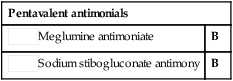
 Pentamidine isethionate
 Rifampin
 Azoles
 Cryotherapy
 Thermotherapy
 Hypertonic sodium chloride
 Doxycycline
 Miltefosine (hexadecylphosphocholine)
 Imiquimod 5%
 Paromomycin ointment
 Photodynamic therapy
 Direct current electrotherapy
 CO2 laser
 Intralesional zinc sulfate
 Pentoxiphylline
 Amphotericin B