[level-membership-for-dermatology-category]
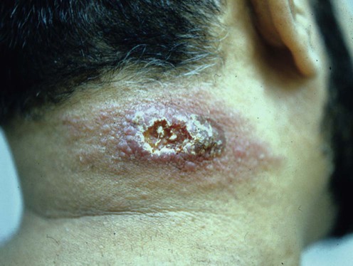
Leishmaniasis
Specific investigations


First-line therapies
 Meglumine antimoniate
 Sodium stibogluconate antimony
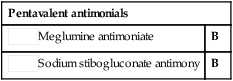
Second-line therapies
 Pentamidine isethionate
 Rifampin
 Azoles
 Cryotherapy
 Thermotherapy
 Hypertonic sodium chloride
 Doxycycline
Third-line therapies
 Miltefosine (hexadecylphosphocholine)
 Imiquimod 5%
 Paromomycin ointment
 Photodynamic therapy
 Direct current electrotherapy
 CO2 laser
 Intralesional zinc sulfate
 Pentoxiphylline
 Amphotericin B
[/level-membership-for-dermatology-category][not-level-membership-for-dermatology-category]
Leishmaniasis
Specific investigations
Genus-specific kinetoplast-DNA PCR and parasite culture for the diagnosis of localized cutaneous leishmaniasis: applications for clinical trials under field conditions in Brazil.
Ampuero J, Rios AP, Carranza-Tamayo CO, Romero GA. Mem Inst Oswaldo Cruz 2009; 104: 992–7.
Buy Membership for Dermatology Category to continue reading. Learn more here
[/not-level-membership-for-dermatology-category]
