[level-membership-for-dermatology-category]
Leiomyoma
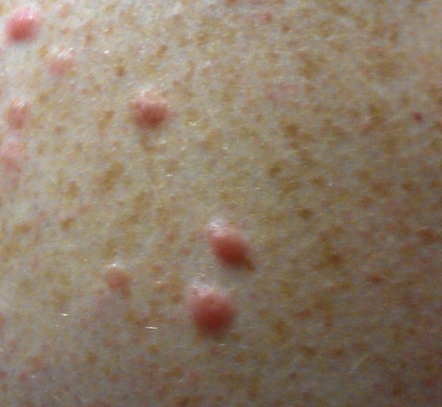
Specific investigations


 Surgical excision
Second-line therapies
 Doxazosin
 Phenoxybenzamine
 Topical hyoscine hydrobromide
 Nifedipine
 Oral glyceryl trinitrate
 Cryotherapy
 Simple analgesics
 Gabapentin
 Duloxetine
 Botulinum toxin A
 CO2 laser
[/level-membership-for-dermatology-category][not-level-membership-for-dermatology-category]
Leiomyoma
Specific investigations
Cutaneous smooth muscle neoplasms: clinical features, histological findings and treatment options.
Holst VA, Junkins-Hopkins JM, Elenitas R. J Am Acad Dermatol 2002; 46: 477–90.
Buy Membership for Dermatology Category to continue reading. Learn more here
[/not-level-membership-for-dermatology-category]
