Legionella

Published on 08/02/2015 by admin

Filed under Basic Science

Last modified 22/04/2025

Print this page

rate 1 star rate 2 star rate 3 star rate 4 star rate 5 star
Your rating: none, Average: 0 (0 votes)

This article have been viewed 1999 times

Legionella

This chapter addresses organisms that will not grow on routine primary plating media and belong to the genus Legionella. Legionella belongs to the family Legionellaceae and includes a single genus, Legionella, comprising approximately 52 species. Legionella pneumophila is the causative agent of Legionnaires’ disease, a febrile and pneumonic illness with numerous clinical presentations. Legionella was discovered in 1976 by scientists at the Centers for Disease Control and Prevention (CDC) who were investigating an epidemic of pneumonia among Pennsylvania State American Legion members attending a convention in Philadelphia. There is retrospective serologic evidence of Legionella infection as far back as 1947. Bacteria resembling Legionella that are capable of living in amoebae have been designated as Legionella-like amoebal pathogens (LLAPs).

General Characteristics

All Legionella spp. are mesophilic (20° to 45° C), obligately aerobic, faintly staining, thin, gram-negative fastidious bacilli that require a medium supplemented with iron and L-cysteine, and buffered to pH 6.9 for optimum growth. The organisms utilize protein for energy generation rather than carbohydrates. The overwhelming majority of Legionella spp. are motile. As of this writing, more than 52 species belong to this genus. Nevertheless, the organism Legionella pneumophila predominates as a human pathogen within the genus and consists of 16 serotypes. In approximately decreasing order of clinical importance are L. pneumophila serotype 1 (about 70% to 90% of the cases of Legionnaires’ disease), L. pneumophila serotype 6, L. micdadei, L. dumoffii, L. anisa, and L. feeleii. Of note, many species of Legionella have only been isolated from the environment or recorded as individual cases. To date, 20 species of Legionella are documented as human pathogens in addition to L. pneumophila. Box 35-1 is an abbreviated list of some of the species of Legionella.

Epidemiology

Legionellae are ubiquitous and widely distributed in the environment. As a result, most individuals are exposed to Legionella spp.; however, few develop symptoms. In nature, legionellae are found primarily in aquatic habitats and thrive at warmer temperatures; these bacteria are capable of surviving extreme ranges of environmental conditions for long periods; studies have shown that L. pneumophila can survive for up to 14 months in water with only a slight loss in viability. Legionella spp. have been isolated from the majority of natural water sources investigated, including lakes, rivers, and marine waters, as well as moist soil. Organisms are also widely distributed in man-made facilities, including air-conditioning ducts and cooling towers; potable water; large, warm-water plumbing systems; humidifiers; whirlpools; and technical-medical equipment in hospitals.

Legionella infections are acquired exclusively from environmental sources; no person-to-person spread has been documented. Inhalation of infectious aerosols (1 to 5 μm in diameter) is considered the primary means of transmission. Exposure to these aerosols can occur in the workplace or in industrial or nosocomial settings; for example, nebulizer’s filled with tap water and showers have been implicated. Infection is acquired through the inhalation of aerosols or microaspiration. Legionnaires’ disease occurs in sporadic, endemic, and epidemic forms. The incidence of disease varies greatly and appears to depend on the geographic area, but it is estimated that Legionella spp. cause less than 1% to 5% of cases of pneumonia.

Pathogenesis and Spectrum of Disease

Legionella spp. can infect and multiply within some species of free-living amoebae (Hartmannella, Acanthamoeba, and Naegleria spp.), as well as within Tetrahymena spp., a ciliated protozoa, or within biofilms (well-organized microcolonies of bacteria usually enclosed in polymer matrices that are separated by water channels that remove wastes and deliver nutrients). This contributes to the organism’s survival in the environment. In addition, L. pneumophila exists in two well-defined, morphologically distinct forms in Hela cells: (1) a highly differentiated, cystlike form that is highly infectious, metabolically dormant, and resistant to antibiotics and detergent-mediated lysis and (2) a replicative intracellular form that is ultrastructurally similar to agar-grown bacteria. The existence of this cystlike form may account for the ability of L. pneumophila to survive for long periods between hosts (amoebae or humans).

Although the exact mechanisms by which L. pneumophila causes disease are not totally delineated, its ability to avoid destruction by the host’s phagocytic cells plays a significant role in the disease process. L. pneumophila is considered a facultative intracellular pathogen. Following infection, organisms are taken up by phagocytosis primarily in alveolar macrophages, where they survive and replicate within a specialized, membrane-bound vacuole by resisting acidification and evading fusion with lysosomes; it is still unknown how Legionella prevent vacuole acidification. Following replication, the organisms will kill the phagocyte releasing them into the lungs and will again be phagocytized by a mononuclear cell, and multiplication of the organism will increase.

The sequestering of legionellae within macrophages also makes it difficult to deliver and accumulate effective antimicrobials. Of significance, studies have shown that although certain antimicrobials can penetrate the macrophage and inhibit bacterial multiplication, L. pneumophila is not killed and, when drugs are removed, the organism resumes replicating. Therefore, a competent cell-mediated immune response is also important for recovery from Legionella infections. Humoral immunity appears to play an insignificant role in the defense against this organism.

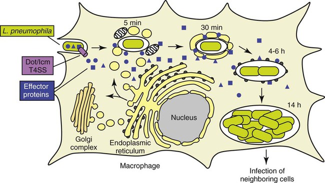
In eukaryotic cells, most proteins secreted or transported inside vesicles to other cellular compartments are synthesized at the endoplasmic reticulum (ER) (Figure 35-1). Many bacterial pathogens use secretion systems as a part of how they cause disease. L. pneumophila possesses genes that are able to “trick” eukaryotic cells into transporting them to the endoplasmic reticulum; these virulence genes are called dot (defective organelle trafficking) or icm (intracellular multiplication). This dot/icm secretion system in L. pneumophila consists of 23 genes and is a type IV secretion system. Bacterial type IV secretion systems are bacterial devices that deliver macromolecules such as proteins across and into cells. After entry but before bacterial replication, L. pneumophila, residing in a membrane-bound vacuole, is surrounded by a ribosome-studded membrane derived from the host cell’s ER and mitochondria. Thus, by exploiting host cell functions, L. pneumophila is able to gain access to the lumen of the ER, which supports its survival and replication where the environment is rich in peptides. A second type II secretion system has also been implicated in the virulence of some strains of Legionella. The type II secretion system carries numerous genes for enzymatic degradation including lipases, proteinases, and a number of novel proteins. Mutations within the type II secretion system results in decreased infectivity of the organism. A number of additional bacterial factors have also been identified as crucial for intracellular infection; some of these are listed in Box 35-2.

image
FIGURE 35-1

(Modified from 2009annualreport.nichd.nih.gov/ump.html.)

Finally, several cellular components and extracellular products of L. pneumophila, such as an extracellular cytotoxin that impairs the ability of phagocytic cells to use oxygen and various enzymes (e.g., phospholipase C), have been purified and proposed as virulence factors. However, their exact role in the pathogenesis of Legionella infections is not completely clear.

Legionella spp. are associated with a spectrum of clinical presentations, ranging from asymptomatic infection to severe, life-threatening diseases. Serologic evidence exists for the presence of asymptomatic disease, because many healthy people surveyed possess antibodies to Legionella spp. Table 35-1 provides a more detailed description of the following three primary clinical manifestations:

TABLE 35-1

Disease Spectrum Associated with Legionella sp.

  Epidemiology Disease
Pneumonia (Legionnaires’ Disease) Community and nosocomial transmission (inhalation of aerosolized particles); immunocompromised patients, particularly in cell-mediated immunity; rarely occurs in children Acute pneumonia indistinguishable from other bacterial pneumonias; clinical syndrome may include nonproductive cough, myalgia, diarrhea, hyponatremia, hypophosphatemia, and elevated liver enzymes
Pontiac Fever Community setting associated with employment (industrial or recreational) or other group Self-limiting, febrile illness; symptoms may include cough, dyspnea, abdominal pain, fever, and myalgia; pneumonia does not occur
Extrapulmonary Rare, metastatic complications from underlying pneumonia; incidents of inoculation into sites via punctures have been identified or therapeutic bathing; highly associated with immunocompromised patients Abscesses have been identified in the brain, spleen, lymph nodes, muscles, surgical wounds, and a variety of tissues and organs

From Mandell GL, Bennett JE, Dolin R: Principles and practices of infectious diseases, ed 7, Philadelphia, 2010, Elsevier.

Individuals at risk for pneumonia are those who are immunocompromised, older than age 60, or heavy smokers. The clinical manifestations following infection with a particular species are primarily caused by differences in the host’s immune response and perhaps by inoculum size; the same Legionella sp. gives rise to different expressions of disease in different individuals.

There are a number of bacteria that grow only within amoebae and are closely related phylogenetically based on 16S rRNA gene sequencing to Legionella species; these organisms are referred to as “Legionella-like amoeba pathogens” (LLAPs). Several LLAPs have been assigned to the Legionella genus. One LLAP has been isolated from the sputum of a patient with pneumonia after the specimen was incubated with the amoeba Acanthamoeba polyphaga. Serologic surveys of patients with community-acquired pneumonia suggest LLAPs may be occasional human pathogens.

Laboratory Diagnosis

Specimen Collection and Transport

Specimens from which Legionella can be isolated include respiratory tract secretions of all types, including expectorated sputum, additional lower respiratory specimens, and pleural fluid; other sterile body fluids, such as blood; and lung, transbronchial, or other biopsy material. Because sputum from patients with Legionnaires’ disease is usually nonpurulent and may appear bloody or watery, the grading system used for screening sputum for routine cultures is not applicable. Patients with Legionnaires’ disease usually have detectable numbers of organisms in their respiratory secretions, even for some time after antibiotic therapy has been initiated. If the disease is present, the initial specimen is often likely to be positive. However, additional specimens should be processed if the first specimen is negative and suspicion of the disease persists. Pleural fluid has not yielded many positive cultures in studies performed in several laboratories, but it may contain organisms. Urine for antigen collection should be collected in a sterile container. The sample should be transported to the laboratory and refrigerated if a delay in processing occurs. Specimens should be transported without holding media, buffers, or saline, which may inhibit the growth of Legionella. The organisms are hardy and are best preserved by maintaining specimens in a small, tightly closed container to prevent desiccation and transporting them to the laboratory within 30 minutes of collection. If a longer delay is anticipated, specimens should be refrigerated. If one cannot ensure that specimens will remain moist, 1 mL of sterile broth may be added.

Specimen Processing

All specimens for Legionella culture should be handled and processed in a class II biologic safety cabinet (BSC). When specimens from nonsterile body sites are submitted for culture, selective media or treatment of the specimen to reduce the numbers of contaminating organisms is proposed. Brief treatment of sputum specimens with hydrochloric acid before culture has been shown to enhance the recovery of legionellae. However, this technique is time consuming and is only recommended on specimens from patients with cystic fibrosis. Respiratory secretions may be held for up to 48 hours at 5° C before culture; if culturing is delayed longer, then the specimen may be frozen.

Tissues are homogenized before smears and cultures are performed, and clear, sterile body fluids are centrifuged for 30 minutes at 4000× g. The sediment is then vortexed and used for culture and smear preparation. Blood for culture of Legionella may be processed with the lysis-centrifugation tube system (Isolator; Wampole Laboratories, Cranbury, New Jersey) and plated directly to buffered charcoal-yeast extract (BCYE) agar. Specimens collected by bronchoalveolar lavage are quite dilute and therefore should be concentrated at least tenfold by centrifugation before culturing.

Direct Detection Methods

Several laboratory methods are used to detect Legionella spp. directly in clinical specimens.

Antigens

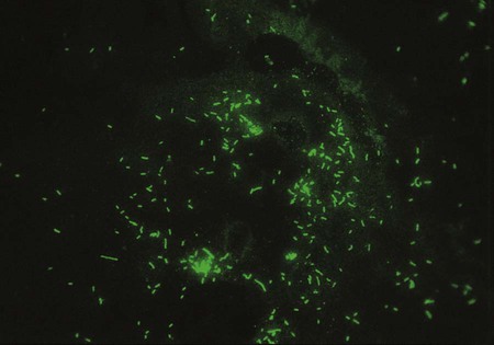
One approach to direct detection of legionellae in clinical specimens is the direct immunofluorescent antibody (DFA) test of respiratory secretions. Polyclonal and monoclonal antisera conjugated with fluorescein are available from several commercial suppliers. Specimens are first tested with pools of antisera containing antibodies to several serotypes of L. pneumophila or several Legionella spp. Those that exhibit positive results are then reexamined with specific conjugated antisera. One reagent made by Genetic Systems Corporation (Seattle, Washington) is a monoclonal antibody directed against a cell wall protein common to L. pneumophila. The manufacturer’s directions should be followed explicitly, and material from commercial systems should never be divided and used separately. Laboratories should decide which serotypes to test for routinely based on the prevalence of isolates in their geographic area. The sensitivity of the DFA test ranges from 25% to 75%, and its specificity is greater than 95%. If positive, organisms appear as brightly fluorescent rods (Figure 35-2). Of importance, cultures always must be performed, because Legionella spp. or serotypes not included in the antisera pool can be recovered. In addition, even in the hands of an experienced microbiologist, false positives may occur. The high complexity of the test and lack of high reproducible sensitivity has reduced the number of laboratories offering DFA testing for Legionella sp.

Rapid detection of Legionella antigen in urine and other body fluids has been accomplished by enzyme immunoassay (EIA) and immunochromatography. Antigen may be present in the prodromal period and by 3 days after the onset of symptoms. Urine should be tested for L. pneumophila, although a drawback of the immunochromatographic urine antigen assay is that it only detects the presence of antigen of L. pneumophila serogroup 1, which constitutes 80% to 90% of all Legionella infections. In addition, false positives may occur in urine in the presence of rheumatoid-like factors, urinary sediment, and freeze-thawing of urine. All positive urine antigen tests should be confirmed. The urine sample should be clarified by brief centrifugation and boiled for 10 minutes to remove rheumatoid-like factors. One Legionella EIA (Biotest, Dreieich, Germany) that utilizes a broadly cross-reactive antibody is available for the detection of all serotypes of L. pneumophila. The relative sensitivity of urine antigen tests in detecting infections ranges from 5% for some serogroups and up to 90% for L. pneumophila serogroup 1 Of note, a comparison of two EIAs demonstrated that the clinical utility for the diagnosis of Legionnaires’ disease differed depending on the category of infection being investigated. Sensitivity (about 45%) for both EIAs was significantly lower for nosocomial cases than for either community-acquired or travel-associated ones. These assays have a sensitivity of 80% in their ability to detect infection caused by L. pneumophila serogroup 1 and are highly specific, although nonspecific false-positive results do occur as a result of excessive urinary sediment and rheumatoid-like factors. Boiling urine for 5 minutes and concentrating urine by centrifugation help increase assay specificity and sensitivity, respectively. Of importance, because bacterial antigen may persist in urine for days to weeks after initiation of antibiotic therapy, these assays may be positive when other diagnostic tests are negative.

Cultivation

Specimens for culture should be inoculated to two agar plates for recovery of Legionella, at least one of which is BCYE without inhibitory agents. This medium contains charcoal to detoxify the medium, remove carbon dioxide (CO2), and modify the surface tension to allow the organisms to proliferate more easily. BCYE is also prepared with ACES buffer (N-(2-Acetoamido)-2-aminoethanesulfonic acid) and the growth supplements cysteine (required by Legionella), yeast extract, α-ketoglutarate, and iron. A second medium, BCYE base with polymyxin B, anisomycin (to inhibit fungi), and cefamandole, is recommended for specimens, such as sputum, that are likely to be contaminated with other flora. These media are commercially available. Several other media, including a selective agar containing vancomycin and a differential agar containing bromthymol blue and bromcresol purple, are also available from Remel (Lenexa, Kansas) and others. Specimens obtained from sterile body sites may be plated to two media without selective agents and may also be inoculated into special blood culture broth without SPS. (Specimens should always be plated to standard media for recovery of pathogens other than Legionella that may be responsible for the disease.)

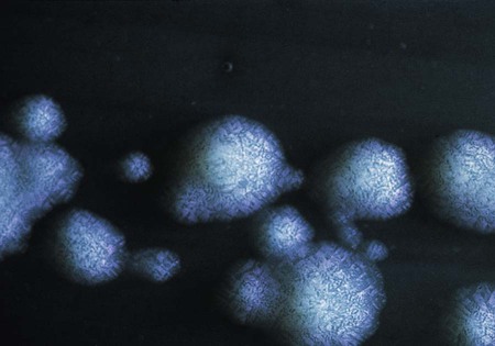
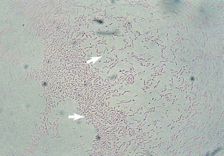
L. pneumophila grows at a temperature range from approximately 20° to 42° C. Plates are typically incubated in a candle jar at the optimal temperature of 35° to 37° C in a humid atmosphere. Some Legionella spp. may be stimulated by increased 2% to 5% concentration of CO2, including L. sainthelensi and L. oakridgensis. The low level of CO2 will not prevent the growth of L. pneumophila. If this concentration is not possible, incubation in air is preferable to 5% to 10% CO2, which may inhibit some legionellae, specifically L. pneumophila. Within 3 to 4 days, colonies should be visible. Plates are held for a maximum of 2 weeks before they are discarded. Blood cultures in biphasic media should be held for 1 month. At 5 days, colonies are 3 to 4 mm in diameter, gray-white to blue-green, glistening, convex, and circular and may exhibit a cut-glass type of internal granular speckling (Figure 35-3). A Gram stain yields thin, gram-negative bacilli (Figure 35-4).

Approach to Identification

Because Legionella spp. are biochemically inert and many tests produce equivocal results, extensive biochemical testing is of little use. Definitive identification requires the facilities of a specialized reference laboratory. Suspect colonies should be Gram stained to determine if bacteria are small to filamentous, gram-negative rods. Colonies should be plated to two media, including a BYCE plate containing L-cysteine and one made without. L. pneumophila will only grow on the BYCE L-cysteine media, providing a more definitive identification. In addition, if only a small amount of growth is present on the primary medium, the growth may be emulsified in sterile water and used for subculturing, staining, and serologic identification. Once the isolate has been determined to be L-cysteine dependent, further identification is completed using serotyping. L. pneumophila spp. can be identified using a monoclonal immunofluorescent stain (Genetic Systems Corporation). Emulsions of organisms from isolated colonies are made in 10% neutral formalin, diluted 1:100 (to produce a very thin suspension), and placed on slides for fluorescent antibody staining. Clinical laboratories probably perform sufficient service to clinicians by indicating the presence of Legionella spp. in a specimen. Serologic typing is a simple method and should provide sufficient characterization. If further identification is necessary, the isolate should be forwarded to an appropriate reference laboratory.

Serodiagnosis

Most patients with legionellosis have been diagnosed retrospectively by detection of a fourfold rise in anti-Legionella antibody with an indirect fluorescent antibody (IFA) test. Serum specimens should be tested no closer than 2 weeks apart. Diagnostic efficacy associated with serologic testing increases with the collection and testing of acute and convalescent paired sera. Convalescent sera should be collected at 4, 6, and 12 weeks following the appearance of the disease. Disease is confirmed by a fourfold rise in titer to more than 128. A single serum with a titer of more than 256 and a characteristic clinical picture may be presumptive for legionellosis; however, because as many as 12% of healthy persons yield titers as high as 1:256, this practice is strongly discouraged. Unfortunately, individuals with Legionnaires’ disease may not exhibit an increase in serologic titers until as long as 10 weeks after the primary illness or they may never display significant antibody titer increases. It is essential to correlate serologic findings with the patient’s clinical presentation because of the variation in antibody response associated with legionellosis. Most patients will develop a classic IgM, IgG, and IgA response. However, some patients may only develop antibodies for a single class (in other words, IgG, IgM, or IgA only). Commercially prepared antigen-impregnated slides for IFA testing are available from numerous suppliers.

Prevention

Although under development, a vaccine against Legionella infections is not currently available. The effectiveness of other approaches to the prevention of Legionella infections, such as the elimination of its presence from cooling towers and potable water, is uncertain.

Chapter Review

1. Legionella can be spread by all of the following except?

3. Which of the following agars should be used for culturing Legionella?

4. What is the specimen of choice for isolating Legionella?

5. Which of the following is acceptable for therapy?

6. Legionella injects proteins into the host cell by:

7. Legionella can be definitively diagnosed by a:

8. Which of the following is not a characteristic of Legionella?

9. All of the following are true of Legionella except:

10. True or False

11. Matching:

image

Advanced Case Study 35-2
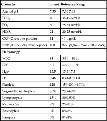
A 78-year-old male retired executive presented to the clinic with a 3- to 5-day illness that consisted of headache and diarrhea. Over-the-counter remedies and hydration were recommended as empiric therapy for viral gastroenteritis. Two days later his wife drove him to the clinic, as the man was too weak to drive. She reported that her husband had become confused and had a high fever for the past 24 hours. In addition, he had developed a dry cough and was complaining of feeling short of breath. The patient has a previous medical history of hypertension and dyslipidemia. He is a former smoker but has no structural lung disease and no history of heart failure.

Physical examination revealed a temperature of 102.5° F, blood pressure of 110/65, a pulse of 110 bpm, a respiratory rate of 26 breaths per minute, and an oxygen saturation of 86%. A lung exam revealed crackles bilaterally in both left and right lungs. There were no signs of heart murmur or cyanosis. The patient had no other significant physical findings. Chest x-ray revealed diffuse pulmonary infiltrates bilaterally. The patient was admitted to the hospital with a provisional diagnosis of pneumonia and potentially HINI influenza. The following laboratory results were obtained:

Laboratory Results

Chemistry Patient Reference Range
Arterial pH 7.35 7.35-7.45
PCO2 40 35-45 mmHg
PO2 60 75-85 mmHg
HCO3 24 20-25 mmol/L
CRP (C reactive protein) 12 <1 mg/dL
BNP (B type natriuretic peptide) 100 9-86 pg/mL (male 75-83 years)
Hematology
WBC 14 5-10 × 109/L
RBC 5.11 5-6 × 10¹²/L
Hgb 15.5 13.5-17.5
Hct 0.46 0.41-0.53 L/L
Platelets 120 150-400 × 109/L
Segmented neutrophils 85% 25%-60%
Lymphocytes 13% 20%-50%
Monocytes 2% 2%-11%
Eosinophils 0% 0%-8%
Basophils 0% 0%-2%

image

Questions

1. Evaluate the laboratory results as presented. Are there any unusual indicators here or in the patient’s history that would signify a predisposition for unusual respiratory infections?

2. What additional laboratory tests would be indicated at this time?

3. Following hospitalization, the patient continued to demonstrate a low sodium level of 120 to 123 (reference range 135-145 mEq/L) despite rehydration efforts. His diarrhea and headache resolved by day 6 of his hospitalization, but his hypoxia increased. He was subsequently intubated and placed on mechanical ventilation for respiratory failure and continued to have a markedly elevated temperature of up to 104.3° F despite antibiotic treatment with cefotaxime and azithromycin. Repeat chest x-ray indicated an increase in infiltrates. His antibiotic treatment was broadened to include fungal and anaerobic antimicrobial agents, and the patient was placed in respiratory isolation. The following serologic test results were obtained: Influenza Rapid Antigen Test—negative; TB skin test—negative, sputum by endotracheal suction for acid-fast bacteria—negative; Mycoplasma IgM <1:16 (negative); Legionella Urine Antigen Test—positive.