CHAPTER 23 LASIK patient evaluation and selection
History
Contact lens (CL) wearers
Naturally, ophthalmic examination should always be complete and meticulous and the results recorded.
Ocular contraindications
Systemic contraindications


Examination
Best corrected visual acuity (BCVA) – manifest refraction
No laser keratorefractive surgery should be performed without assessing the cornea with topography. Corneal topography and aberrometry are essential preoperatively and Chapter 1 of this book is dedicated to this subject. Contrast sensitivity testing is not usually routinely assessed but used only for academic purposes.
Ocular motility
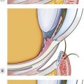
Postoperative ptosis3 has been reported and, according to various studies, is in the range of 0.4%. It is usually temporary and is caused by prolonged use of steroids, usually after PRK. Should the patient present with any ptosis preoperatively, photos should be taken and kept on file for medicolegal reasons.
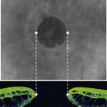
The pupil
It is always preferable that the laser ablation zone covers the scotopic pupil, as it is best measured with the automated infrared scotopic pupillometry, e.g. procyon pupillometer. The Zywave wavefront sensor with the fixating target turned off using the study settings and light conditions provided measurements of scotopic pupil diameter that were closest to the reference values (Procyon). With other devices (Colvard pupillometer, Zywave aberrometer with the fixating target switched on, Wasca aberrometers and Orbscan topographer), the difference was statistically significant4. When small ablation zones, i.e. those not sufficiently covering the scotopic pupil, are used, the prismatic effects of the reshaped corneal optics are liable to induce postoperative glare and halo phenomena.
Schirmer test
Schirmer testing is performed to avoid treating cases with xerophthalmia, something that may delay healing. It is important both in PRK5,7 and in LASIK6,7. However, some preservatives used in drugs (even artificial tears) following LASIK may accentuate symptoms of xerophthalmia. The patient should be aware that artificial tears may be needed in excess of 3–6 months postoperatively and that, in some cases, permanent plugs may be required.
Intraocular pressure measurement (IOP)
Following PRK or LASIK, the gold-standard Goldmann applanation tonometer (GAT) underestimates the IOP. Glaucoma is a vision-threatening disease and diagnosis may be missed in a patient who has undergone LASIK. Currently, it seems that the Pascal dynamic contour tonometer8 (PDCT) is the only tonometer that accurately records the IOP in post-LASIK or -PRK eyes. Its measurement is not affected by corneal curvature or thickness. Corneal abnormalities seem to influence GAT IOP measurements more than PDCT IOP measurements9.
Decision making
Amount of refractive error – limits of LASIK
Despite the patient’s eyes being eligible for LASIK surgery, i.e. normal topography, adequate corneal thickness, small scotopic pupil, there is a general trend to lower the limits of keratorefractive surgery. Many surgeons post their upper limit to as low as −6.0 D for myopia and +4.0 D for hyperopia, suggesting alternative techniques (phakic IOLs or clear lensectomy10,11) for higher errors.
Informed consent
The informed consent form should contain at least the following:
The patient should understand the possibility of:
The consent should never be signed with the patient under cycloplegia.
1 Smith RJ, Maloney RK. Laser in situ keratomileusis in patients with autoimmune diseases. J Cataract Refract Surg. 2006;32(8):1292-1295.
2 Kohnen T. Excimer laser refractive surgery in autoimmune diseases (editorial). J Cataract Refract Surg. 2006;32(8):1241.
3 Loewenstein A, Lipshitz I, Varssano D, et al. Complications of excimer laser photorefractive keratectomy for myopia. J Cataract Refract Surg. 1997;23(8):1174-1176.
4 Kohnen T, Terzi E, Kasper T, et al. Correlation of infrared pupillometers and CCD-camera imaging from aberrometry and videokeratography for determining scotopic pupil size. J Cataract Refract Surg. 2004;30(10):2116-2123.
5 Siganos DS, Popescu CN, Siganos CS, et al. Tear secretion following spherical and astigmatic excimer laser photorefractive keratectomy. J Cataract Refract Surg. 2000;26(11):1585-1589.
6 Siganos DS, Popescu CN, Siganos CS, et al. Tear secretion following excimer laser in situ keratomileusis. J Refract Surg. 2002;18(2):124-126.
7 Lee JB, Ryu CH, Kim J, et al. Comparison of tear secretion and tear film instability after photorefractive keratectomy and laser in situ keratomileusis. J Cataract Refract Surg. 2000;26(9):1326-1331.
8 Siganos DS, Papastergiou GI, Moedas C. Assessment of the Pascal dynamic contour tonometer in monitoring intraocular pressure in unoperated eyes and eyes after LASIK. J Cataract Refract Surg. 2004;30(4):746-751.
9 Papastergiou GI, Kozobolis V, Siganos DS. Assessment of the pascal dynamic contour tonometer in measuring intraocular pressure in keratoconic eyes. J Glaucoma. 2008;17(6):484-488.
10 Siganos DS, Siganos CS, Pallikaris IG. Clear lens extraction and intraocular lens implantation in normally sighted hyperopic eyes. J Refract Corneal Surg. 1994;10(2):117-121. discussion 122–4
11 Siganos DS, Pallikaris IG. Clear lensectomy and intraocular lens implantation for hyperopia from +7 to +14 diopters. J Refract Surg. 1998;14(2):105-113.