CHAPTER 24 Laser technology (excimer and femto)
Introduction
The interaction of the excimer laser with corneal tissue, which was initially described in early 1980s, is the fundamental aspect of refractive surgery. Corneal photoablation with excimer laser is a safe and effective technique for the modification of corneal curvature through tissue removal in order to achieve refraction changes1,2. The latest evolution in laser refractive surgery is the use of femtosecond laser technology, which is capable of creating cuts with high precision in the corneal stroma and replaces the use of the blade in several applications of corneal surgery. Both laser technologies, excimer and femtosecond, are commercially available with platforms created to fit the current standards of refractive surgery practice.
Basic features of laser devices
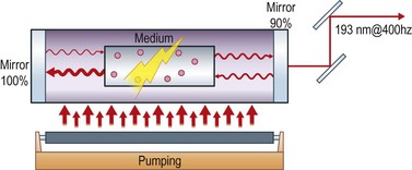
A laser device consists of three fundamental elements: the optical cavity, the gain medium, and the pumping system (Fig. 24.1). The optical cavity is an arrangement of mirrors which allows the light to oscillate within it. An optical cavity can be created by two oppositely placed plane or concave mirrors. The gain medium is the material in which the process of stimulated emission takes place. It can be gas, solid, liquid, or semiconductor, and the critical properties of the emitted laser radiation depend on the material used. The gain medium generates and amplifies the radiation that travels through it guided by the mirrors of the optical cavity. The pumping system provides the necessary energy for the amplification of the laser radiation. Pumping can be either optical or electrical.
Interaction of laser with ophthalmic tissues
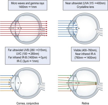
There are three main types of laser tissue interaction that are utilized in ophthalmology. The thermal effect of laser radiation is used to treat retinal tissue in photocoagulation. In photoablation the photochemical effect of laser radiation is used to remove corneal tissue with high accuracy. In photodisruption, the effect of photoionization is used to create plasma inside ophthalmic tissue and thus create cuts such as in YAG laser capsulotomy and in corneal surgery applications of femtosecond lasers3. Each type of effect takes place with different laser applications depending on several laser radiation properties such as the absorbance of the wavelength by the tissue, the pulse energy and duration (Fig. 24.2).
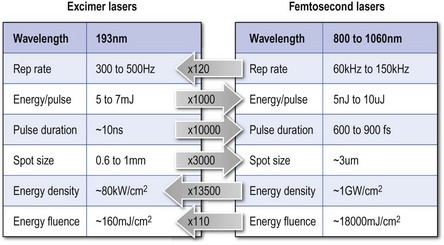
Excimer lasers
Concurrent effects of excimer laser energy
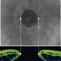
One of the important side effects of the laser radiation is the collateral thermal damage. It is caused by the direct absorption of laser radiation with energy lower than the ablation threshold by tissue surrounding the ablation, and also by thermal energy deriving by the rapidly moving splinters of collagen. It has been shown that the amount of thermal damage is related with haze formation after photorefractive keratectomy (PRK). The local rise of temperature in the tissue surrounding the ablation is higher when ablation is repeated in the same area with a high frequency. Due to this fact, lower repetition rate lasers are more suitable for PRK than high repetition rate lasers4. In modern devices each pulse targets a different position than the previous, and the incidence of subsequent pulses in the same point is avoided.
Femtosecond lasers
The laser radiation features when it reaches the corneal surface are: a pulse duration of a few hundred femtoseconds, repetition rate of up to 120 kHz, pulse energy of a few microjoules, and wavelength in the near infrared region (1053 nm). These parameters may differ between the commercially available femtosecond laser units (Fig. 24.3).
Advantages of femtosecond lasers in corneal surgery
Currently, in refractive surgery femtosecond lasers are used for flap creation in LASIK, pocket creation for inlay insertion, and other applications such as sutureless anterior lamellar keratoplasty, astigmatic keratotomy, diagnostic corneal biopsy, anterior lamellar corneal staining-tattooing, and intrastromal vision correction5–10. In keratoplasty applications, these devices are able to create several customized patterns of incisions. Commercially available devices are capable of controlling the spot size, pulse energy, separation, and position of pulse placement. Therefore they can create precise cuts of almost any possible geometry within the cornea.
1 Trokel SL, Srinivasan R, Braren B. Excimer laser surgery of the cornea. Am J Ophthalmol. 1983;96(6):710-715.
2 Munnerlyn C, Koons S, Marshall J. J Cataract Refract Surg. 1988:14.
3 Krauss JM, Puliafito CA. Lasers in ophthalmology. Lasers Surg Med. 1995;17(2):102-159.
4 Kymionis GD, Diakonis VF, Kounis G, et al. Effect of excimer laser repetition rate on outcomes after photorefractive keratectomy. J Cataract Refract Surg. 2008;34(6):916-919.
5 Soong HK, Malta JB. Femtosecond lasers in ophthalmology. Am J Ophthalmol. 2009;147(2):189. 197.e2
6 Coskunseven E, Kymionis GD, Tsiklis NS, et al. One-year results of intrastromal corneal ring segment implantation (KeraRing) using femtosecond laser in patients with keratoconus. Am J Ophthalmol. 2008;145(5):775. 779.e1
7 Kymionis GD, Ide T, Galor A, et al. Femtosecond-assisted anterior lamellar corneal staining-tattooing in a blind eye with leukocoria. Cornea. 2009;28(2):211-213.
8 Kymionis GD, Yoo SH, Ide T, et al. Femtosecond-assisted astigmatic keratotomy for post-keratoplasty irregular astigmatism. J Cataract Refract Surg. 2009;35(1):11-13.
9 Yoo SH, Kymionis GD, Koreishi A, et al. Femtosecond laser-assisted sutureless anterior lamellar keratoplasty. Ophthalmology. 2008;115(8):1303. 1307.e1
10 Yoo SH, Kymionis GD, O’Brien TP, et al. Femtosecond-assisted diagnostic corneal biopsy (FAB) in keratitis. Graefe’s Arch Clin Exp Ophthalmol. 2008;246(5):759-762.