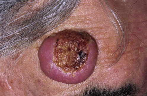
Keratoacanthoma

Published on 16/03/2015 by admin

Filed under Dermatology

Last modified 16/03/2015

Print this page

rate 1 star rate 2 star rate 3 star rate 4 star rate 5 star
Your rating: none, Average: 0 (0 votes)

This article have been viewed 1472 times

Keratoacanthoma

Caroline M. Owen and Nicholas R. Telfer

Evidence Levels:  A Double-blind study  B Clinical trial ≥ 20 subjects  C Clinical trial < 20 subjects  D Series ≥ 5 subjects  E Anecdotal case reports

image

Keratoacanthoma (KA) is a distinctive tumor with characteristic histopathologic and clinical features. Classically, it presents as a rapidly proliferating, firm, dome-shaped, crateriform nodule. It shares many features of a squamous cell carcinoma (SCC), but is self-healing and may be considered a form of aborted malignancy. Although eventually self-limiting, its growth can be unpredictable and locally destructive, causing problems in management. Lesions are usually solitary and mainly affect sun-exposed sites in patients of middle age and older. Less commonly, lesions may be large (giant KA), progressive with central clearing (KA centrifugum marginatum), or multiple (e.g., multiple KA of Ferguson–Smith type, generalized eruptive KA of Grzybowski). KA may arise in tattoos (especially red ink), is a recognized feature of Muir Torre syndrome and may occur in eruptive form in patients receiving the RAF-inhibitor (sorafenib) and BRAF-inhibitor (vemurafenib) therapy for solid tumours.

Management strategy

Initial management is directed at making an accurate diagnosis of KA, in particular differentiating it from SCC. Although much research has been directed at the investigation of the differential cytokine profile or ultrastructure of KA and SCC, in practice the diagnosis of KA relies on the correlation between clinical and histopathologic criteria as follows.

A KA classically has three clinical stages:

The five most relevant histopathologic criteria when differentiating KA from SCC have been shown to be:

Accurate assessment of these histopathologic criteria relies on the provision of an adequate histologic specimen. This should be deep enough to include subcutaneous fat, either by total excision or by wedge or transverse biopsy through the center of the lesion.

Once the diagnosis of KA has been established, the aims of treatment are to hasten resolution, prevent destruction of important structures, prevent recurrence, and obtain as favorable a cosmetic outcome as possible. Management therefore depends on the type of KA, its site, its rate of growth, and whether or not it has previously been treated.

In general, excision is the treatment of choice for a small solitary KA because this will remove the lesion, provide an adequate specimen for histologic examination, and generally give a good cosmetic result. An alternative to this is incisional biopsy to provide an adequate histologic specimen, followed by curettage or blunt dissection of the residual lesion.

If one is confident of the diagnosis and the lesion has been documented as being static or already starting to involute, then observation is an option. Obviously, any concern that the lesion is not following an involutional pattern makes treatment mandatory.

Recently, topical photodynamic therapy (PDT) has been used with success. Intralesional methotrexate, topical 5-fluorouracil, topical imiquimod and (now out-dated) argon laser ablation have also been shown to be effective.

Large proliferating lesions for which surgical management is not possible or is likely to result in a poor cosmetic outcome can be treated with radiotherapy, intralesional methotrexate, or intralesional 5-fluorouracil. More recently, successful treatment using intralesional interferon-α has been reported. Intralesional methotrexate has also been used to reduce the size of KA lesions prior to excision.

Multiple KAs have been successfully treated with oral retinoids, intralesional 5-fluorouracil and, more recently, using the epidermal growth factor receptor inhibitor erlotinib.

Recurrent KA has been shown to respond well to radiotherapy, but oral retinoids have also been used with success. The multiple KA and KA-like lesions induced by RAF- and BRAF-inhibitor therapy will spontaneously resolve following cessation or completion of therapy, and topical PDT has been used to palliate the lesions, allowing treatment to be continued.

Keratoacanthoma centrifugum marginatum is often difficult to treat, although there have been recent case reports of successful treatment using topical 5-fluorouracil and oral erlotinib.

Generally, the level of evidence in the literature for the treatment of KA is poor, consisting mainly of small case series. Without a control group it is very difficult to be sure of the success of any intervention, particularly with a lesion that has a natural history of spontaneous involution.