[level-membership-for-dermatology-category]
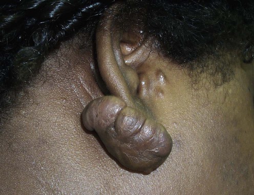
Keloids

First-line therapies
 Intralesional corticosteroids
 Compression
 Occlusive dressing
 Intralesional 5-fluorouracil
Second-line therapies
 Intralesional interferon-α2b
 Cryosurgery
 Radiation
Third-line therapies
 Laser surgery
 Imiquimod
 Mitomycin C
 Intralesional interferon-γ
 Topical retinoic acid
 Intralesional bleomycin
 Verapamil
 Surgery
[/level-membership-for-dermatology-category][not-level-membership-for-dermatology-category]
Keloids
First-line therapies
Buy Membership for Dermatology Category to continue reading. Learn more here
[/not-level-membership-for-dermatology-category]
