Published on 19/03/2015 by admin
Filed under Dermatology
Last modified 22/04/2025
This article have been viewed 2076 times
Ranon Mann, Adam Friedman and Adam Wulkan
Evidence Levels: A Double-blind study B Clinical trial ≥ 20 subjects C Clinical trial < 20 subjects D Series ≥ 5 subjects E Anecdotal case reports
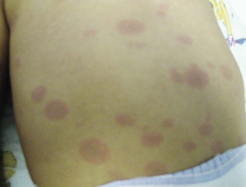
Kawasaki disease (KD), seen primarily in infants and children, is an acute febrile multiorgan vasculitic process well known for its mucocutaneous and nodal involvement. Although KD was described in Japan nearly 50 years ago, its pathogenesis has yet to be clarified. Increasing evidence supports an infectious etiology.
The primary goal when treating a patient with KD is the prevention of cardiac complications, including coronary artery disease, aneurysm formation, myocardial infarction, and even sudden death. The mainstay of therapy has for years been the use of high-dose salicylates (e.g., aspirin) and intravenous γ-globulin (IVIG) in order to manage the acute, intense, inflammatory characteristics of this condition and to prevent the aforementioned cardiovascular sequelae.
In the acute inflammatory phase, aspirin, a potent inhibitor of prostaglandin synthesis, is initially dosed at 80–100 mg/kg/day orally, divided four times per day for 2 weeks. After the patient is afebrile for 48 hours, the dose is reduced to 3–5 mg/kg orally daily and continued for 6 to 8 weeks until the erythrocyte sedimentation rate (ESR) and platelet count normalize. Multiple trials have failed to elucidate the benefit of high-dose aspirin therapy with regards to outcomes; therefore, current evidence is insufficient to support the use of salicylates as an integral component of first-line therapy in KD.
IVIG is a known first-line therapy in the treatment of KD. IVIG, which can lead to rapid defervescence, is able to neutralize circulating myelin antibodies and to downregulate proinflammatory cytokines, including interferon-γ (INF-γ). The pediatric dosing, which is equivalent to the adult dosing, is 400 mg/kg/day intravenously over 2 hours as a single daily infusion for 4 consecutive days, or alternatively (and apparently more efficaciously) a single dose of 2 g/kg intravenously infused over 12 hours. Failure of IVIG therapy has been linked to a gene polymorphism in the plasma platelet-activating factor acetylhydrolase. No specific regimen has been assigned for these non-responding cases.
Laboratory examination: ESR, complete blood count + platelet count, lactate dehydrogenase
Multi-detector CT
Echocardiography
Pinna GS, Kafetzis D, Tselkas O, Skevaki CL. Curr Opin Infect Dis 2008; 21: 263–70.
Echocardiography, stress imaging, angiography, MRI, and ultrafast computed tomography (CT) scans have been useful in the diagnosis of coronary aneurysms, occlusions, and stenosis. Echocardiography is recommended both at the time of diagnosis and after 2 to 6 weeks. It has recently been demonstrated that multi-detector CT is preferable to transthoracic echocardiography or MRI. CT can detect calcification and estimate soft plaques, offer rapid data collection and simple interpretation of images, all of which serve as an advantage over other diagnostic modalities. Conversely, MRI cannot offer rapid capture of images, thus prolonging the time under anesthesia and its associated risks. Transthoracic echocardiography can only image the proximal arteries and therefore cannot reliably detect stenosis.
Royle J, Burgner D, Curtis NJ. Pediatr Child Health 2005; 41: 87–93.
These guidelines highlight the difficulties in the diagnosis of KD. A meta-analysis of recent data offers insight that may assist in the early recognition of this important pediatric disease. The clinical features of KD are common to many other childhood illnesses, and therefore the diagnostic criteria are not highly sensitive. Blood serologies and chemistries may be helpful, but none is diagnostic and most have a low specificity. The echocardiogram should not be used as a diagnostic test. A normal echo does not exclude KD, as coronary lesions generally occur in the convalescent phase and may develop as late as 6 to 8 weeks after the onset of fever. Because there is no specific diagnostic test for KD, increased awareness of the epidemiology and the spectrum of clinical presentation is essential for early recognition and optimal management.
Kuo HC, Yang KD, Chang WC, Ger LP, Hsieh KS. Pediatr Neonatol 2012; 53: 4–11.
This review provides an update on the various treatment modalities available for KD, including methylprednisolone, TNF-α antagonists, statins, plasma exchange, and cytotoxic agents. It concludes that high-dose aspirin does not appear to decrease the incidence of coronary artery lesions, although further studies are required.
Treating KD refractory to IVIG is a therapeutic challenge. Recent studies have shown associations of age, platelet count, ESR, hemoglobin, C-reactive protein (CRP), eosinophil count, LDH, albumin, and alanine aminotransferase (ALT) with failure of primary treatment with IVIG. Because IVIG failure has an increased risk of coronary artery lesions, it is essential to treat such patients with a second dose of IVIG, intravenous methylprednisolone, or a TNF-α inhibitor.
Tremoulet AH, Best BM, Song S, Wang S, Corinaldesi E, Eichenfield JR, et al. J Pediatr 2008; 153: 117–21.
IVIG treatment for the acute stage of KD has shown to be effective and safe. However, it is known that 10–20% of patients are resistant to initial therapy (2 g/kg IV). These patients are at increased risk for the development of coronary artery abnormalities. Using demographic and laboratory information on IVIG-resistant cases, this retrospective study attempted to develop a scoring system to help identify future resistant cases among patients in San Diego County. This would perhaps indicate the need for secondary therapies early in the treatment of these patients. Unfortunately, the diversity of the patient population did not allow for the development of an accurate and clinically useful scoring system.
Uehara R, Belay ED, Maddox RA, Holman RC, Nakamura Y, Yashiro M, et al. Pediatr Infect Dis J 2008; 27: 155–60.
Some KD patients do not respond to initial treatment with IVIG. The purpose of this study was to determine potential risk factors associated with IVIG non-response among KD patients in Japan. The results emphasize that physicians should consider IVIG non-response in patients with recurrent KD, as well as in KD patients diagnosed and treated before the fifth day of illness who continue to have laboratory values associated with non-response, such as low platelet count, elevated ALT and CRP. These patients may benefit from the administration of a second-line treatment early during the illness in addition to the initial IVIG treatment.
Hsieh KS, Weng KP, Lin CC, Huang TC, Lee CL, Huang SM. Pediatrics 2004; 114: 689–93.
In North America, high-dose aspirin (80–100 mg/kg/day orally) is widely used during the acute phase of KD. However, the necessity of this therapy has yet to be elucidated. This study indicated that treatment in the acute stage of KD without aspirin had no effect on the response rate of IVIG therapy, duration of fever, or incidence of coronary abnormalities. This response was seen when children were treated with high-dose (2 g/kg) IVIG as a single infusion, regardless of whether treatment was commenced before or after day 5 of illness. Therefore, the available data show no appreciable benefit of aspirin in preventing IVIG non-response, aneurysm formation, or shortening of fever duration.
Kuo HC, Wang CL, Liang CD, et al. 6th Asian Society of Pediatric Research, Taipei, Taiwan. 2010.
In this Taiwanese study involving 609 KD patients from two medical centers, patients were randomized to receive either high-dose aspirin (n = 274) or no aspirin (n = 335). This study found that there were no significant differences between the two groups when comparing white blood cell count, hemoglobin, platelets, or CRP levels both before and after IVIG treatment.
Kobayashi T, Saji T, Otani T, Takeuchi K, Nakamura T, Arakawa H. Lancet 2012; 379: 1613–20.
This multicenter, prospective, randomized trial took place at 74 hospitals in Japan between 2008 and 2010. Individuals with severe KD were randomized to receive either current standard of care IVIG (2 g/kg/day given over 24 hours) and aspirin (30 mg/kg/day) or IVIG, and aspirin (same dose as control) plus prednisolone (2 mg/kg/day with 15-day taper after normalization of CRP). There were 125 subjects assigned to the intervention group and 123 that received only IVIG. The study found that the incidence of coronary artery lesions was significantly decreased in the intervention arm with intravenous prednisolone (four patients) versus the control arm (28 patients). Because this study was performed in Japan, larger studies involving individuals from various ethnic backgrounds are needed.
Furukawa T, Kishiro M, Akimoto K, Nagata S, Shimizu T, Yamashiro Y. Arch Dis Child 2008; 93: 142–6.
In this non-randomized study, the effectiveness of intravenous methylprednisone (IVMP) was compared with that of additional IVIG (2 g/kg 12–24 hours) and aspirin (30 mg/kg/day) as second-line therapy for KD. Fever was rapidly alleviated after IVMP administration (30 mg/kg/day for 3 consecutive days with heparin infusion 10–20 U/kg/h) in all IVIG-resistant patients in the study; 77% recovered without recurrence of KD, and did not develop coronary artery aneurysms. The findings suggested that early IVMP treatment in IVIG-resistant patients is as effective as treatment with additional IVIG as second-line therapy.
Muta H, Ishii M, Furui J, Nakamura Y, Matsuishi T. Acta Paediatr 2006; 95: 189.
The goal of this study, using data from a nationwide survey in Japan, was to identify the characteristics of patients who needed retreatment with IVIG. Elevated ESR, anemia, and high LDH are known predictive values for necessitating IVIG re-treatment. In this study, male gender, incomplete and recurrent cases of KD, and treatment with IVIG at a dose of 1 g/kg or less within 4 days of illness onset, were identified as independent risk factors associated with the need for re-treatment. Identification of these risk factors would be beneficial to predict which patients might need IVIG re-treatment. This could help physicians to initially create a strategy to prevent cardiovascular complications in these patients.
Suzuki H, Terai M, Hamada H, Honda T, Suenaga T, Takeuchi T, et al. Pediatr Infect Dis J 2011; 30: 871–6.
In this trial involving 28 individuals who failed IVIG therapy, cyclosporine A (CsA), 4–8 mg/kg/day oral administration, was shown to be a safe, well tolerated, and efficacious option. Of the 28 treated with CsA, 18 were afebrile within 3 days of therapy, four were afebrile within 4 to 5 days, and the remaining six failed to respond to treatment.
Raman V, Kim J, Sharkey A. Pediatr Infect Dis J 2001; 20: 635–7.
A case of aggressive and protracted KD with coronary aneurysms, myocarditis, pericarditis, and valvular insufficiency, despite repeated administration of IVIG, responded to combination therapy with pulse and high-dose corticosteroids and CsA (3 mg/kg/day divided into three administrations).
Kanai T, Ishiwata T, Kobayashi K, Sato H, Takizawa M, Kawamura Y, et al. Circulation 2011; 124: 2822–8.
This is a retrospective study comparing 369 patients with KD who were treated with a combination of ulinastatin (UTI) at 15 000 U/kg/day divided into three doses, aspirin (30 mg/kg/day) and IVIG (1–2 g/kg) versus 1178 patients treated with IVIG and aspirin. Those treated in the UTI group experienced fewer CALs (coronary artery lesions) (3%) than those in the control group (7%). Thus, treatment with UTI was associated with fewer rescue treatments and coronary artery lesions.
Son MB, Gauvreau K, Burns JC, Corinaldesi E, Tremoulet AH, Watson VE, et al. J Pediatr 2011; 158: 644–9.
This retrospective study performed between 2000 and 2008 assessed fever duration and coronary artery dimensions in patients refractory to IVIG therapy, who subsequently received either infliximab (5 mg/kg) or additional IVIG (2 g/kg). Individuals treated with infliximab experienced decreased fever duration, shorter hospitalization, yet similar coronary artery dimensions, when compared to the control group.
Sauvaget E, Bonello B, David M, Chabrol B, Dubus JC, Bosdure E. J Pediatr 2012; 160: 875–6.
This is a case of a 6-year-old boy with KD refractory to IVIG and systemic steroids successfully treated with rituximab (15 mg/kg/day), an anti-CD20 monoclonal antibody, which was initiated on day 20 of disease onset. Within 2 days of rituximab therapy, the child’s fever had dissipated and the echocardiography revealed improvement of the CALs.
Hokosaki T, Mori M, Nishizawa T, Nakamura T, Imagawa T, Iwamoto M, Yokota S. Pediatr Int 2012; 54: 99–103.
This is a retrospective study involving 125 patients with KD refractory to IVIG therapy treated with plasma exchange therapy (PE). The success of plasma exchange therapy was dependent on both the presence of CALs prior to plasma exchange and on what day of disease onset the plasma exchange therapy was initiated. CALs remained in 2.8% of individuals treated with PE when PE was initiated prior to day 9 of the onset of KD. For those with CALs in whom PE was started after day 9 of KD onset, sequelae remained 15% of the time. For the 105 patients in whom the coronary arteries were normal prior to PE initiation, none of them went on to develop CALs. Thus, plasma exchange therapy is optimal for refractory KD when used prior to the development of CALs.
Suda K, Kudo Y, Sugawara Y. Nippon Rinsho 2008; 66: 355–9.
To prevent coronary thrombosis in KD, long-term antithrombotic therapy using antiplatelet drugs such as aspirin, dipyridamole, ticlopidine, clopidogrel, and abciximab, with or without warfarin, is recommended by official guidelines.
Yamauchi H, Ochi M, Fujii M, Hinokiyama K, Ohmori H, Sasaki T, et al. J Nippon Med Sch 2004; 71: 279–86.
The authors studied 21 patients with KD and coronary complications who underwent coronary artery bypass grafting (CABG) over a 12-year period. They conclude that CABG is successful when completed shortly after the acute onset of disease.
Treatment of Skin Disease Comprehensive Therapeutic Strategies 4e
WhatsApp us


 Immunoglobulin
 Aspirin (acetylsalicylic acid)
 Corticosteroids
 Re-treatment with immunoglobulin
 Cyclosporin A
 Ticlopidine
 Pentoxifylline
 Ulinastatin
 Infliximab
 Plasma exchange
 Rituximab
 Dipyridamole
 Cardiac transplantation
 Coronary artery bypass grafting
