[level-membership-for-dermatology-category]
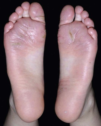
Juvenile plantar dermatosis

 Await spontaneous resolution
Second-line therapies
 Change to non-occlusive footwear
 Sport avoidance
 Emollients
 Topical corticosteroids
 Rotation of topical agents
 Topical tacrolimus
 Zinc oxide/impregnated bandages
 Bed rest/footwear avoidance
[/level-membership-for-dermatology-category][not-level-membership-for-dermatology-category]
Juvenile plantar dermatosis
[level-membership-for-dermatology-category]
[/level-membership-for-dermatology-category][not-level-membership-for-dermatology-category]
