Published on 19/03/2015 by admin
Filed under Dermatology
Last modified 22/04/2025
This article have been viewed 4094 times
Joanna E. Gach
Evidence Levels: A Double-blind study B Clinical trial ≥ 20 subjects C Clinical trial < 20 subjects D Series ≥ 5 subjects E Anecdotal case reports
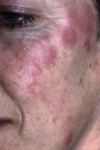
Jessner’s lymphocytic infiltrate (JLI) is a chronic inflammatory condition presenting with erythematous or reddish brown papules, annular or arciform plaques which can expand peripherally and sometimes develop central healing. The lesions are usually seen in adults and affect the face, neck, or upper trunk. Although they are frequently asymptomatic, some patients report itch or burning. The lesions can persist from weeks to years and disappear without sequelae, but may recur.
JLI runs a waxing and waning course marked by intermittent improvement and subsequent exacerbations, which makes the evaluations of therapeutic effectiveness difficult. Patients demand treatment because they find the lesions disfiguring or itchy.
Potent topical steroids applied once or twice daily for 4 weeks are the first-line treatment for many dermatologists. Unfortunately, the results of treatment are variable, and, most importantly, only short-lasting. If the response is inadequate, injection of intralesional corticosteroids into localized lesions or the use of potent topical steroid under occlusive dressing may be beneficial, but is associated with a greater risk of skin atrophy. Topical tacrolimus may be a safer alternative.
In cases where ultraviolet exposure has been reported to induce or exacerbate lesions, additional therapy with sunscreen may be needed. This group of patients may respond to antimalarials, in particular hydroxychloroquine.
A variety of other therapies have been reported to be effective in the management of JLI. Thalidomide, oral gold, and retinoids proved to be helpful in some cases, but their use may be limited by the adverse effects, which may be difficult for the patient and physician to accept, especially as JLI is harmless.
Many other treatments, including bismuth sub-salicylate injections, nicotinamide, vitamin E, phenindamine, para-aminobenzoic acid, penicillin, chlortetracycline, minocycline, dapsone, quinacrine (mepacrine), and radiotherapy, have been tried unsuccessfully.
Skin biopsy
Lesional skin for direct immunofluorescence
Antinuclear antibodies/extractable nuclear antigen
The diagnosis can be made on clinical grounds. The investigations are helpful in differentiating the condition from discoid lupus erythematosus, lupus erythematous tumidus, and cutaneous lymphoma.
Cerio R, Oliver GF, Jones EW, Winkelmann RK. J Am Acad Dermatol 1990; 23: 63–7.
Skin biopsy of the lesion shows normal epidermis and a moderate, dense sleeve-like perivascular and periadnexal infiltrate in the middle dermis. This consists of normal-looking lymphocytes with the B cells grouped in close proximity to the superficial vessels and T cells at the periphery, and occasional plasma cells.
Lipsker D, Mitschler A, Grosshans E, Cribier B. Dermatology 2006; 213: 15–22.
Immunofluorescence studies of lesional skin showed lupus band with IgG, IgM ± C3 in 9.5% of cases of JLI. Antinuclear antibodies were present in 45% of cases.
Toonstra J, Wildschut A, Boer J, Smeenk G, Willemze R, van der Putte SC, et al. Arch Dermatol 1989; 125: 1525–30.
JLI can coexist with polymorphic light eruption, and can evolve into discoid lupus erythematosus.
A series of 100 patients with JLI assessing clinical manifestations, duration, clinical course, and treatments. Forty-three of 91 patients treated with topical steroids had an excellent (n=13) or good (n=30) response, with temporary regression of the lesions. Many patients had no response.
Higgins CR, Wakeel RA, Cerio R. Br J Dermatol 1994; 131: 99–101.
A case report of an 11-year-old boy with JLI responding to intralesional corticosteroids.
Volden G. Tidsskr Nor Laegeforen 1992; 112: 1272–4.
JLI was among other chronic skin diseases that were treated once a week with clobetasol propionate lotion left under the completely occlusive hydrocolloid dressing Duoderm. The lesions cleared within a few weeks.
Tzung TY, Wu JC. Br J Dermatol 2005; 152: 383–4.
Tacrolimus 0.1% ointment twice daily produced an improvement after 2 weeks but had to be withdrawn because of diffuse erythema and desquamation that occurred 2 weeks later. The same effect occurred on rechallenge and settled with a single 5 mg dose of dexamethasone IM. Subsequently, pimecrolimus 1% cream cleared the rash within 6 weeks, with no relapse 2 months later.
Farrell AM, McGregor JM, Staughton RC, Bunker CB. Clin Exp Dermatol 1999; 24: 500.
Intramuscular injection of 40 mg Kenalog led to temporary improvement in this patient before oral gold was used successfully.
Antimalarial drugs (chloroquine sulfate and hydroxychloroquine sulfate) were used in 15 patients, with a good response in six. There were no details given regarding doses or treatment regimens.
Hydroxychloroquine 200 mg once or twice a day, or chloroquine 200 mg daily were used in other case reports.
Morgan J, Adams J. Br J Dermatol 1990; 122: 570.
One case of JLI was treated with etretinate 50 mg daily for 3 months and 25 mg daily for another 3 months. The rash subsided within 4 weeks of treatment and did not recur for 9 months after stopping treatment.
Isotretinoin 20 mg daily was used in other case reports, but was not helpful.
Park KY, Kim HK, Li K, Kim BJ, Seo SJ, Kim MN, et al. Clin Exp Dermatol 2012; 3: 235–7.
Remission was observed after two sessions of methyl 5-aminolevulinic acid cream photodynamic therapy with Aktilite 1 lamp, 630 nm.
Laurinaviciene R, Clemmensen O, Bygum A. Acta Derm Venereol 2009; 89: 542–3.
Oral methotrexate 15 mg weekly for 6 months produced clearance. No recurrence 2 years after withdrawal of the drug.
Borges da Costa J, Boixeda P, Moreno C. J Eur Acad Dermatol Venereol 2009; 23: 595–6.
Single treatment with 595 nm pulsed-dye laser cleared all lesions. New lesions were treated 2 months later.
Johansson EA. Dermatologica 1987; 174: 117–21.
Eight of the 15 patients treated with proquazone [1-isopropyl-4-phenyl-7-methyl-2(IH)] 600 mg daily healed completely after 1 to 2 months of treatment. Recurrence of the rash in two cases responded to 600 mg daily for 3 to 4 days, alternate months. Two further patients from among the initial responders showed a partial remission. Indometacin (50–75 mg daily) and ibuprofen (1200 mg daily) were tried, without effect.
A marked improvement within 3 weeks of starting auranofin 3 mg twice daily. This case was unresponsive to chloroquine, hydroxychloroquine, minocycline, dapsone, and isotretinoin. Monitoring of the therapy includes monthly full blood count for thrombocytopenia and urinalysis to detect nephritis.
Guillaume JC, Moulin G, Dieng MT, Poli F, Morel P, Souteyrand P, et al. Arch Dermatol 1995; 131: 1032–5.
Twenty of 27 patients achieved complete remission after 2 months of therapy with thalidomide 100 mg daily. Sixteen (59%) patients were in complete remission 1 month after stopping therapy. Side effects included sleepiness, constipation, dry mouth, and sensorimotor neuropathy in two patients.
Treatment of Skin Disease Comprehensive Therapeutic Strategies 4e
WhatsApp us


 Potent topical/intralesional corticosteroids
 Topical immunomodulators
 Systemic corticosteroids
 Antimalarials
 Retinoids
 Photodynamic therapy
 Methotrexate
 Laser therapy
 Proquazone
 Auranofin
 Thalidomide